[Preprint]. 2024 Jul 7:2024.07.05.602260.
doi: 10.1101/2024.07.05.602260.

Frequent transitions in self-assembly across the evolution of a central metabolic enzyme

Affiliations

Frequent transitions in self-assembly across the evolution of a central metabolic enzyme

Franziska L Sendker et al. bioRxiv. .

Update in

Abstract

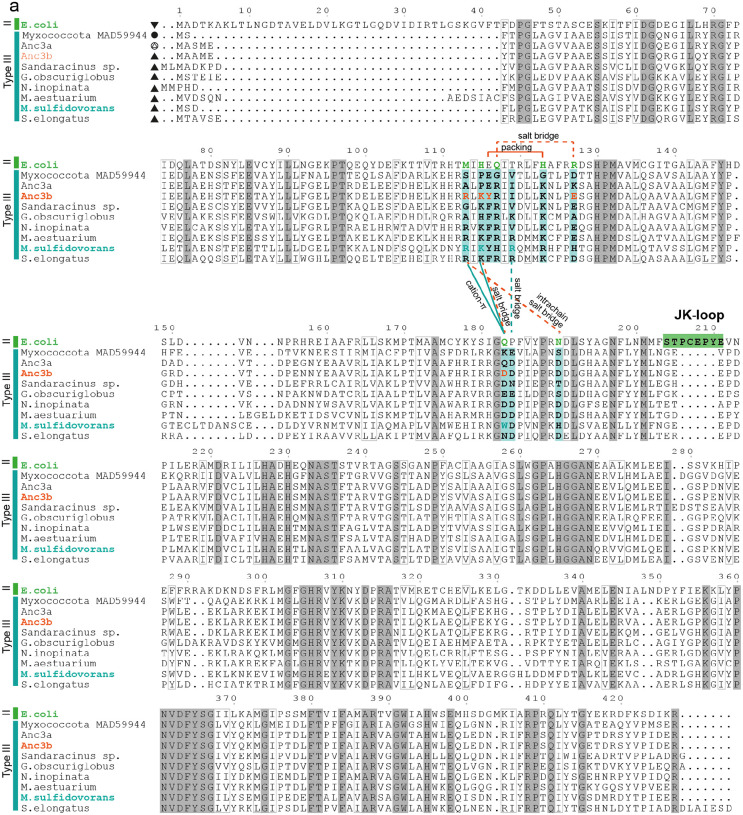
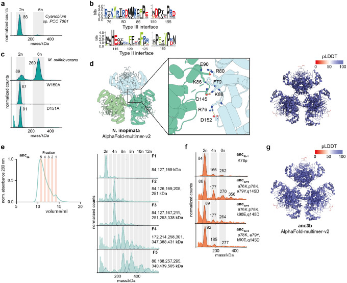
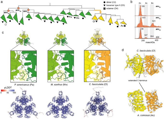
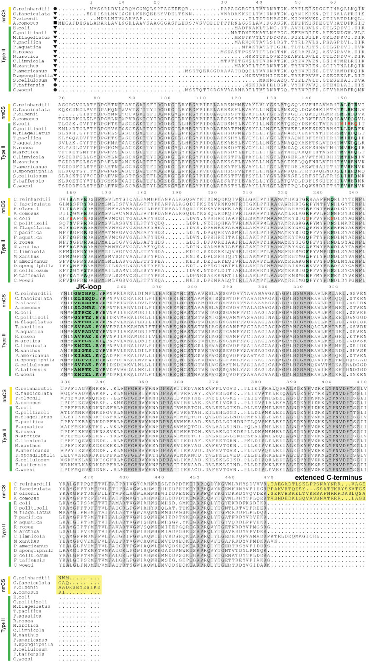
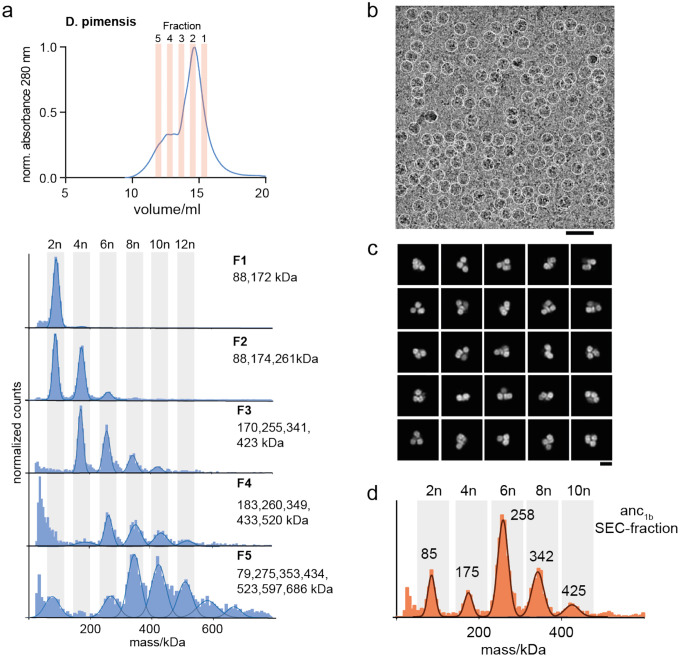
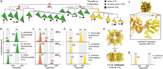
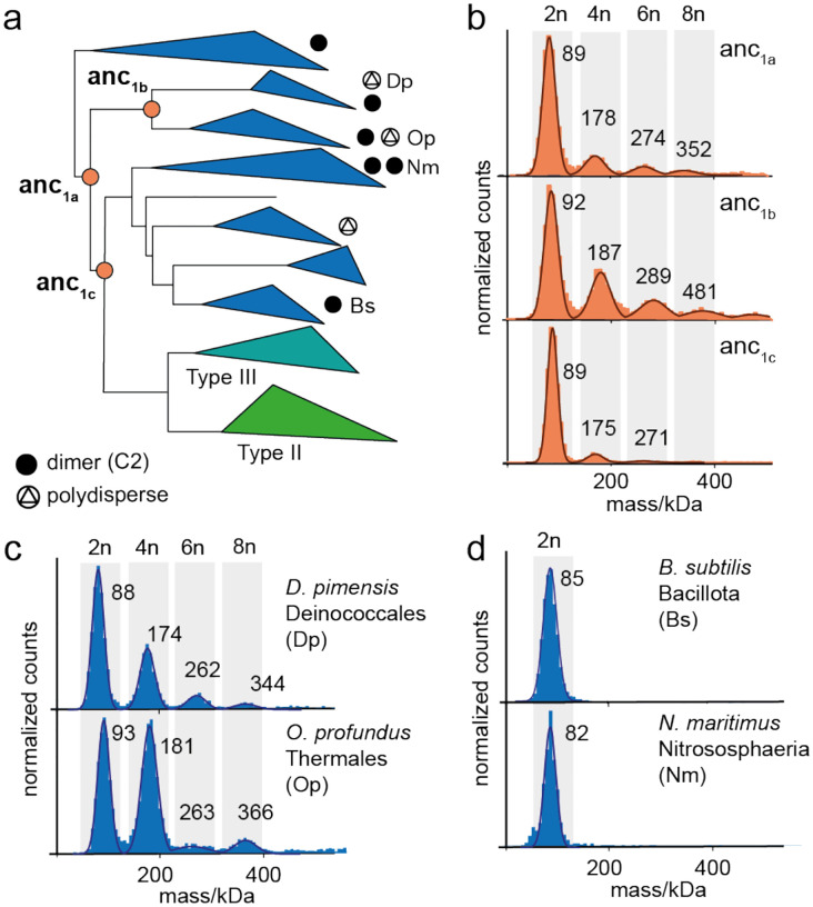
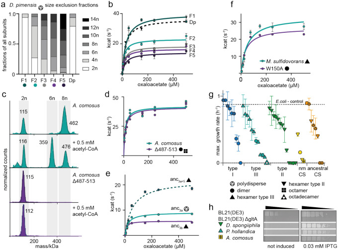
Many enzymes assemble into homomeric protein complexes comprising multiple copies of one protein. Because structural form is usually assumed to follow function in biochemistry, these assemblies are thought to evolve because they provide some functional advantage. In many cases, however, no specific advantage is known and, in some cases, quaternary structure varies among orthologs. This has led to the proposition that self-assembly may instead vary neutrally within protein families. The extent of such variation has been difficult to ascertain because quaternary structure has until recently been difficult to measure on large scales. Here, we employ mass photometry, phylogenetics, and structural biology to interrogate the evolution of homo-oligomeric assembly across the entire phylogeny of prokaryotic citrate synthases - an enzyme with a highly conserved function. We discover a menagerie of different assembly types that come and go over the course of evolution, including cases of parallel evolution and reversions from complex to simple assemblies. Functional experiments in vitro and in vivo indicate that evolutionary transitions between different assemblies do not strongly influence enzyme catalysis. Our work suggests that enzymes can wander relatively freely through a large space of possible assemblies and demonstrates the power of characterizing structure-function relationships across entire phylogenies.


Conflict of interest statement

Competing interests: The authors declare no other competing interests.

Figures

Extended Data Fig. 1
Extended Data Fig. 1. Conservation of residues inducing hexameric assembly in type III CS.
Alignment of characterized type III CS, ancestors anc3a, anc3b and the type II CS from E. coli. Residues marked in teal are part of the hexamer interface in type III enzymes. Connecting lines indicate direct interactions between residues in structures (teal=M. sulfidovorans, orange=anc3b). Residues that take part in the hexamer formation in E. coli CS are colored in green.
Extended Data Fig. 2
Extended Data Fig. 2. Important residues for type III hexamer formation
(a) MP measurement of the dimeric type III CS from Cyanobium sp. PCC 7001. (b) Sequence logo of amino acid residues that are found within the interface of type II or III CS, demonstrating strong differences. Homologous sites are aligned, shift in site numbers results from a longer N-terminus in type II CS. (c) MP measurement of interface variants of the type III CS from M. sulfidovorans. (d) Model of the hexameric type III CS from N. inopinata inferred with AlphaFold-multimer-v2 with close up of the interface between dimers. Right: structural model colored according to the predicted local distance difference test (pLDDT) (e) SEC trace of the polydisperse anc3a and MP measurements of the isolated fractions. (f) MP measurements of variants of anc3a with different combinations of substitutions that emerged in the interval to anc3b (small and capital letters indicate the ancestral and derived amino acid, respectively). No combination of 4 was sufficient to induce hexameric assembly. The significance of the substitution p78K was shown by a reversal (K78p) in the hexameric anc3b. (g) AlphaFold-multimer model of Anc3b color-coded according to the site-wise pLDDT score.
Extended Data Fig. 3
Extended Data Fig. 3. Inconclusive inference of the emergence of type II hexamers but conserved involvement of the JK-loop
(a) Schematic representation of part of the CS phylogeny displaying the quaternary structures of characterized type II CS. Nodes corresponding to resurrected ancestral CS are indicated. (b) MP measurements of anc2a and its descendants anc2c and anc2d. (c) Upper: AlphaFold-Multimer predictions of hexameric type II CS and nmCS with close ups on the interface that display the involvement of the JK-loop for all of them. Lower: pLDDT-colored structures of the same models. (d) Comparison of the extended C-terminus in the hexameric structure from C. fasiculata (AlphaFold-Multimer) and octameric structure from A. comosus (cryo-EM). Only two dimers are displayed for both structures to highlight the cross-connection by the C-terminus between adjacent dimers.
Extended Data Fig. 4
Extended Data Fig. 4. Conservation of residues inducing oligomeric assembly in type II CS and nmCS.
Alignment of the characterized type II CS and nmCS. Residues marked in green take part in the hexamer formation in the hexamer from E. coli. The extended C-terminus of nmCS is colored in yellow.
Extended Data Fig. 5
Extended Data Fig. 5. Polydisperse type I enzymes form multiples of dimers
(a) SEC trace of polydisperse CS from D. pimensis and MP measurements of the isolated fractions. (b) Exemplary cryo-EM micrograph of oligomeric complexes of polydisperse anc1a, low-pass filtered at 5 Å. Particle picks are depicted as white circles (diameter = 180 Å). Scale bar = 40 nm. (c) 2D class averages of polydisperse anc1a. Scale bar = 10 nm. (d) MP measurement of the SEC-fraction used for cryo-EM.
Extended Data Fig. 6
Extended Data Fig. 6. Kinetic measurements of CS variants with saturated oxaloacetate
Michaelis-Menten kinetics of (a) extant CS from A. comosus and a variant that disrupts interfaces between dimeric subcomplexes and prevent assembly into larger oligomers (Δ487–513). Error bars = SD, n = 3 technical replicates; (b) the ancestral CS bracketing the emergence of hexamers within type III enzymes (anc3a, anc3b) and the minimal substitution construct to yield hexameric complexes (anc3a+5). Error bars = SD, n = 3 technical replicates; (c) extant CS from M. sulfidovorans and a variant that disrupts interfaces between dimeric subcomplexes and prevent assembly into larger oligomers (W150A). Error bars = SD, n = 3 technical replicates W150A
Extended Data Fig. 7
Extended Data Fig. 7. Protein production and complementation on solid media
(a) Western Blots depicting the CS protein production of all tested extant and ancestral CS enzymes in E. coli using the same conditions as for the growth curves. (b) Spot-assays of complemented E. coli BL21(DE3) ΔgltA strains. Cultures were spotted in a five-step serial dilution using a ratio of 1:5 for each step and incubated on M9-solid media supplemented with glucose and different IPTG concentrations. One representative plate is shown for each experiment, out of a total of three replicates for each plate.
Fig. 1.
Fig. 1.. Assembly of CS across the phylogenetic tree
(a) Phylogenetic tree of CS in Bacteria, Archaea and eukaryotic nmCS (full phylogeny in supplementary Fig. 1) and classification into type I, II, III, and nmCS. Brackets indicate the number of sequences within each clade. Branch supports values are shown for important nodes as Felsenstein’s bootstrap values. The transfer into Eukaryotes is well supported by phylogenetic and experimental results (see below) but the branching order of major eukaryotic lineages is poorly supported and disagrees with known relationships. Symbols (●, ▲, ▼etc.) indicate the quaternary structure of characterized CS within the respective clade: White symbols represent solved structures that have been deposited to the PDB and black symbols correspond to assemblies that were characterized in this study. Representative structures of known CS assemblies are shown. (b) Cartoon representation of the amino acid sequence structure of type I-III and nmCS. (c) Mass photometry (MP) measurements of purified CS displaying different forms of assembly.
Fig. 2.
Fig. 2.. Parallel evolution of hexameric CS
(a) MP measurements of two type III CS. (b) Schematic representation of the CS phylogeny displaying the quaternary structures of characterized type III CS. All MP spectra and species names for the characterized quaternary structures are found in supplementary Fig 1+2. Nodes corresponding to resurrected ancestral CS are indicated. (c) X-ray structure of hexameric type III CS from M. sulfidovorans. (d) Comparison of the interface area that connects dimers into hexamers in the type III structure and the type II CS from E. coli. (e) MP measurements of resurrected ancestral CS along the evolutionary trajectory of hexameric assembly within type III enzymes. (f) Location of historical substitutions (orange sites) within the type III interface in a modelled structure of anc3b using AlphaFold-multimer-v2. (g) MP measurement a variant of anc3a with a set of 5 historical substitutions shown in (f) that are sufficient to trigger formation of hexamers and loss of polydisperse behavior (anc3+5). Bar graph displays the fraction of all active sites in dimers vs hexamers for anc3+5.
Fig. 3
Fig. 3. Widespread hexameric assembly in type II CS and evolution of novel octamers
(a) Schematic representation of part of the CS phylogeny displaying the quaternary structures of characterized type II CS and nmCS. All MP spectra and species names for the characterized quaternary structures are found in supplementary Fig. 1 and 2. Nodes corresponding to resurrected ancestral CS are indicated. (b-d) MP measurements of extant type II CS (b), ancestral type II CS (c) and nmCS (d). (e) Cryo-EM structure of octameric nmCS from A. comosus. (f) Focus on the extended C-terminus of nmCS within the Cryo-EM density of A. comosus which folds over from one dimeric subcomplex to an adjacent one. (g) MP measurement of a variant of A. comosus CS in which the extended C-terminus was cut off (Δ487–513).
Fig. 4
Fig. 4. Polydisperse assemblies early in the evolution of CS
(a) Schematic representation of the CS phylogeny displaying the quaternary structures of characterized type I CS. All MP spectra and species names for the characterized quaternary structures are found in supplementary Fig. 1 and 2. Nodes corresponding to resurrected ancestral CS are indicated. (b-d) MP measurements of ancestral CS (b), dimeric (c) and polydisperse (d) type I CS.
Fig. 5
Fig. 5. Effects on catalytic function by changes in quaternary structure
(a) Fraction of CS subunits within the different oligomeric states for each SEC-fraction determined by MP for the CS from D. pimensis (Dp) (F1-F5, see also Extended Data Fig. 5a). (b) Michaelis-Menten kinetics of the different fractions shown in (a) and the unseparated sample of D. pimensis CS (Dp). Error bars = SD, n = 3 technical replicates. (c) MP measurements of CS from A. comosus and its truncation variant Δ487–513 in the absence and presence of acetyl-CoA. (d) Michaelis-Menten kinetics of CS from A. comosus and its Δ487–513 variant. Error bars = SD, n=2 technical replicates. (e) Michaelis-Menten kinetics of ancestral CS bracketing the emergence of hexamers within type III enzymes (anc3a, anc3b) and the minimal substitution construct to yield hexameric complexes (anc3a+5). Error bars = SD, n = 3 technical replicates. (f) Michaelis-Menten kinetics of extant CS from M. sulfidovorans and a variant that disrupts the interface between dimers (W150A). Error bars = SD, n = 3 technical replicates. (g) Maximum growth rates on M9 media of an E. coli strain lacking the native CS gene (BL21(DE3) ΔgltA) complemented with a plasmid encoding for CS genes from all characterized extant and ancestral enzyme, error bars = SD, n = 3 biological replicates. The positive control is the KO-strain complemented with the native CS from E. coli which displayed the same growth behaviour as the wild-type BL21(DE3) strain. (h) Spot assays on solid M9 media using either leaky expression or induction by IPTG, shown for strains that did not complement in (g), showing that increased CS production leads to complementation. Cultures were spotted in a five-step serial dilution using a ratio of 1:5 for each step and incubated on M9-solid media supplemented with glucose and different IPTG concentrations. One representative plate is shown for each experiment, out of a total of three replicates for each plate. Full data for all strains shown in Extended Data Fig. 7.

Similar articles

References

    1. Goodsell D. S. & Olson A. J. Structural symmetry and protein function. Annu. Rev. Biophys. Biomol. Struct. 29, 105–153 (2000). - PubMed
    1. Levy E. D. & Teichmann S. Structural, evolutionary, and assembly principles of protein oligomerization. in Progress in Molecular Biology and Translational Science vol. 117 25–51 (Elsevier B.V., 2013). - PubMed
    1. Matthews J. M. & Sunde M. Dimers, oligomers, everywhere. Advances in Experimental Medicine and Biology vol. 747 1–18 at 10.1007/978-1-4614-3229-6_1 (2012). - DOI - PubMed
    1. Perica T. et al. The emergence of protein complexes: quaternary structure, dynamics and allostery. Biochem. Soc. Trans. 40, 475–491 (2012). - PubMed
    1. Marianayagam NJ, Sunde M, M. J. The power of two: protein dimerization in biology. Trends Biochem. Sci. 29, 618–625 (2004). - PubMed

Publication types

LinkOut - more resources