[Preprint]. 2024 Jul 2:2024.07.01.601619.
doi: 10.1101/2024.07.01.601619.

APCCdh1-mediated degradation of Cdh1 is necessary for faithful meiotic chromosome segregation in S. cerevisiae

Affiliations

APCCdh1-mediated degradation of Cdh1 is necessary for faithful meiotic chromosome segregation in S. cerevisiae

Denis Ostapenko et al. bioRxiv. .

Abstract

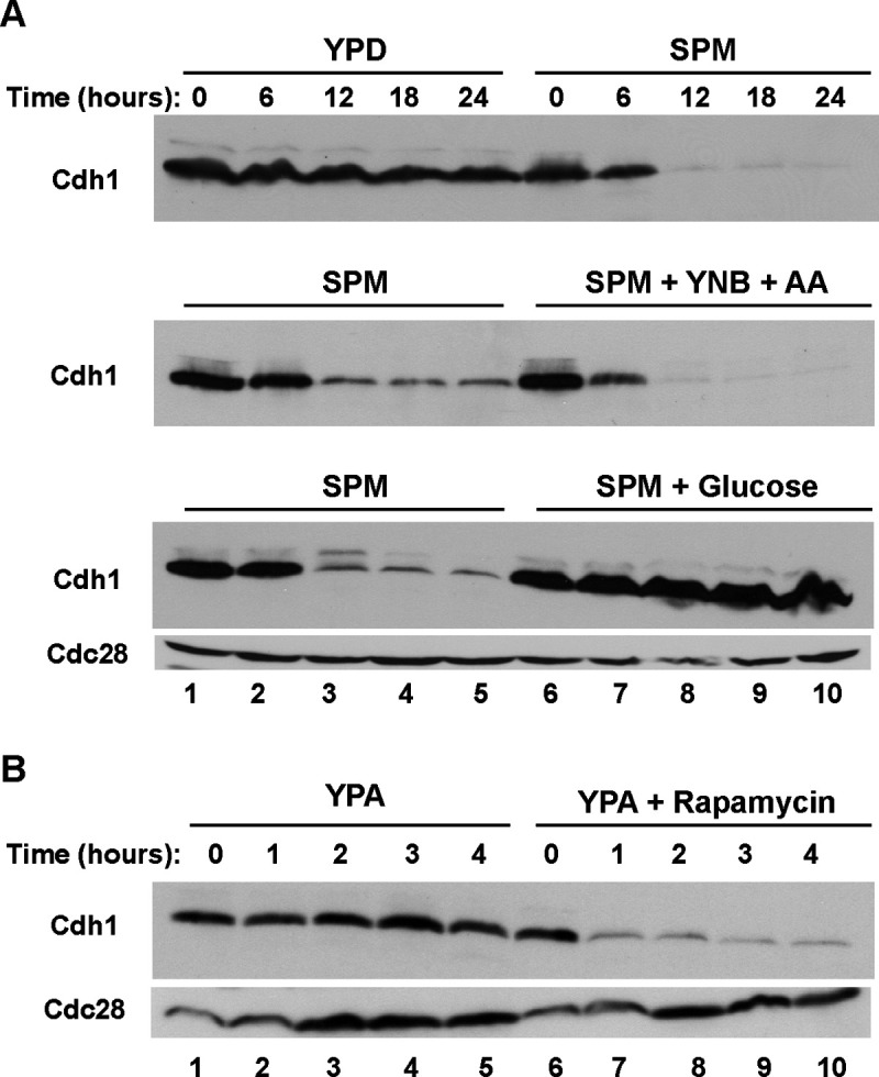
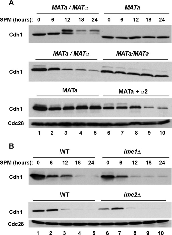
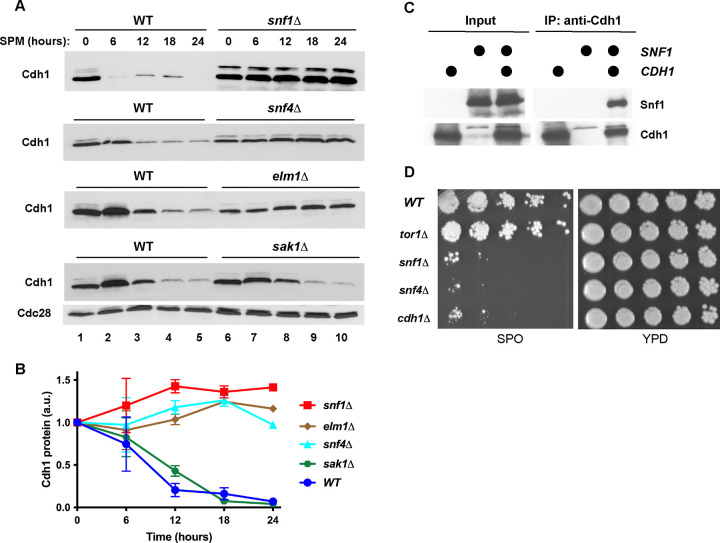
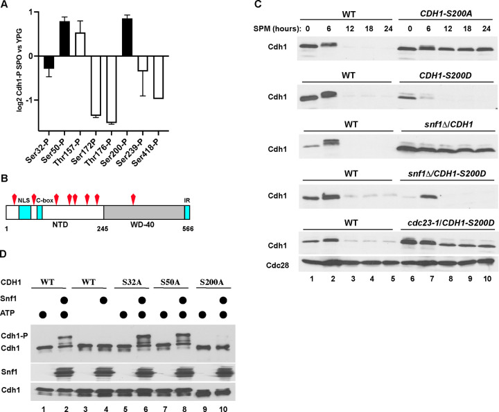
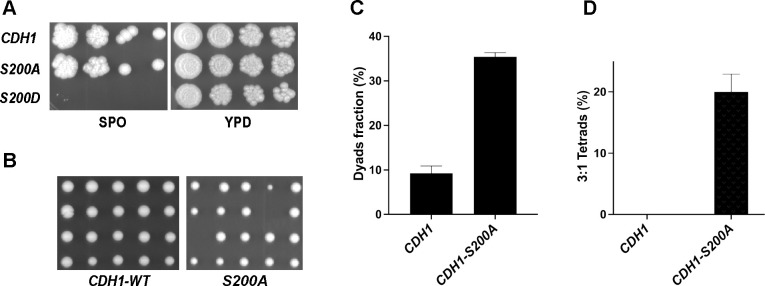
The Anaphase-Promoting Complex/Cyclosome (APC/C) is a ubiquitin ligase that promotes the ubiquitination and subsequent degradation of numerous cell cycle regulators during mitosis and in G1. Proteins are recruited to the APC/C by activator proteins such as Cdh1. During the cell cycle, Cdh1 is subject to precise regulation so that substrates are not degraded prematurely. We have explored the regulation of Cdh1 during the developmental transition into meiosis and sporulation in the budding yeast S. cerevisiae. Transition to sporulation medium triggers the degradation of Cdh1. Cdh1 degradation is mediated by the APC/C itself in a "trans" mechanism in which one molecule of Cdh1 recruits a second molecule of Cdh1 to the APC/C for ubiquitination. Degradation requires an intact glucose-sensing SNF1 protein kinase complex (orthologous to the mammalian AMPK nutritional sensor), which directly phosphorylates Cdh1 on Ser-200 within an unstructured N-terminal region. In the absence of phosphorylation, expression of a Cdh1-S200A mutant is fully stabilized, leading to chromosome instability and loss of viability. We hypothesize that Cdh1 degradation is necessary for the preservation of cell cycle regulators and chromosome cohesion proteins between the reductional and equational meiotic divisions, which occur without the intervening Gap or S phases found in mitotic cell cycles.


Figures

Figure 1.
Figure 1.
Cdh1 protein levels decline during sporulation. (A) (top) Cdh1 protein in extracts from an asynchronous population of diploid yeast cells (strain DOY2361) grown in rich medium (YPD, lanes 1–5) or cells transferred from YPA to sporulation medium (SPM, lanes 6–10). Samples were withdrawn at the indicated times and monitored for the presence of Myc-Cdh1 by immunoblotting with 9E10 antibodies. (middle) As above with cells transferred to sporulation medium (SPM, lanes 1–5) or to sporulation medium supplemented with 0.67% yeast nitrogen base, 0.2% amino acid mixture (SPM + YNB + AA, lanes 6–10). (bottom) As above with cells transferred to sporulation medium (SPM, lanes 1–5) or to sporulation medium supplemented with 1% glucose (lanes 6–10). As a loading control, the membrane was re-probed with anti-PSTAIR antibodies to detect Cdc28. (B) Asynchronous cultures of diploid yeast cells (strain DOY2361) pre-grown in acetate-containing medium (YPA) were mock-treated (lanes 1–5) or incubated with 1 μg/ml rapamycin (lanes 6–10). Samples were withdrawn at the indicated times and monitored for the presence of Cdh1 by immunoblotting with anti-Myc antibodies.
Figure 2.
Figure 2.
Cdh1 degradation depends on heterozygosity of the mating type locus but not on cell ploidy per se. (A) (top) Asynchronous diploid MATa/MATα cells (strain DOY2361) (lanes 1–5) and haploid MATa cells (strain DOY2282) (lanes 6–10) were transferred from YPA to sporulation medium as in Figure 1A. Samples were withdrawn at the indicated times and processed for immunoblotting to detect Myc-tagged Cdh1. (middle) The same time courses as above were performed using two isogenic diploid strains that differ only in the configuration at the mating-type loci: MATa/MATα (strain DOY2780) (lanes 1–5) and MATa/MATa (strain DOY2783) (lanes 6–10). Only the MATa/MATα cells were able to sporulate and produce tetrads. (bottom) The same time courses were performed using a haploid MATa strain carrying either an empty vector (lanes 1–5) or a vector expressing the α2 transcription factor (strain DOY2786) normally expressed in MATa/MATα diploid cells, thus making these cells equivalent to MATa/MATα cells at the mating-type locus but otherwise haploid (lanes 6–10). The MATa + α2 cells could not undergo sporulation. Anti-Cdc28 probing of the same filter was used as a loading control. (B) Expression of Inducer of MEiosis genes is not required for Cdh1 degradation. Homozygous diploid strains that were wild-type (strain DOY2361) or deleted for IME1 (strain DOY2554), or IME2 (strain DOY2555) were transferred to sporulation medium, and tested at the indicated times for Cdh1 expression. Mutation of either IME gene prevents transcription of early meiotic genes and yeast sporulation. Samples were processed for immunoblotting with 9E10 antibodies to detect Myc-Cdh1 protein. The membrane was re-probed with anti-PSTAIR antibodies to detect Cdc28 as a loading control.
Figure 3.
Figure 3.
Cdh1 is degraded in an APC/C-dependent manner during sporulation. (A) pdr5Δ homozygous diploid cells (which are permeable to the proteasome inhibitor MG132; strain DOY2989) were transferred to sporulation medium in the absence (lanes 1–5) or presence of 20 μM MG132 to inhibit the proteasome. Samples were withdrawn at the indicated times and processed to detect Myc-Cdh1 as in Figure 1A. Anti-Cdc28 was used as a loading control. (B) Wild-type and isogenic homozygous diploid strains carrying a conditional allele of the ubiquitin-conjugating enzyme of the SCF complex (cdc34–2, top panel; strain DOY2542), or an APC/C core subunit (cdc23–1, second panel; strain DOY2540) were grown at permissive conditions and transferred to sporulation medium at 35°C to inactivate the temperature-sensitive proteins. Of note, transfer to 35°C did not interfere with the ability of wild-type cells to form tetrads but arrested cell cycle progression of the cdc34–2 and cdc23–1 strains. (Third panel) Wild type CDH1 (strain DOY2719) and cdh1Δ mutant (strain DOY2725) strains carried an endogenous Myc-CDH1-ΔIR (“Cdh1-ΔIR”) allele, which lacks the C-terminal two amino acid residues of Cdh1 necessary for binding to the APC/C core. Therefore, the Cdh1-ΔIR protein is nonfunctional in promoting the degradation of APC/C substrates, but can serve as a substrate itself. Samples were processed to detect Myc-Cdh1-ΔIR protein levels as in Figure 1A. (Bottom panel) A wild-type and an isogenic homozygous diploid ama1Δ strain (strain DOY2602), which carries a homozygous deletion of the meiosis-specific APC/C activator, were transferred to sporulation-inducing medium and incubated at 30°C for the indicated times. Samples were processed to detect Myc-Cdh1-ΔIR protein levels as in Figure 1A. (C) The relative stability of Cdh1 in wild-type and the indicated mutant strains was quantified by ImageJ software (http://www.ImageJ.net). The initial level of Cdh1 in each individual strain at time 0 was set to 100%. Data represent the means and standard deviations from three independent experiments. (D) All strains expressed the non-functional “Cdh1-ΔIR” reporter described in (C) and either a wild-type copy of CDH1 (strain DOY2970;), or a form of Cdh1 that has been mutated so that it can no longer recognize Destruction Boxes (cdh1-dbr; strain DOY2971; top), KEN boxes (cdh1-mkr; strain DOY2972; middle), or the ABBA motif (cdh1-amr; strain DOY2973; bottom). All strains were transferred to sporulation medium and incubated for the indicated times. Samples were withdrawn and immunoblotted with 9E10 antibodies to detect Myc-Cdh1-ΔIR. Immunoblotting with an anti-Cdc28 antibody was used as a loading control.
Figure 4.
Figure 4.
The glucose-sensing AMP-dependent protein kinase (AMPK) pathway is required for Cdh1 turnover and cell survival during sporulation. (A) A wild-type strain (strain DOY2361) and isogenic mutant strains carrying homozygous deletions of the AMPK/SNF1 alpha subunit (snf1Δ strain DOY2603), the SNF1 regulatory subunit (snf4Δ strain DOY2655), or the Snf1-activating kinases Elm1 (elm1Δ strain DOY2676) or Sak1 (sak1Δ strain DOY2675) were grown in YPA and transferred to sporulation medium. Samples were processed to detect Myc-Cdh1 protein as in Figure 1A. (B) The relative abundance of Cdh1 in the wild-type strain and in the indicated mutant strains in (A) were quantified using ImageJ software and normalized relative to the level of Cdc28 protein from the same sample. The initial amount of Cdh1 at time 0 was set at 100% and the fractions of the protein remaining in the samples at subsequent times were plotted relative to this starting level. (C) Cdh1 interacts with SNF1 in vivo. Yeast strains carrying endogenous SNF1-TAP and galactose-inducible GST-CDH1 and GST expression vectors, as indicated on the figure, were activated by galactose for four hours. GST-Cdh1 was immunoprecipitated from cell extracts with glutathione beads and probed for the presence of Snf1-TAP by immunoblotting with PAP antibodies. The levels of GST-Cdh1 was determined by immunoblotting anti-Cdh1 antibodies. (D) Wild type (strain DOY2361) and three isogenic diploid strains carrying homozygous deletions of TOR1 (strain DOY2512), SNF1 (strain DOY2603), SNF4 (strain DOY2655), and CDH1 (strain DOY2589) were pre-incubated in sporulation-inducing medium (SPO) for 7 days at 30°C, serially diluted, and spotted on a plate containing rich medium (YPD). Asynchronous control cultures of the same strains grown in rich medium (YPD) were diluted and plated in the same order; both plates were grown at 30°C for three days.
Figure 5.
Figure 5.
Snf1-mediated phosphorylation regulates Cdh1 stability. (A) Wild-type diploid cells carrying GST-CDH1 (strain DOY3384) were grown in YP-galactose (YPG) at 30°C or transferred to sporulation medium (SPO) for 6 hours at 30°C. GST-Cdh1 was affinity purified and subjected to mass-spectrometry to detect phospho-peptides. The phosphorylated peptides corresponding to indicated Cdh1 sites were quantified using Scaffold5 software (https://www.proteomesoftware.com), normalized relative to the total peptide counts, log2 transformed, and compared between SPO and YPG cultures. Open bars within the graph represent known CDK phosphorylation sites, and closed bars indicate sites that resemble the SNF1 consensus phosphorylation site. (B) A cartoon of Cdh1 indicating the phosphorylation sites shown in (A). Most of the differentially-phosphorylated sites are located within the unstructured N-terminal domain (NTD), including a cluster of six CDK sites (Thr157, Ser169, Thr173, Thr176, Ser227, Ser239). Of note, peptides corresponding to three other CDK sites (Thr12, Ser16, and Ser42) were not detected by mass-spectrometry. The second cluster of phospho-sites (Ser32, Ser50, Se200) is located within the region that regulates Cdh1 localization and its interaction with the core APC (Hockner et al., 2016). The locations of an NLS (nuclear localization signal), the C-box, and the IR motifs are indicated. (C) Phosphorylation of Ser-200 regulates Cdh1 stability during meiosis. Ser-200 was mutated to either alanine or aspartic acid. (top two panels) Wild type CDH1 (strain DOY2899) and two heterozygous diploid strains, CDH1-S200A (strain DOY4043) and CDH1-S200D (strain DOY4011) (carrying an untagged copy of wild-type CDH1) were tested after shift to sporulation medium as in Figure 1A. (third and fourth panels) snf1Δ CDH1 (strain DOY3459) and snf1Δ CDH1-S200D (strain DOY4244) strains were grown and analyzed for Myc-Cdh1 expression. (bottom) cdc23–1 CDH1-S200D cells (strain DO4246) were pre-grown under permissive conditions and transferred to sporulation medium at 35°C to inactivate Cdc23. Samples were collected at the indicated times and processed to detect Myc-Cdh1 protein as in Figure 1A. (D) Snf1 phosphorylates Cdh1 Ser-200 in vitro. Constitutively active GST-SNF1-G53R was expressed in yeast cells grown in YP-galactose (YPG) (strain DOY4195). The SNF1 protein kinase complex was affinity purified and incubated with recombinant His6-Cdh11−250 in the absence and presence of ATP, as indicated. Three putative Snf1 consensus sites (Ser32, Ser50, Ser-200) were individually mutated to alanine. The products of the assay were resolved on a Phos™-tag gel (top panel), conventional SDS-PAGE (bottom panel) and probed with anti-Cdh1 antibodies. Snf1 was detected by immunoblotting with anti-GST antibodies (middle panel).
Figure 6
Figure 6
Stabilized Cdh1-S200A increases the rate of chromosome mis-segregation during sporulation (A) Diploid wild-type CDH1 (strain DOY2899), CDH1-S200A (strain DOY4191), and CDH1-S200D (strain DO3970) cells were incubated in sporulation-inducing medium (SPO) for 7 days at 30°C, serially diluted, and spotted on YPD plates (left). Asynchronous cultures of the same strains grown in rich medium (YPD) to early stationary phase were similarly diluted and spotted (right); both plates were grown at 30°C for three days. (B) Following incubation in sporulation-inducing medium, tetrads derived from wild type CDH1 (strain DOY2899) and CDH1-S200A (strain DO4191) cells were dissected, and individual spores were spotted on YPD plates. The resulting spores germinated into colonies of haploid cells for 3 days at 30°C. Of note, strain CDH1-S200D (strain DOY3970) did not form any tetrads in sporulation-inducing medium and, thus, was impossible to dissect. (C) Incomplete meiosis or sporulation leads to the appearance of dyads, composed of only two spores. The fraction of dyads in 500 random tetrads from wild type CDH1 (strain DOY2899) and isogenic CDH1-S200A (strain DO4191) strains is shown. (D) Under optimal sporulation conditions, yeast diploid cells produce 4 viable haploid spores. Wild type CDH1 (strain DOY2899) and isogenic CDH1-S200A (strain DO4191) cells were sporulated for 2 days at 30°C, approximately 100 asci were dissected, and the percentage of tetrads with only 3 viable spores (3:1 Tetrads) were plotted. Examples of such tetrads are shown in panel B.

Similar articles

References

    1. Barford D (2011) Structure, function and mechanism of the anaphase promoting complex (APC/C). Q Rev Biophys 44: 153–190 - PubMed
    1. Brandeis M, Hunt T (1996) The proteolysis of mitotic cyclins in mammalian cells persists from the end of mitosis until the onset of S phase. EMBO J 15: 5280–5289 - PMC - PubMed
    1. Brown NG, VanderLinden R, Watson ER, Qiao R, Grace CR, Yamaguchi M, Weissmann F, Frye JJ, Dube P, Ei Cho S et al. (2015) RING E3 mechanism for ubiquitin ligation to a disordered substrate visualized for human anaphase-promoting complex. Proc Natl Acad Sci U S A 112: 5272–5279 - PMC - PubMed
    1. Burton JL, Solomon MJ (2000) Hsl1p, a Swe1p inhibitor, is degraded via the anaphase-promoting complex. Mol Cell Biol 20: 4614–4625 - PMC - PubMed
    1. Cardozo T, Pagano M (2004) The SCF ubiquitin ligase: insights into a molecular machine. Nature Reviews Molecular Cell Biology 5: 739–751 - PubMed

Publication types

LinkOut - more resources