[Preprint]. 2024 Jul 9:2024.07.06.601241.
doi: 10.1101/2024.07.06.601241.

Co-option of mitochondrial nucleic acid sensing pathways by HSV-1 UL12.5 for reactivation from latent Infection

Affiliations

Co-option of mitochondrial nucleic acid sensing pathways by HSV-1 UL12.5 for reactivation from latent Infection

Sean R Cuddy et al. bioRxiv. .

Update in

Abstract

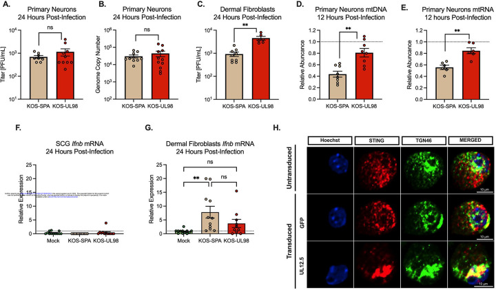
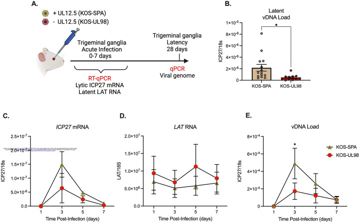
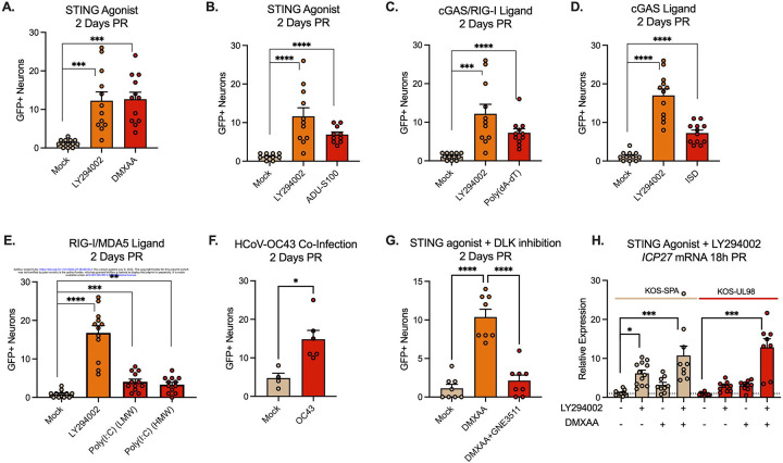
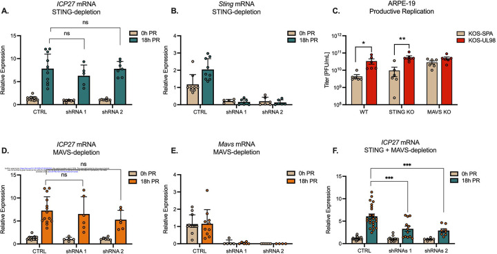
Although viruses subvert innate immune pathways for their replication, there is evidence they can also co-opt anti-viral responses for their benefit. The ubiquitous human pathogen, Herpes Simplex Virus-1 (HSV-1), encodes a protein (UL12.5) that induces the release of mitochondrial nucleic acid into the cytosol, which activates immune sensing pathways and reduces productive replication in non-neuronal cells. HSV-1 establishes latency in neurons and can reactivate to cause disease. We found that UL12.5 is required for HSV-1 reactivation in neurons and acts to directly promote viral lytic gene expression during initial exit from latency. Further, the direct activation of innate immune sensing pathways triggered HSV reactivation and compensated for a lack of UL12.5. Finally, we found that the induction of HSV-1 lytic genes during reactivation required intact RNA and DNA sensing pathways, demonstrating that HSV-1 can both respond to and active antiviral nucleic acid sensing pathways to reactivate from a latent infection.


Figures

Figure 1
Figure 1. UL12.5 does not affect lytic replication in peripheral neurons.
(A &B) Primary neurons isolated from the superior cervical ganglia (SCG) of newborn mice were infected at an MOI of 10 PFU/cell. Viral titer (A) and viral genome copy number quantified by qPCR (B) were measured at 24 hours post-infection with either KOS-SPA (WT) or KOS-UL98 (UL12.5 null). (C) Dermal fibroblasts isolated from newborn mice were infected at an MOI of 3 PFU/ml and viral titer quantified at 24 hours post-infection. (D & E) Relative abundance of (D) mtDLoop1 mtDNA and (E) mtCOX2 mtRNA transcripts measured by (RT-) qPCR 12 hours after infection with KOS-SPA or KOS-UL98. (F & G) Relative Ifnb mRNA expression 24 hours after infection of (F) primary sympathetic neurons or (G) dermal fibroblasts (H) Representative immunofluorescence images of STING/pIRF3 at 3-days post-transduction with either GFP or UL12.5 expressing lentiviral vector. All in A-G data are plotted as biological replicates from at least three independent experiments, shown as mean +/− SEM. Statistical comparisons were made using a t-test or one-way ANOVA with Tukey’s multiple comparison test. *p<0.05, **p<0.01
Figure 2
Figure 2. UL12.5 supports initial de-repression of lytic HSV genes following in vivo infection.
(A) Schematic of the in vivo model of HSV-1 latent infection. (B) Quantification of the latent viral genome copy number measured by qPCR at 28 days after infection with KOS-SPA or KOS-UL98. The copy number of viral DNA was normalized to host 18s rDNA. (C & D) ICP27 (C) or LAT (D) RNA copy numbers normalized to host 18S rRNA quantified by RT-qPCR during the acute infection period. (E) Viral genome copy number measured by qPCR over 7 days after infection with KOS-SPA or KOS-UL98 during the acute infection period. All data in B-E are plotted as biological replicates from at least three independent mouse experiments; shown as mean +/− SEM. Individual biological replicates are plotted in B. Statistical comparisons were made using a t-test (B) or one-way ANOVA with Tukey’s multiple comparison test (C-D). All statistical comparisons for time points in (C-E) are n.s. between viral strains except where otherwise noted. *p<0.05
Figure 3
Figure 3. UL12.5 is required for Phase I lytic gene expression during HSV-1 reactivation.
(A) Schematic of the in vitro HSV latent infection and reactivation model. Neuronal infection was carried out in the presence of acyclovir (ACV; 50 μM). Reactivation was induced using LY294002 (20 μM) in the presence of WAY-150138 (20 μM) to limit cell-to-cell spread. (B) Quantification of relative viral genome in latently infected (0 hours) and reactivated (60 hours) neurons infected with either KOS-SPA and KOS-UL98 measured by qPCR. (C) Quantification of lytic mRNAs and LAT in neurons latently infected with either KOS-SPA and KOS-UL98 measured by RT-qPCR for ICP27 (IE), UL30 (E), VP16 (L), and gC (L). (D) Quantification of lytic mRNAs at 18 hours post-reactivation (Phase I). (E) Quantification of lytic mRNAs at 48 hours post-reactivation (Phase II). (F) Quantification of ICP27 mRNA during Phase I reactivation in neurons latently infected with F/L or the recue virus; F/LR. (G) Quantification of ICP27 mRNA during Phase I reactivation in neurons latently infected with AN-1 or the wild-type strain KOS. All in B-G data are plotted as biological replicates from at least three independent experiments; shown as mean +/− SEM. Statistical comparisons were made using a t-test (F-G) or one-way ANOVA with Tukey’s multiple comparison test. *p<0.05, **p<0.01, ***p<0.001
Figure 4
Figure 4. UL12.5 is expressed and functions during Phase I of HSV-1 reactivation.
(A-B) Neurons were latently infected with HSV-1 Stayput-GFP and reactivated in the presence of cycloheximide (CHX; 10 μg/ml) added at either the same time as LY294002 or 10 hours later. Relative ICP27 (A) and gC (B) mRNA was quantified by RT-qPCR. (C) Quantification of mtDNA abundance by qPCR of mtDLoop1 at 18 hours after reactivation post-reactivation. (D) Neurons latently infected with either KOS-SPA or KOS-UL98 were transduced with lentivirus expressing either GFP or UL12.5. The relative expression of ICP27 mRNA was measured by RT-qPCR at 18 hours post-reactivation. (E) Representative immunofluorescence images of UL12/UL12.5 and TOM20 following reactivation. (F) Quantification of UL12.5 expressing neurons as a percentage of positive neurons per field of view at 0-, 4-, and 8 hours after reactivation. All in A-F data are plotted as biological replicates from at least three independent experiments; shown as mean +/− SEM. Statistical comparisons were made using a t-test (C) or one-way ANOVA with Tukey’s multiple comparison test. *p<0.05, **p<0.01, ***p<0.001, ****p<0.0001
Figure 5
Figure 5. Activation of innate immune signaling induces HSV-1 reactivation from latent infection.
(A-E) Neurons latently infected with Stayput-GFP were treated with ligands for the DNA/RNA sensing pathways using DMXAA (50 μg/mL; A), ADU-S100 (10 μg/mL; B), Poly(dA-dT)/LyoVec (5 μg/mL; C), ISD (5μg/mL; D) or Poly(I:C) (HMW), or Poly(I:C) (LMW) (10 μg/mL; E). (F) Neurons latently infected with Stayput-GFP were co-infected with the HCoV-OC43 at an MOI of 3 PFU/cell. Reactivation was quantified based on the numbers of Us11-GFP expressing neurons at 2 days post co-infection. (G) Neurons latently infected with Stayput-GFP were reactivated with DMXAA in the presence or absence of the DLK inhibitor GNE-3511 (4 μM). (G) Neurons latently infected with KOS-SPA and KOS-UL98 were treated with either LY294002 or DMXAA or LY294002, followed 2.5 hours later by DMXAA. All in A-F data are plotted as biological replicates from at least three independent experiments; shown as mean +/− SEM. Statistical comparisons were made using a t-test (F) or one-way ANOVA with Tukey’s multiple comparison test. *p<0.05, **p<0.01, ***p<0.001, ****p<0.0001
Figure 6
Figure 6. Intact STING and MAVS signaling is required for Phase I HSV-1 gene expression.
(A & B) Latently infected neurons were transduced with vectors expressing shSTING or shCTRL during latency. Quantification of ICP27 (A) and Sting (B) transcripts by RT-qPCR at 0 and 18 hours post-reactivation (PR) with LY294002 (20μM). (C) Titers of infectious virus at 24 hours post-infection of Wild-type, STING KO, or MAVS KO ARPE-19 cells infected with KOS-SPA or KOS-UL98 at an MOI of 5 PFU/cell. (D & E) Latently infected neurons were transduced with vectors expressing shMAVS or shCTRL during latency. Quantification of ICP27 (D) and Mavs (E) transcripts by RT-qPCR at 0 and 18 hours post-reactivation (PR) (F) Quantification of ICP27 mRNA at 18 hours post-reactivation following depletion of both STING and MAVS. All in A-F data are plotted as biological replicates from at least three independent experiments; shown as mean +/− SEM. Statistical comparisons were made using a one-way ANOVA with Tukey’s multiple comparison test. *p<0.05, **p<0.01, ***p<0.001

Similar articles

References

    1. Pandey S., Kawai T., and Akira S. (2014). Microbial sensing by Toll-like receptors and intracellular nucleic acid sensors. Cold Spring Harb Perspect Biol 7, a016246. 10.1101/cshperspect.a016246. - DOI - PMC - PubMed
    1. Gong T., Liu L., Jiang W., and Zhou R. (2020). DAMP-sensing receptors in sterile inflammation and inflammatory diseases. Nature Reviews Immunology 20, 95–112. 10.1038/s41577-019-0215-7. - DOI - PubMed
    1. Aarreberg L.D., Esser-Nobis K., Driscoll C., Shuvarikov A., Roby J.A., and Gale M. Jr. (2019). Interleukin-1beta Induces mtDNA Release to Activate Innate Immune Signaling via cGAS-STING. Mol Cell 74, 801–815 e806. 10.1016/j.molcel.2019.02.038. - DOI - PMC - PubMed
    1. Aguirre S., Luthra P., Sanchez-Aparicio M.T., Maestre A.M., Patel J., Lamothe F., Fredericks A.C., Tripathi S., Zhu T., Pintado-Silva J., et al. (2017). Dengue virus NS2B protein targets cGAS for degradation and prevents mitochondrial DNA sensing during infection. Nat Microbiol 2, 17037. 10.1038/nmicrobiol.2017.37. - DOI - PMC - PubMed
    1. Moriyama M., Koshiba T., and Ichinohe T. (2019). Influenza A virus M2 protein triggers mitochondrial DNA-mediated antiviral immune responses. Nature Communications 10, 4624. 10.1038/s41467-019-12632-5. - DOI - PMC - PubMed

Publication types

LinkOut - more resources