Case Reports
. 2024 Jun 28:12:1423858.
doi: 10.3389/fped.2024.1423858. eCollection 2024.

Case Report: Common variable immunodeficiency phenotype and granulomatous-lymphocytic interstitial lung disease with a novel SOCS1 variant

Affiliations
Case Reports

Case Report: Common variable immunodeficiency phenotype and granulomatous-lymphocytic interstitial lung disease with a novel SOCS1 variant

María Soledad Caldirola et al. Front Pediatr. .

Abstract

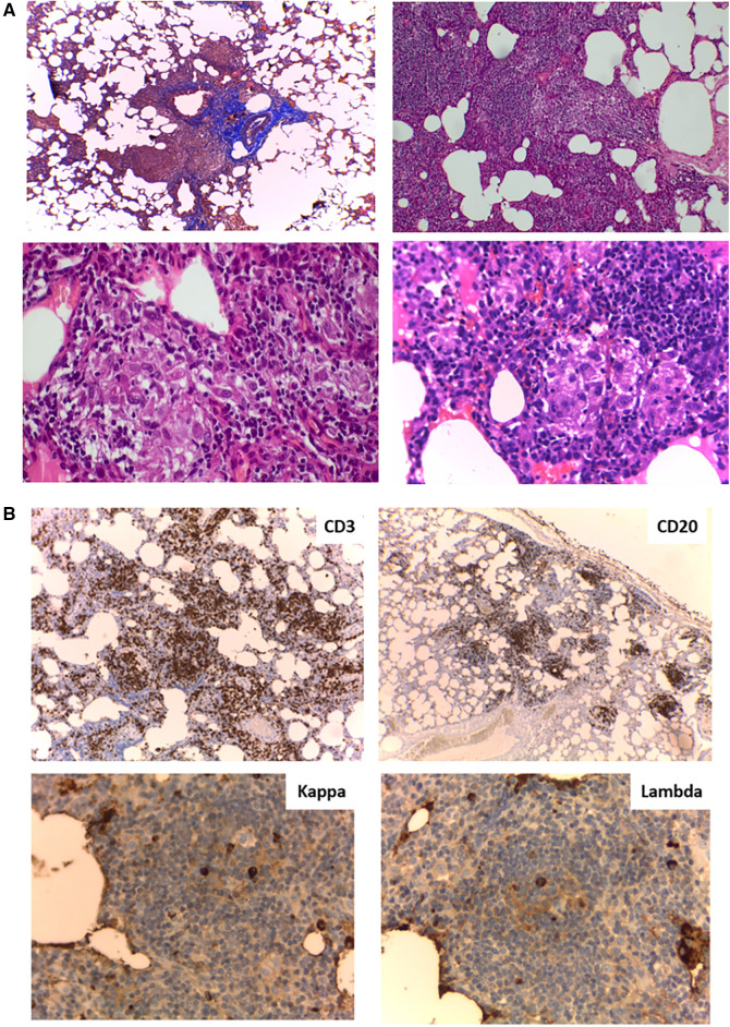
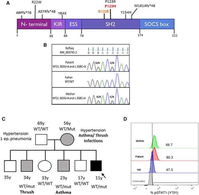
Common variable immunodeficiency is a heterogeneous symptomatic group of inborn errors of immunity that mainly affects antibodies production and/or function, predisposing patients to recurrent and severe infections. More than half of them usually develop autoimmunity, lymphoproliferation, enteropathy, and malignancies. Among these conditions, chronic lung disease such as granulomatous-lymphocytic interstitial lung disease is one of the leading causes of death in these patients. Recently, many genes that play a key role in B and T cells' development, maintenance, and/or cytokines signaling pathways have been implicated in the pathogenesis of the disease. Here, we describe the first Argentinian patient presenting with common variable immunodeficiency and granulomatous-lymphocytic interstitial lung disease, harboring two in cis heterozygous variants in the SOCS1 gene.

Keywords: GLILD; SOCS1; common variable immunodeficiency; granulomatous–lymphocytic interstitial lung disease; inborn errors of immunity.


Conflict of interest statement

The authors declare that the research was conducted in the absence of any commercial or financial relationships that could be construed as a potential conflict of interest.

Figures

Figure 1
Figure 1
(A) Patient abdominal CT scan. Homogeneous craniocaudal splenomegaly measuring 176 mm associated with increased diameter of splenic vascular structures and increased hepatic hilar vasculature. No evidence of free fluid. (B) Patient chest CT scan. Bilateral diffusely distributed nodules, including solid, subsolid, and ground-glass opacities, predominantly in the lung bases with the largest measuring 12 mm.
Figure 2
Figure 2
(A) Lung biopsy histology. (Upper left) Masson's trichrome stain (4×) peribronchiolar mononuclear inflammatory infiltrate; (Upper right) Hematoxylin and eosin (H&E) stain (10×) Interstitial expansion of lymphocytes and histiocytes; (Lower left and right) H&E stain (40×) Presence of poorly formed non-necrotizing granulomas. (B) Lung biopsy immunohistochemistry. (Upper left) (4×) CD3+ lymphocytes at the interstitial level; (Upper right) (4×) Aggregates of CD20+ lymphocytes; (Lower left and right) (40×) Kappa and lambda chains in similar proportions.
Figure 3
Figure 3
(A) SOCS1 protein domains and locations of the variants. The kinase inhibitory region (KIR) functions as a pseudo-substrate that can inhibit the tyrosine kinase activity of JAK proteins. The SRC-homology 2 (SH2) domain binds the activation loop of the JAK proteins’ catalytic domain. The SOCS box recruits the ubiquitin-transferase system and initiates the proteasomal degradation of JAK proteins. Mutations are described in the literature. Our patient's mutations are in red. (B) Pedigrees of the family with SOCS1 variants. Squares: males; circles: females; black: affected mutation carriers; gray: unaffected mutation carriers. WT, wild-type SOCS1 allele. (C) Sanger sequencing analysis. Sanger sequencing of the exon 1 of SOCS1 in the proband and his parents confirmed the maternal origin of: c.365G>A (p.G122E) and c.368C>A (p.P123H) in the same allele of SOCS1 gene. (D) STAT1 phosphorylation assay. Increased phosphorylation of monocytes (CD14+) after 15 min with IFN-γ stimulation in the patient and his mother compared with a healthy control (HD).

References

    1. Bonilla FA, Barlan I, Chapel H, Costa-Carvalho BT, Cunningham-Rundles C, De La Morena MT, et al. International Consensus Document (ICON): common variable immunodeficiency disorders. J Allergy Clin Immunol Pract. (2016) 4:38–59. 10.1016/j.jaip.2015.07.025 - DOI - PMC - PubMed
    1. Sullivan K. Stiehm’s Immune Deficiencies. Waltham, MA: Elsevier; (2020).
    1. Gereige JD, Maglione PJ. Current understanding and recent developments in common variable immunodeficiency associated autoimmunity. Front Immunol. (2019) 10:2753. 10.3389/fimmu.2019.02753 - DOI - PMC - PubMed
    1. Ameratunga R, Woon S-T. Perspective: evolving concepts in the diagnosis and understanding of common variable immunodeficiency disorders (CVID). Clinic Rev Allerg Immunol. (2020) 59:109–21. 10.1007/s12016-019-08765-6 - DOI - PubMed
    1. Patel S, Anzilotti C, Lucas M, Moore N, Chapel H. Interstitial lung disease in patients with common variable immunodeficiency disorders: several different pathologies? Clin Exp Immunol. (2019) 198:212–23. 10.1111/cei.13343 - DOI - PMC - PubMed

Publication types

LinkOut - more resources