. 2024 Jul 10:12:e17722.
doi: 10.7717/peerj.17722. eCollection 2024.

Ginkgetin attenuates bone loss in OVX mice by inhibiting the NF-κB/IκBα signaling pathway

Affiliations

Ginkgetin attenuates bone loss in OVX mice by inhibiting the NF-κB/IκBα signaling pathway

GeJin Wei et al. PeerJ. .

Abstract

Background: Osteoporosis is a disease associated with bone resorption, characterized primarily by the excessive activation of osteoclasts. Ginkgetin is a compound purified from natural ginkgo leaves which has various biological properties, including anti-inflammation, antioxidant, and anti-tumor effects. This study investigated the bone-protective effects of ginkgetin in ovariectomized (OVX) mice and explored their potential signaling pathway in inhibiting osteoclastogenesis in a mouse model of osteoporosis.

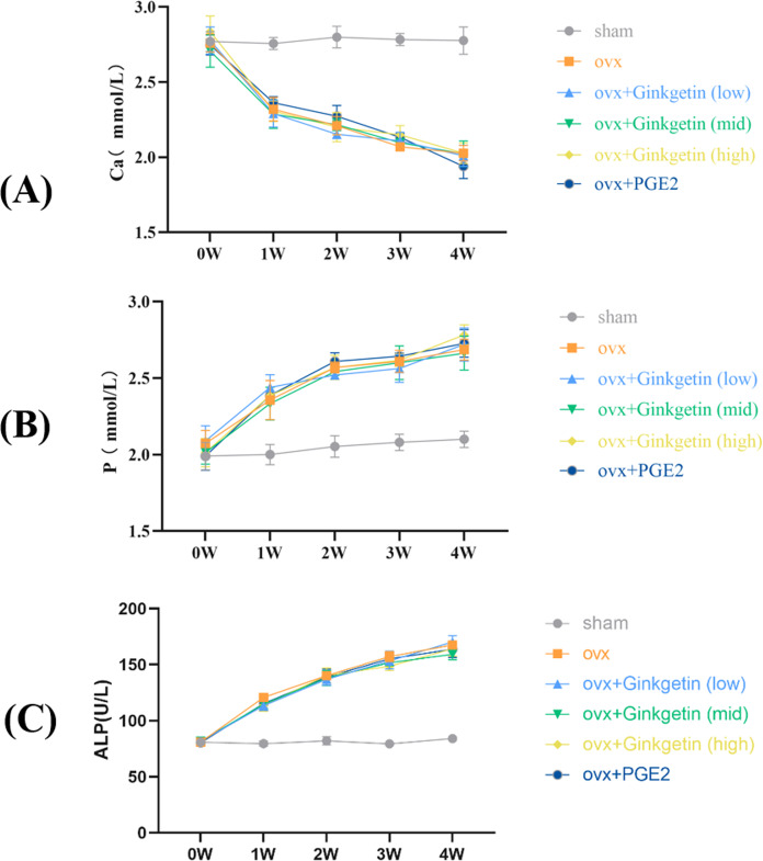
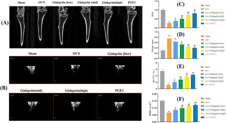
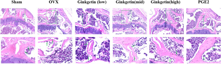
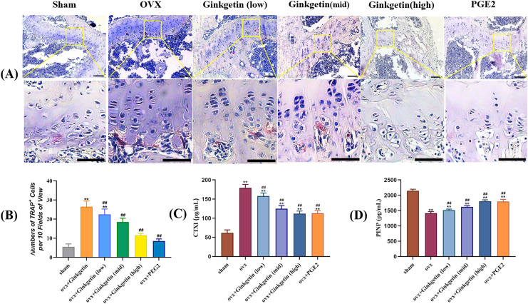
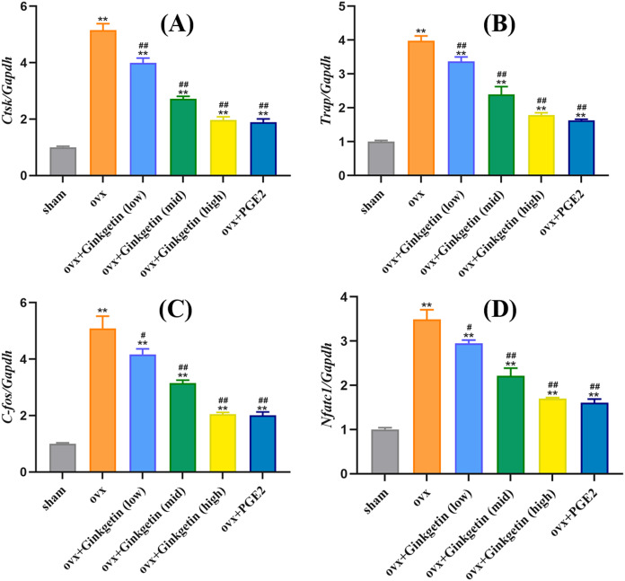
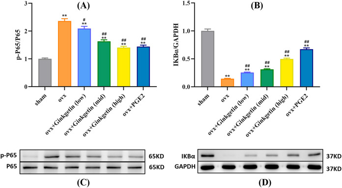
Methods: Biochemical assays were performed to assess the levels of Ca, ALP, and P in the blood. Micro CT scanning was used to evaluate the impact of ginkgetin on bone loss in mice. RT-PCR was employed to detect the expression of osteoclast-related genes (ctsk, c-fos, trap) in their femoral tissue. Hematoxylin and eosin (H&E) staining was utilized to assess the histopathological changes in femoral tissue due to ginkgetin. The TRAP staining was used to evaluate the impact of ginkgetin osteoclast generation in vivo. Western blot analysis was conducted to investigate the effect of ginkgetin on the expression of p-NF-κB p65 and IκBα proteins in mice.

Results: Our findings indicate that ginkgetin may increase the serum levels of ALP and P, while decreasing the serum level of Ca in OVX mice. H&E staining and micro CT scanning results suggest that ginkgetin can inhibit bone loss in OVX mice. The TRAP staining results showed ginkgetin suppresses the generation of osteoclasts in OVX mice. RT-PCR results demonstrate that ginkgetin downregulate the expression of osteoclast-related genes (ctsk, c-fos, trap) in the femoral tissue of mice, and this effect is dose-dependent. Western blot analysis results reveal that ginkgetin can inhibit the expression of p-NF-κB p65 and IκBα proteins in mice.

Conclusion: Ginkgetin can impact osteoclast formation and activation in OVX mice by inhibiting the NF-κB/IκBα signaling pathway, thereby attenuating bone loss in mice.

Keywords: Ginkgetin; NF-κB; Osteoclasts; Osteoporosis.


Conflict of interest statement

The authors declare that they have no competing interests.

Figures

Figure 1
Figure 1. Effects of ginkgetin on serum ALP, P, and Ca levels measured by the biochemical instrument at 1-, 2-, 3- and 4-week respectively in OVX mice.
(A) The effects of different concentrations of ginkgetin on serum Ca were measured and the levels of serum Ca were significantly decreased. (B) The effects of different concentrations of ginkgetin on serum P were measured and the levels of serum p were significantly increased. (C) The effects of different concentrations of ginkgetin on serum ALP were measured and the levels of serum ALP were significantly increased.
Figure 2
Figure 2. Ginkgetin has a protective effect on bone loss in OVX C57BL/6J mice.
(A) Representative 2D‐μCT images of tibias in different groups of mice. (B) Representative 3D‐μCT images of tibias in different groups of mice. (C) Comparative analysis of trabecular bone number (Tb.N) that were significantly improved after ginkgetin administration. (D) Comparative analysis of trabecular bone separation (Tb.Sp, mm) that were significantly decreased after ginkgetin administration. (E) Comparative analysis of bone volume fraction (BV/TV, %) that were significantly improved after ginkgetin administration. (F) Comparative analysis of bone mineral density (BMD, in g/mm3) that were significantly improved after ginkgetin administration. n = 6. All bar charts are presented as the mean ± SD. *P < 0.05, ∗∗P < 0.01 compared with the sham group. #P < 0.05, ##P < 0.01 compared with the OVX group.
Figure 3
Figure 3. Effects of ginkgetin on the morphology of OVX mice bone tissue.
Representative H&E staining femoral sections of mice in different groups. H&E staining indicated that the thickness of the tibial trabecula surface was more maintained in the ginkgetin-treated groups than in the OVX group in a dose-dependent manner (scale bar = 100 μm). The green arrows refer to the absorption bay.
Figure 4
Figure 4. (A–D) Effects of ginkgetin on the generation of osteoclasts and the expression of bone turnover biomarkers such as CTXI and PINP.
(A) Representative TRAP staining images of osteoclasts in femoral sections treated without or with different concentration ginkgetin. TRAP-positive multinucleated cells (nuclei >3) were regarded as osteoclasts. The original images scale bar = 100 µm, the enlarged images scale bar = 50 µm. (B) Quantitative analysis of TRAP-positive Cells. TRAP staining showing the inhibitory effect of ginkgetin on osteoclastogenesis in a dose-dependent manner. (C) Quantitative analysis of serum CTXI expression. The expression of CTXI was significantly reduced, after ginkgetin intervention with different concentrations. (D) Quantitative analysis of serum PINP expression. The expression of PINP was significantly increased after ginkgetin intervention with different concentrations. ∗∗P < 0.01 compared with the sham group. ##P < 0.01 compared with the OVX group.
Figure 5
Figure 5. Ginkgetin suppresses the expression of osteoclast-related genes in OVX mice.
(A) Quantitative analysis of mRNA expression of CTSK. (B) Quantitative analysis of mRNA expression of TRAP. (C) Quantitative analysis of mRNA expression of c-fos. (D) Quantitative analysis of mRNA expression of NFATc1. ∗∗P < 0.01 compared with the sham group. #P < 0.05, ##P < 0.01 compared with the OVX group.
Figure 6
Figure 6. Different doses of ginkgetin (low, 25 mg/kg; mid, 50 mg/kg; high, 100 mg/kg) suppress the expression of p-P65 and IκBα proteins in C57BL/6 mice.
(A) Histogram showing that ginkgetin inhibits the expression of p-P65 proteins. (B) Histogram showing that ginkgetin inhibits the expression of IκBα proteins. (C) Typical Western blot images of ginkgetin inhibiting the expression of p-P65 proteins. (D) Typical Western blot images of ginkgetin inhibiting the expression of IκBα proteins. ∗∗P < 0.01 compared with the sham group. n = 5, #P < 0.05, ##P < 0.01 compared with the OVX group.

References

    1. Alharbi KS, Fuloria NK, Fuloria S, Rahman SB, Al-Malki WH, Javed Shaikh MA, Thangavelu L, Singh SK, Rama Raju Allam VS, Jha NK, Chellappan DK, Dua K, Gupta G. Nuclear factor-kappa B and its role in inflammatory lung disease. Chemico-Biological Interactions. 2021;345(Suppl 3):109568. doi: 10.1016/j.cbi.2021.109568. - DOI - PubMed
    1. Arceo-Mendoza RM, Camacho PM. Postmenopausal osteoporosis: latest guidelines. Endocrinology and Metabolism Clinics of North America. 2021;50(2):167–178. doi: 10.1016/j.ecl.2021.03.009. - DOI - PubMed
    1. Black DM, Abrahamsen B, Bouxsein ML, Einhorn T, Napoli N. Atypical femur fractures: review of epidemiology, relationship to bisphosphonates, prevention, and clinical management. Endocrine Reviews. 2019;40(2):333–368. doi: 10.1210/er.2018-00001. - DOI - PubMed
    1. Brinkley TE, Lovato JF, Arnold AM, Furberg CD, Kuller LH, Burke GL, Nahin RL, Lopez OL, Yasar S, Williamson JD, Ginkgo Evaluation of Memory (GEM) Study Investigators Effect of ginkgo biloba on blood pressure and incidence of hypertension in elderly men and women. American Journal of Hypertension. 2010;23(5):528–533. doi: 10.1038/ajh.2010.14. - DOI - PMC - PubMed
    1. Cheng J, Li Y, Kong J. Ginkgetin inhibits proliferation of HeLa cells via activation of p38/NF-κB pathway. Cellular and Molecular Biology. 2019;65(4):79–82. doi: 10.14715/cmb/2019.65.4.13. - DOI - PubMed

LinkOut - more resources