Case Reports
. 2022 Aug 4;12(2):104-107.
doi: 10.4103/jmau.jmau_85_21. eCollection 2024 Apr-Jun.

Role of p63 in Determining the Histogenesis of Low-Grade Neoplasms versus Cystic Lesion

Affiliations
Case Reports

Role of p63 in Determining the Histogenesis of Low-Grade Neoplasms versus Cystic Lesion

Dipanshu Aggarwal et al. J Microsc Ultrastruct. .

Abstract

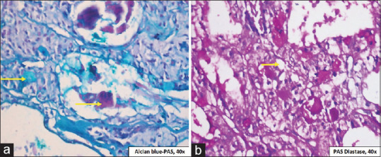
The biological nature of salivary gland neoplasms and the overlapping characteristics that result from the heterogeneity of the cells of origin make diagnosis difficult. Hence, we intend to present a case of low grade mucoepidermoid carcinoma (MEC) on the palate and to understand the importance of biomarker such as p63 in the early diagnosis of tumor as it also has a role in its histogenesis. A 53-year-old female reported with a unilateral swelling for 3 months on posterolateral palatal region of the right side. Clinical differentials for such palatal swellings include a varied spectrum of lesions such as reactive, benign, and malignant lesions. Based on the incisional and excisional biopsy, histopathological findings and immunohistochemical examination with p63 the case were diagnosed with low grade MEC. The tumor cell differentiation in MEC could be the result of multiplicity of differentiation pathways leading to the formation of various histological patterns. This case report highlights the complexity of salivary gland pathology diagnosis and role of specific tumor marker such as p63 as an early marker for differentiation of salivary gland tumor such as low grade MEC from other cystic lesions occurring on the palate.

Keywords: Immunohistocytochemistry; mucoepidermoid carcinoma; salivary gland neoplasm.


Conflict of interest statement

There are no conflicts of interest.

Figures

Figure 1
Figure 1
(a) Clinical examination showing a diffuse solitary nodular swelling 4 cm × 5 cm in size extending from canine posteriorly on posterolateral aspect of the palate. (b) Macroscopic examination of the incisional biopsy shows two soft-tissue bits without evidence of any cystic areas. (c) Histopathological examination showing cystic lining predominantly composed of large cells overlying a fibrocellular stroma (H and E, ×4 magnification). (d) Focal area of stroma shows ductal structure/nest lined by cuboidal to columnar cells having eosinophilic cytoplasm and hyperchromatic nucleus. Hyalinization of stroma is also evident (H and E, ×4 magnification). (e) Photomicrograph shows large cells in the lining epithelium consists of amorphous eosinophilic material suggestive of foamy cytoplasm and nucleus located at periphery (H and E, ×40 magnification). (f) Photomicrograph shows focal areas of cuboidal and polygonal cells with hyperchromatic nucleus and prominent intercellular junction and positivity with PAS (g). Mucicarmine (h) and Alcian blue (i) Stain suggestive of the heterogenous nature of the secretory material and confirming the presence of mucous cells. (H and E, ×40 magnification)
Figure 2
Figure 2
(a) Gross examination showing multiple tissue sections which were harvested from the hemimaxillectomy specimen. (b) Figure showing sites from where multiple tissue sections were harvested from the hemimaxillectomy specimen. (c) Stereomicroscopic examination revealed revealing nodular proliferation inside the cystic lesion. (d) Specimen radiograph revealed that the tumor was expansile with destructive and infiltrative margins. (e) Photomicrograph showing cystic lining of variable thickness having intraluminal papillary proliferation (H and E, ×4 magnification). (f) Photomicrograph showing lining epithelium consisting of cuboidal and polygonal cells with hyperchromatic nucleus and prominent intercellular junction along with mucous cells having foamy cytoplasm (H and E, ×40 magnification). (g) Photomicrograph showing lining epithelium with intramural proliferation (H and E, ×4 magnification). (h) Photomicrograph showing intramural area with mucous cells having amorphous eosinophilia material (H and E, ×40 magnification). (i) Photomicrograph showing multiple cystic structure of variable thickness having amorphous eosinphilic material (H and E, ×10 magnification). (j) Photomicrograph showing large cystic space lined by mucous cells and focal area of clear cell differentiation (H and E, ×10 magnification). (k) Photomicrograph showing stroma with areas of sclerosis and lymphocytic infiltrate under the influence of spilled mucin (H and E, ×10 magnification). (l) Photomicrograph showing nuclear immunoexpression in intermediate and squamous cells of the solid growth areas (IHC, ×40)
Figure 3
Figure 3
(a) Photomicrograph showing positivity with Alcian blue PAS which is suggestive of the presence of both neutral and sialomucin (Periodic acid Schiff-Alcian blue, 40 × magnification). (b) Photomicrograph showing Periodic acid Schiff–Diastase staining suggesting that clear cells are glycogen-rich cells

Similar articles

Cited by

References

    1. Iyer J, Hariharan A, Cao UM, Mai CT, Wang A, Khayambashi P, et al. An overview on the histogenesis and morphogenesis of salivary gland neoplasms and evolving diagnostic approaches. Cancers (Basel) 2021;13:3910. - PMC - PubMed
    1. Dardick I, Burford-Mason AP. Current status of histogenetic and morphogenetic concepts of salivary gland tumorigenesis. Crit Rev Oral Biol Med. 1993;4:639–77. - PubMed
    1. Auclair PL, Goode RK, Ellis GL. Mucoepidermoid carcinoma of intraoral salivary glands. Evaluation and application of grading criteria in 143 cases. Cancer. 1992;69:2021–30. - PubMed
    1. Goode RK, Auclair PL, Ellis GL. Mucoepidermoid carcinoma of the major salivary glands: Clinical and histopathologic analysis of 234 cases with evaluation of grading criteria. Cancer. 1998;82:1217–24. - PubMed
    1. Cipriani NA, Lusardi JJ, McElherne J, Pearson AT, Olivas AD, Fitzpatrick C, et al. Mucoepidermoid carcinoma: A comparison of histologic grading systems and relationship to MAML2 rearrangement and prognosis. Am J Surg Pathol. 2019;43:885–97. - PMC - PubMed

Publication types

LinkOut - more resources