

Case Reports
doi: 10.1016/j.jaccas.2024.102410.
eCollection 2024 Jul 17.
Successful Percutaneous Closure of Gerbode Defect and Right Atrial-Aortic Fistula Following Infective Endocarditis
Affiliations
- PMID: 39006409
- PMCID: PMC11246054
- DOI: 10.1016/j.jaccas.2024.102410
Item in Clipboard
Case Reports
Successful Percutaneous Closure of Gerbode Defect and Right Atrial-Aortic Fistula Following Infective Endocarditis
JACC Case Rep.
.
Abstract
We report a case of infective endocarditis with a septal abscess that was complicated with abnormal blood flow from the left ventricle to the right atrium (Gerbode defect) along with abnormal blood flow from the aorta to the right atrium (atrial-aortic fistula). This is the first reported case of successful correction of both defects by a percutaneous approach.
Keywords: Gerbode defect; atrial-aortic fistula; infective endocarditis; percutaneous approach; septal defects.
© 2024 The Authors.
Conflict of interest statement
The authors have reported that they have no relationships relevant to the contents of this paper to disclose.
Figures


Transesophageal Echocardiographic Biplane Image at the Aortic Valve Level (A) Short-axis view. (B) Long-axis view. Images show a large vegetation attached to the right coronary cusp of aortic valve prolapsing to left ventricular outflow tract (arrows). There is coaptation of aortic valve leaflets.
Transesophageal Echocardiographic Biplane Image at the Aortic Valve Level With Color Flow Doppler During Diastole (A) Short-axis view. (B) Long-axis view. The arrows demonstrate severe, anteriorly directed aortic regurgitation. LA = left atrium; LV = left ventricle; RA = right atrium.

Transesophageal Echocardiographic Biplane Images at the Midesophageal Level With Color Flow Doppler (A and B) The arrows demonstrate abnormal flow between the aorta and the right atrium (RV; fistula between the aorta and the right atrium). Abbreviations as in Figure 2.

Transesophageal Echocardiographic Image at the Midesophageal Long Axis With Color Flow Doppler Demonstrating Flow Between Right Coronary Cusp to the RA The arrow demonstrates abnormal flow between right coronary cusp to right atrium. Abbreviations as in Figure 2.

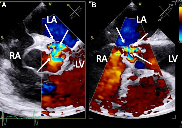
Transesophageal Echocardiographic images (A) Midesophageal 4-chamber view showing a Gerbode defect with color flow between the left ventricle (LV) and the right atrium (RA), with systolic color flow. (B) Long-axis view with color flow Doppler showing abnormal flow between the left ventricle and the right atrium. Abbreviations as in Figures 2 and 3.

Transesophageal Echocardiographic Short-Axis View at the Aortic Valve Level With Color Flow Doppler Showing Successful Closure of the Right Atrial-Aortic Fistula The arrow shows an occluder device. Abbreviations as in Figures 2 and 3.

Transesophageal Echocardiographic Images Showing Gerbode Defect Closure (A) Without Doppler. (B) With color Doppler. These images did not reveal any flow between the left ventricle (LV) and the right atrium (RA), and they show successful closure of the Gerbode defect. Abbreviations as in Figures 2 and 3.
References
-
- Silbiger J.J., Kamran M., Handwerker S., Kumar N., Marcali M. The Gerbode defect: left ventricular to right atrial communication—anatomic, hemodynamic, and echocardiographic features. Echocardiography. 2009;26(8):993–998. - PubMed
-
- Amat-Santos I.J., Rojas P., Stella P.R., et al. Intracardiac shunts following transcatheter aortic valve implantation: a multicentre study. EuroIntervention. 2018;13:1995–2002. - PubMed
-
- Kelle A.M., Young L., Kaushal S., Duffy C.E., Anderson R.H., Backer C.L. The Gerbode defect: the significance of a left ventricular to right atrial shunt. Cardiol Young. 2009;19(suppl 2):96–99. - PubMed
Publication types
LinkOut - more resources
Full Text Sources
