. 2024 Jun 15:19:e00545.
doi: 10.1016/j.ohx.2024.e00545. eCollection 2024 Sep.

Open-source and low-cost miniature microscope for on-site fluorescence detection

Affiliations

Open-source and low-cost miniature microscope for on-site fluorescence detection

Michio Kawai et al. HardwareX. .

Abstract

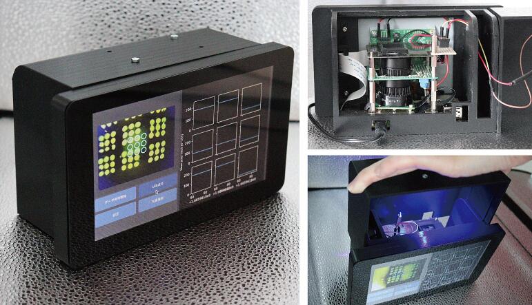
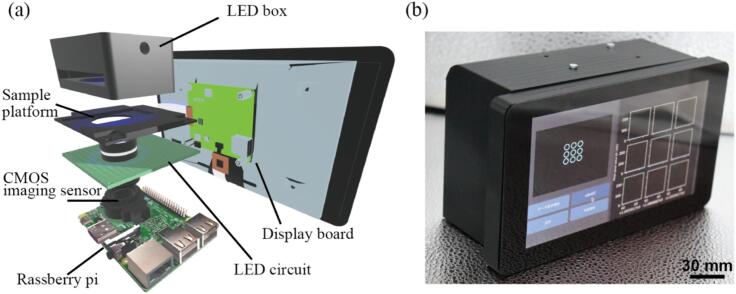
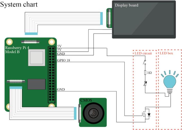
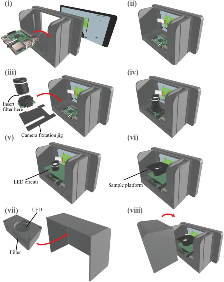
The development of a compact and affordable fluorescence microscope can be a formidable challenge for growing needs in on-site testing and detection of fluorescent labeled biological systems, especially for those who specialize in biology rather than in engineering. In response to such a situation, we present an open-source miniature fluorescence microscope using Raspberry Pi. Our fluorescence microscope, with dimensions of 19.2 × 13.6 × 8.2 cm3 (including the display, computer, light-blocking case, and other operational requirements), not only offers cost-effectiveness (costing less than $500) but is also highly customizable to meet specific application needs. The 12.3-megapixel Raspberry Pi HQ Camera captures high-resolution imagery, while the equipped wide-angle lens provides a field of view measuring 21 × 15 mm2. The integrated wireless LAN in the Raspberry Pi, along with software-controllable high-powered fluorescence LEDs, holds potential for a wide range of applications. This open-source fluorescence microscope offers biohybrid sensor developers a versatile tool to streamline unfamiliar mechanical design tasks and open new opportunities for on-site fluorescence detections.

Keywords: Biohybrid sensor; Fluorescence microscope; Open-source; Raspberry Pi.


Conflict of interest statement

The authors declare that they have no known competing financial interests or personal relationships that could have appeared to influence the work reported in this paper.

Figures

None
Graphical abstract
Fig. 1
Fig. 1
(a) Mechanical design of our proposed device (b) Outlook of our proposed device.
Fig. 2
Fig. 2
Connection diagram of the system.
Fig. 3
Fig. 3
Assembly steps of our proposed device.
Fig. 4
Fig. 4
Evaluation of the radial distortion of the wide-angle lens (a) The fluorescent image of the grid line taken by our proposed device. (b) Grid intersections are shown in blue dots. (c) Arrows indicating the displacement of each grid after distortion. (d) Heatmap representing the amount of displacement of each grid after distortion. Scale bar, 1 mm. (For interpretation of the references to colour in this figure legend, the reader is referred to the web version of this article.)
Fig. 5
Fig. 5
Comparison of the performance between our proposed device and the fluorescence microscope (a)The fluorescence image of the array of fluorescent dye solutions with different concentrations. The image on the left was taken by our proposed device, and the image on the right was taken by the fluorescent microscope. (b)The relationship between the fluorescent intensity calculated from the taken image of our proposed device and the concentration of the fluorescent dye. (c)The comparison of fluorescent intensity measured by our proposed device and the fluorescent microscope. (d)Brand-Altman plot comparing the performance of our proposed device and the fluorescent microscope. Scale bars, 1 mm.
Fig. 6
Fig. 6
Demonstration of the device as a biohybrid sensor using sensor cells (a) Image of the device during measurement (b) Fluorescence intensity change of sensor cells in response to odorant. Scale bar, 10 mm.
Fig. 7
Fig. 7
Observation of red fluorescence by exchanging the filter (a) Image of the device used for observation. (b) Observation of red fluorescent beads through the replaced filter. Scale bars, 1 mm. (For interpretation of the references to colour in this figure legend, the reader is referred to the web version of this article.)

Similar articles

References

    1. Zhang J., Campbell R.E., Ting A.Y., Tsien R.Y. Creating new fluorescent probes for cell biology. Nat. Rev. Mol. Cell Biol. 2002;3:906–918. doi: 10.1038/nrm976. - DOI - PubMed
    1. Moter A., Göbel U.B. Fluorescence in situ hybridization (FISH) for direct visualization of microorganisms. J. Microbiol. Methods. 2000;41:85–112. doi: 10.1016/s0167-7012(00)00152-4. - DOI - PubMed
    1. Golm S.K., Hübner W., Müller K.M. Fluorescence Microscopy in Adeno-Associated Virus Research. Viruses. 2023;15:1174. doi: 10.3390/v15051174. - DOI - PMC - PubMed
    1. Shi J., Zhang Y., Yang M. Recent development of microfluidics-based platforms for respiratory virus detection. Biomicrofluidics. 2023;17 doi: 10.1063/5.0135778. - DOI - PMC - PubMed
    1. Gunasekaran R., Lalitha P., Megia-Fernandez A., Bradley M., Williams R.L., Dhaliwal K., Prajna N.V., Mills B. Exploratory Use of Fluorescent SmartProbes for the Rapid Detection of Microbial Isolates Causing Corneal Ulcer. Am. J. Ophthalmol. 2020;219:341–350. doi: 10.1016/j.ajo.2020.06.014. - DOI - PubMed