

COVLIAS 3.0: cloud-based quantized hybrid UNet3+ deep learning for COVID-19 lesion detection in lung computed tomography
- PMID: 39006802
- PMCID: PMC11240867
- DOI: 10.3389/frai.2024.1304483
COVLIAS 3.0: cloud-based quantized hybrid UNet3+ deep learning for COVID-19 lesion detection in lung computed tomography
Abstract
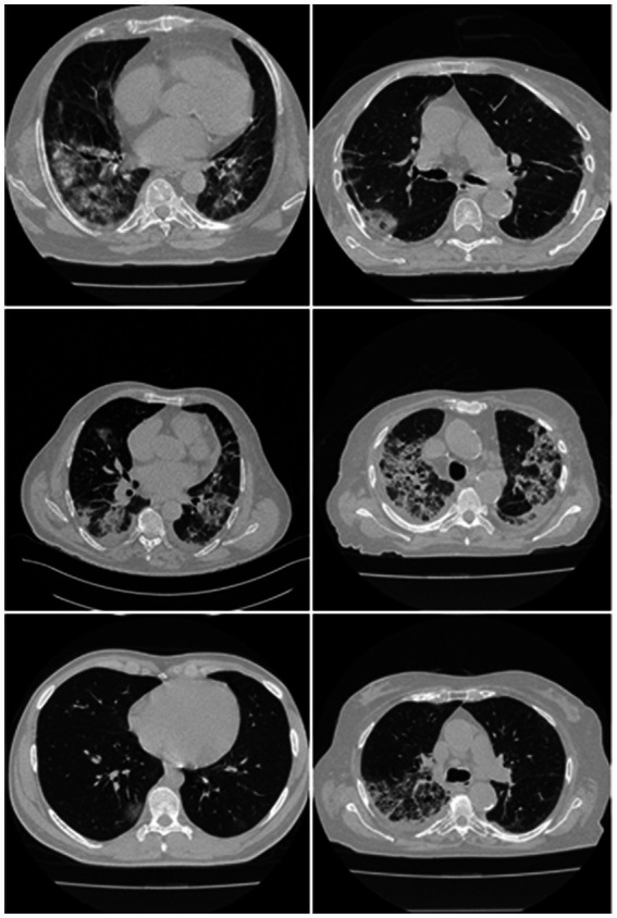
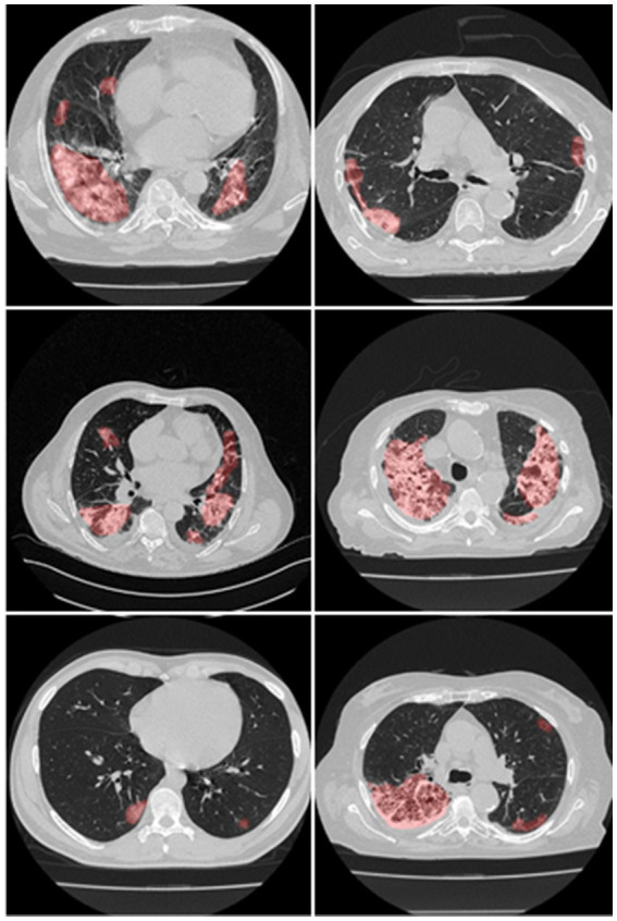
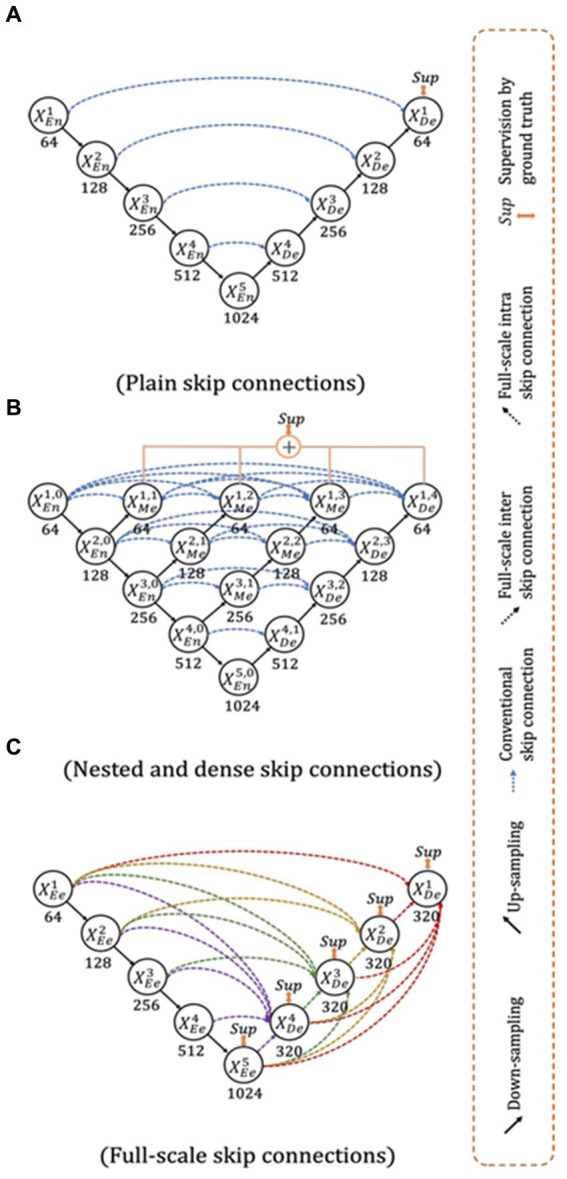
Background and novelty: When RT-PCR is ineffective in early diagnosis and understanding of COVID-19 severity, Computed Tomography (CT) scans are needed for COVID diagnosis, especially in patients having high ground-glass opacities, consolidations, and crazy paving. Radiologists find the manual method for lesion detection in CT very challenging and tedious. Previously solo deep learning (SDL) was tried but they had low to moderate-level performance. This study presents two new cloud-based quantized deep learning UNet3+ hybrid (HDL) models, which incorporated full-scale skip connections to enhance and improve the detections.
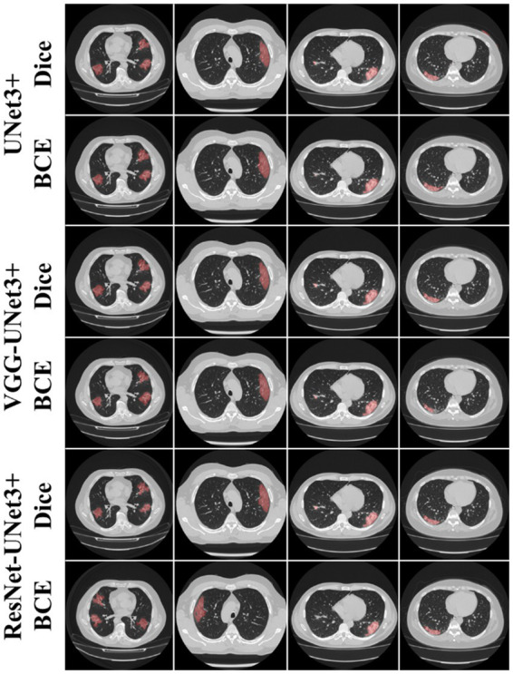
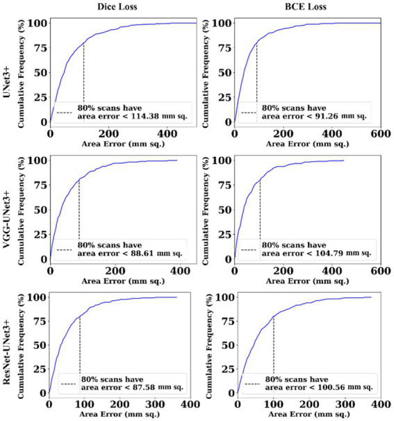
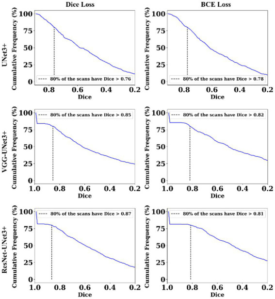
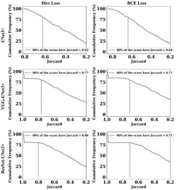
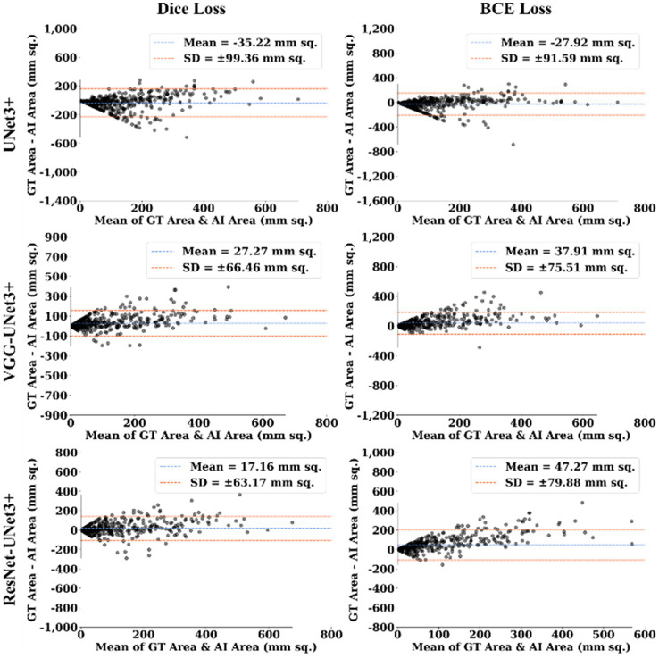
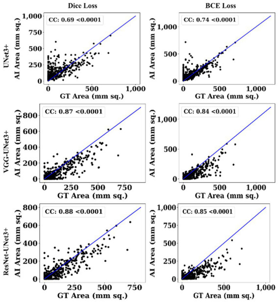
Methodology: Annotations from expert radiologists were used to train one SDL (UNet3+), and two HDL models, namely, VGG-UNet3+ and ResNet-UNet3+. For accuracy, 5-fold cross-validation protocols, training on 3,500 CT scans, and testing on unseen 500 CT scans were adopted in the cloud framework. Two kinds of loss functions were used: Dice Similarity (DS) and binary cross-entropy (BCE). Performance was evaluated using (i) Area error, (ii) DS, (iii) Jaccard Index, (iii) Bland-Altman, and (iv) Correlation plots.
Results: Among the two HDL models, ResNet-UNet3+ was superior to UNet3+ by 17 and 10% for Dice and BCE loss. The models were further compressed using quantization showing a percentage size reduction of 66.76, 36.64, and 46.23%, respectively, for UNet3+, VGG-UNet3+, and ResNet-UNet3+. Its stability and reliability were proved by statistical tests such as the Mann-Whitney, Paired t-Test, Wilcoxon test, and Friedman test all of which had a p < 0.001.
Conclusion: Full-scale skip connections of UNet3+ with VGG and ResNet in HDL framework proved the hypothesis showing powerful results improving the detection accuracy of COVID-19.
Keywords: COVID lesions; COVID-19; computed tomography; glass ground opacities; hybrid deep learning; quantization; segmentation.
Copyright © 2024 Agarwal, Saxena, Carriero, Chabert, Ravindran, Paul, Laird, Garg, Fatemi, Mohanty, Dubey, Singh, Fouda, Singh, Naidu, Viskovic, Kukuljan, Kalra, Saba and Suri.
Conflict of interest statement
SA was employed at GBTI, United States. JS was employed by AtheroPoint™, United States. The remaining authors declare that the research was conducted in the absence of any commercial or financial relationships that could be construed as a potential conflict of interest.
Figures
References
-
- Acharya U. R., Mookiah M. R. K., Vinitha Sree S., Afonso D., Sanches J., Shafique S., et al. (2013c). Atherosclerotic plaque tissue characterization in 2D ultrasound longitudinal carotid scans for automated classification: a paradigm for stroke risk assessment. Med. Biol. Eng. Comput. 51, 513–523. doi: 10.1007/s11517-012-1019-0, PMID: - DOI - PubMed
-
- Acharya U. R., Saba L., Molinari F., Guerriero S., Suri J. S. (2012b). “Ovarian tumor characterization and classification: a class of GyneScan™ systems” in 2012 Annual International Conference of the IEEE Engineering in Medicine and Biology Society. IEEE, pp. 4446–4449. - PubMed
-
- Acharya U. R., Vinitha Sree S., Mookiah M. R. K., Yantri R., Molinari F., Zieleźnik W., et al. (2013a). Diagnosis of Hashimoto’s thyroiditis in ultrasound using tissue characterization and pixel classification. Proc. Inst. Mech. Eng. H J. Eng. Med. 227, 788–798. doi: 10.1177/0954411913483637, PMID: - DOI - PubMed
LinkOut - more resources
Full Text Sources