. 2024 Jun;2(2):80-87.
doi: 10.1016/j.pccm.2024.04.002. Epub 2024 Jun 12.

Mechanisms of pulmonary endothelial barrier dysfunction in acute lung injury and acute respiratory distress syndrome

Affiliations

Mechanisms of pulmonary endothelial barrier dysfunction in acute lung injury and acute respiratory distress syndrome

Yunchao Su et al. Chin Med J Pulm Crit Care Med. 2024 Jun.

Abstract

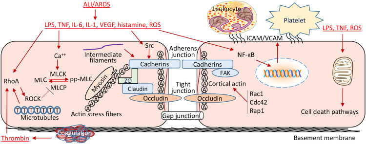
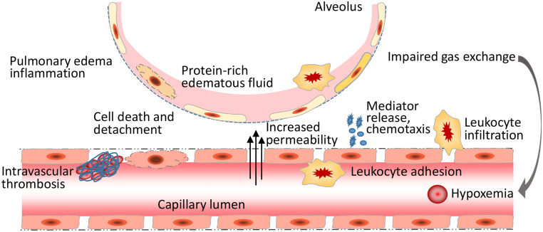
Endothelial cells (ECs) form a semi-permeable barrier between the interior space of blood vessels and the underlying tissues. Pulmonary endothelial barrier integrity is maintained through coordinated cellular processes involving receptors, signaling molecules, junctional complexes, and protein-regulated cytoskeletal reorganization. In acute lung injury (ALI) or its more severe form acute respiratory distress syndrome (ARDS), the loss of endothelial barrier integrity secondary to endothelial dysfunction caused by severe pulmonary inflammation and/or infection leads to pulmonary edema and hypoxemia. Pro-inflammatory agonists such as histamine, thrombin, bradykinin, interleukin 1β, tumor necrosis factor α, vascular endothelial growth factor, angiopoietin-2, and platelet-activating factor, as well as bacterial toxins and reactive oxygen species, cause dynamic changes in cytoskeletal structure, adherens junction disorganization, and detachment of vascular endothelial cadherin (VE-cadherin) from the actin cytoskeleton, leading to an increase in endothelial permeability. Endothelial interactions with leukocytes, platelets, and coagulation enhance the inflammatory response. Moreover, inflammatory infiltration and the associated generation of pro-inflammatory cytokines during infection cause EC death, resulting in further compromise of the structural integrity of lung endothelial barrier. Despite the use of potent antibiotics and aggressive intensive care support, the mortality of ALI is still high, because the mechanisms of pulmonary EC barrier disruption are not fully understood. In this review, we summarized recent advances in the studies of endothelial cytoskeletal reorganization, inter-endothelial junctions, endothelial inflammation, EC death, and endothelial repair in ALI and ARDS, intending to shed some light on the potential diagnostic and therapeutic targets in the clinical management of the disease.

Keywords: Acute lung injury; Acute respiratory distress syndrome; Endothelium; Lung; Pulmonary edema.


Conflict of interest statement

The authors declare that they have no known competing financial interests or personal relationships that could have appeared to influence the work reported in this paper.

Figures

Fig 1
Fig. 1
Mechanisms of pulmonary endothelial barrier disruption in ALI/ARDS. Endothelial barrier disruption is resulted from actin cytoskeletal reorganization due to actin–myosin interaction after MLC-phosphorylation, which is regulated by MLCK and MLCP in ALI/ARDS. RhoA activation induced by pro-inflammatory mediators potentiates MLC phosphorylation by inhibiting MLC phosphatase activity leading to the increased formation of actin stress fibers and the dispersal of cortical actin bundles. Ratcheting of myosin heads against actin microfilaments generates a contractile tension, which pulls VE-cadherin inward and forces VE-cadherin to dissociate from its adjacent partner causing inter-endothelial gaps and endothelial barrier disruption. Src activation induced by pro-inflammatory mediators induces VE-cadherin phosphorylation leading to disorganization of AJ proteins and detachment of VE-cadherin from the actin cytoskeleton. Rac1, Cdc42, and Rap1 contribute to an intact barrier function by modifying the formation of cortical F-actin. FAK, a non-receptor tyrosine kinase, regulates the turnover of focal adhesion formation by binding to focal adhesion proteins as well as enhancing AJ formation. NF-κB activation promotes the expression of various leukocyte adhesion molecules including ICAM-1 and VCAM-1 and selectins, which mediate endothelium–leukocyte and endothelium–platelet interactions. Initiation of coagulation leads to proteolytic cleavage of prothrombin and release of thrombin. Bacterial infection and inflammatory infiltration and cytokines cause EC death, resulting in the compromise of structural integrity of lung endothelial barrier. AJ: Adherens junction; ALI: Acute lung injury; ARDS: Acute respiratory distress syndrome; Cdc42: Cell division cycle 42; EC: Endothelial cells; FAK: Focal adhesion kinase; ICAM: Intercellular adhesion molecule 1; IL: Interleukin; LPS: Lipopolysaccharide; MLC: Myosin light chain; MLCK: MLC kinase; MLCP: MLC phosphatase; NF-κB: Nuclear factor-κB; Rac1: Ras-related C3 botulinum toxin substrate 1; Rap1: Ras-related protein 1; RhoA: Ras homolog family member A; ROCK: Rho kinase; ROS: Reactive oxygen species; TNF: Tumor necrosis factor; VCAM: Vascular cell adhesion molecule 1; VE-cadherin: Vascular endothelial cadherin; VEGF: Vascular endothelial growth factor; ZO: Zonula occludens.
Fig 2
Fig. 2
The loss of endothelial barrier integrity leads to pulmonary edema and hypoxemia in ALI/ARDS. The inflammatory mediator, bacterial toxins, and ROS cause dynamic changes in the cytoskeletal structure and inter-endothelial junctions, leading to an increase in endothelial permeability. Endothelial interactions with leukocytes, platelets, and coagulation enhance inflammatory response. Bacterial infection and inflammatory infiltration and cytokines induce EC death, causing the compromise of the structural integrity of lung endothelial barrier, eventually resulting in pulmonary edema, impaired gas exchange, and hypoxemia. ALI: Acute lung injury; ARDS: Acute respiratory distress syndrome; EC: Endothelial cells; ROS: Reactive oxygen species.

References

    1. Millar FR, Summers C, Griffiths MJ, Toshner MR, Proudfoot AG. The pulmonary endothelium in acute respiratory distress syndrome: insights and therapeutic opportunities. Thorax. 2016;71:462–473. doi: 10.1136/thoraxjnl-2015-207461. - DOI - PubMed
    1. Kellner M, Noonepalle S, Lu Q, Srivastava A, Zemskov E, Black SM. ROS Signaling in the pathogenesis of acute lung injury (ALI) and acute respiratory distress syndrome (ARDS) Adv Exp Med Biol. 2017;967:105–137. doi: 10.1007/978-3-319-63245-2_8. - DOI - PMC - PubMed
    1. Vassiliou AG, Kotanidou A, Dimopoulou I, Orfanos SE. Endothelial damage in acute respiratory distress syndrome. Int J Mol Sci. 2020;21:8793. doi: 10.3390/ijms21228793. - DOI - PMC - PubMed
    1. Wongsawat M, Glaharn S, Srisook C, et al. Immunofluorescence study of cytoskeleton in endothelial cells induced with malaria sera. Malar J. 2024;23:10. doi: 10.1186/s12936-023-04833-7. - DOI - PMC - PubMed
    1. Ni J, Dong Z, Han W, Kondrikov D, Su Y. The role of RhoA and cytoskeleton in myofibroblast transformation in hyperoxic lung fibrosis. Free Radic Biol Med. 2013;61:26–39. doi: 10.1016/j.freeradbiomed.2013.03.012. - DOI - PMC - PubMed

LinkOut - more resources