Review
. 2024 Jul 11:10:96.
doi: 10.1038/s41378-024-00692-7. eCollection 2024.

Architecture design and advanced manufacturing of heart-on-a-chip: scaffolds, stimulation and sensors

Affiliations
Review

Architecture design and advanced manufacturing of heart-on-a-chip: scaffolds, stimulation and sensors

Feng Xu et al. Microsyst Nanoeng. .

Abstract

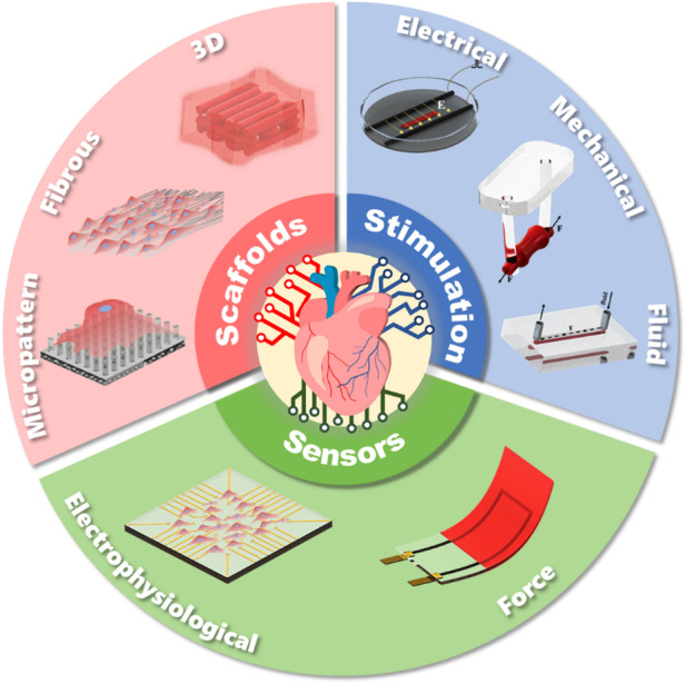
Heart-on-a-chip (HoC) has emerged as a highly efficient, cost-effective device for the development of engineered cardiac tissue, facilitating high-throughput testing in drug development and clinical treatment. HoC is primarily used to create a biomimetic microphysiological environment conducive to fostering the maturation of cardiac tissue and to gather information regarding the real-time condition of cardiac tissue. The development of architectural design and advanced manufacturing for these "3S" components, scaffolds, stimulation, and sensors is essential for improving the maturity of cardiac tissue cultivated on-chip, as well as the precision and accuracy of tissue states. In this review, the typical structures and manufacturing technologies of the "3S" components are summarized. The design and manufacturing suggestions for each component are proposed. Furthermore, key challenges and future perspectives of HoC platforms with integrated "3S" components are discussed. Architecture design concepts of scaffolds, stimulation and sensors in chips.

Keywords: Biosensors; Electrical and electronic engineering; Microfluidics.


Conflict of interest statement

Conflict of interestThe authors declare no competing interests.

Figures

None
Architecture design concepts of scaffolds, stimulation and sensors in chips.
Fig. 1
Fig. 1
Major milestones in cardiac tissue scaffolds, stimulation, and sensors architectures and applications–,,–
Fig. 2
Fig. 2. 2D scaffolds.
Point contact constraint structures: a Schematic diagram of point contact constraint; b PDMS micropillar arrays; c PDMS micropillar arrays coated with fibronectin; Some groove structures: d Schematic diagram of groove constraint; e Silicon substrates containing micron-sized notches; f PEG nanogrooves; Fibers constraint structures: g Schematic diagram of fiber constrained structure; h Disordered PCL fibrous networks; i Ordered nanofiber
Fig. 3
Fig. 3. 3D scaffolds.
Typical porous scaffolds: a Schematic diagram of porous scaffolds; b Porous poly(octamethylene maleate (anhydride) citrate) (POMaC) scaffolds; c Accordion-like honeycombs scaffolds; d POMaC scaffolds with rhombic pores; e Ventricles-like fibrous scaffolds; 3D scaffolds with volumetric structure: f Schematic diagram of volumetric structure; g DIW 3D microfibrous hydrogel scaffolds; h Gold Nanocomposite Bioink; i 3D Printing of Personalized Thick and Perfusable Cardiac Patches and Hearts; j 3D bioprinting of collagen to rebuild components of the human heart
Fig. 4
Fig. 4. Typical structures for electrical stimulation.
Show typical structures of rod electrodes: a Schematic diagram of rod electrodes; b Biowire with rod electrodes; c Arrayed rod electrodes,; Show integrated rod electrodes structures: d Schematic diagram of integrated electrical stimulation on chip; e Integrated patterned counter electrodes on chip; f Integrated stainless steel rods as electrical stimulation on chip; Conductive scaffolds assist Rod electrodes structures: g Schematic diagram of conductive scaffolds; h Carbon-Nanotube-Embedded Hydrogel as conductive scaffolds; i Conductive nanofibrous meshes; 2D patterned electrodes: j Schematic diagram of patterned electrodes; k Surface-patterned electrode bioreactor; l Micropatterned SU-8 cantilever integrated with metal electrode for enhanced electromechanical stimulation
Fig. 5
Fig. 5
. Typical structures for passive stimulation, a Schematic diagram of flexible frame constrain; b Engineered cardiac tissue patch; c Schematic diagram of flexible columns constrain; d Typical flexible column structures; e Schematic diagram of cantilever beam; f Cantilever beam structures
Fig. 6
Fig. 6
Active mechanical stimulation structures, a,c Two typical stretch structures; b Anisotropic stretch structure; d Isotropic stretch structure; e Schematic diagram of squeeze structure; f Integrated mechanical squeeze stimulation chip
Fig. 7
Fig. 7. Force sensor structures.
Show some typical sensor structures for thin tissue: a Schematic diagram of thin film force sensor; b CellDrum; c Cantilever beam force sensor; Sensor structures for thick tissue: d Schematic diagram of thick film force sensor; e Force sensing of flexible columns; f A platform with flexible columns sensors
Fig. 8
Fig. 8. MEA structure diagram.
Typical 2D MEA structures: a Schematic diagram of 2D MEA; b Gold mushroom-shaped microelectrodes; c Microelectrode arrays on soft materials; d Nanobranched microelectrode array; e Flexible Graphene Multielectrode Arrays; 3D MEA structures: f Schematic diagram of 3D MEA; g 3D transistor arrays; h 3D self-rolled biosensor array; i Flexible 3D printed microwires

Similar articles

Cited by

References

    1. Zhuang RZ, Lock R, Liu B, Vunjak-Novakovic G. Opportunities and challenges in cardiac tissue engineering from an analysis of two decades of advances. Nat. Biomed. Eng. 2022;6:327–338. doi: 10.1038/s41551-022-00885-3. - DOI - PubMed
    1. Wang X, et al. Engineered cardiac tissues: a novel in vitro model to investigate the pathophysiology of mouse diabetic cardiomyopathy. Acta Pharmacol. Sin. 2021;42:932–941. doi: 10.1038/s41401-020-00538-8. - DOI - PMC - PubMed
    1. Dou W, et al. Microengineered platforms for characterizing the contractile function of in vitro cardiac models. Microsyst. Nanoeng. 2022;8:26. doi: 10.1038/s41378-021-00344-0. - DOI - PMC - PubMed
    1. Abulaiti M, et al. Establishment of a heart-on-a-chip microdevice based on human iPS cells for the evaluation of human heart tissue function. Sci. Rep. 2020;10:19201. doi: 10.1038/s41598-020-76062-w. - DOI - PMC - PubMed
    1. Zhang F, et al. Design and fabrication of an integrated heart-on-a-chip platform for construction of cardiac tissue from human iPSC-derived cardiomyocytes and in situ evaluation of physiological function. Biosens. Bioelectr. 2021;179:113080. doi: 10.1016/j.bios.2021.113080. - DOI - PubMed

LinkOut - more resources