

Modified TI-RADS Coupled with BRAFV600E Enhances Diagnostic Efficiency in Papillary Thyroid Carcinoma: Prospective Study
- PMID: 39006910
- PMCID: PMC11246655
- DOI: 10.2147/IJGM.S456820
Modified TI-RADS Coupled with BRAFV600E Enhances Diagnostic Efficiency in Papillary Thyroid Carcinoma: Prospective Study
Abstract
Background: Thyroid disorders, relatively common diseases of the endocrine system, have risen gradually in recent years. Early detection and accurate diagnosis of thyroid cancer hold exceptional importance. This study aimed to determine the efficacy of a modified TI-RADS and BRAFV600E mutation testing for thyroid cancer (PTC) diagnosis.
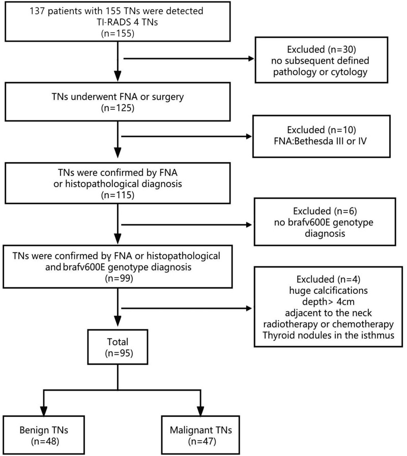
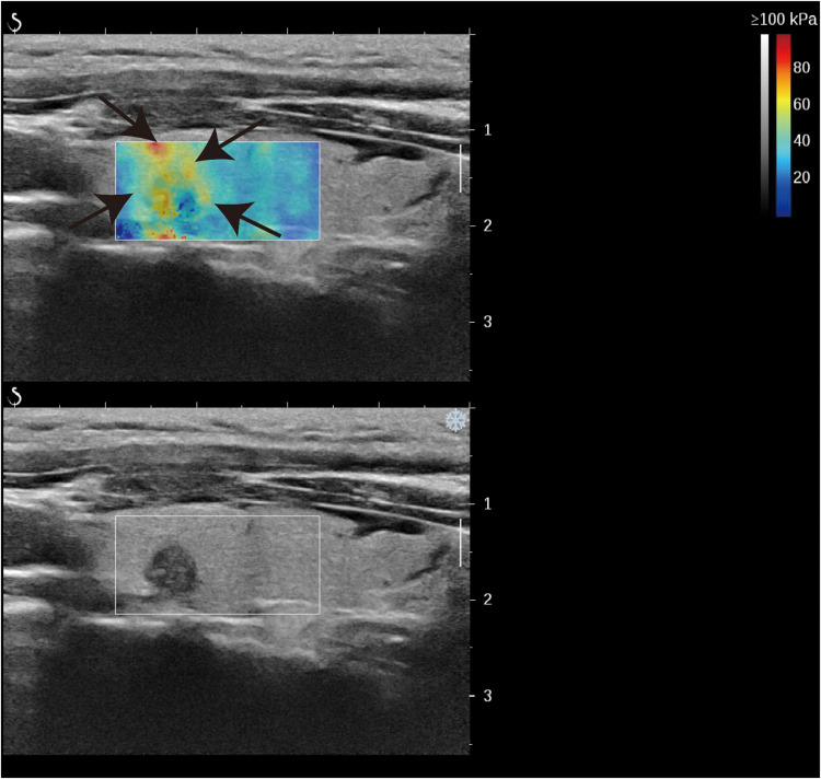
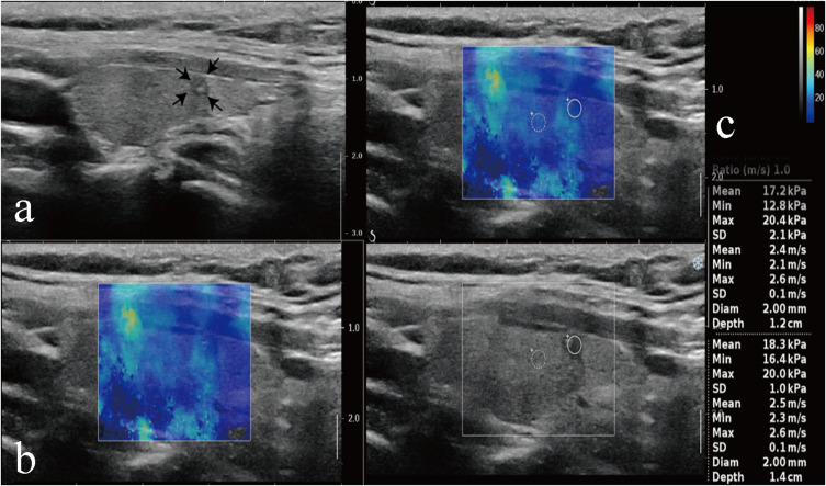
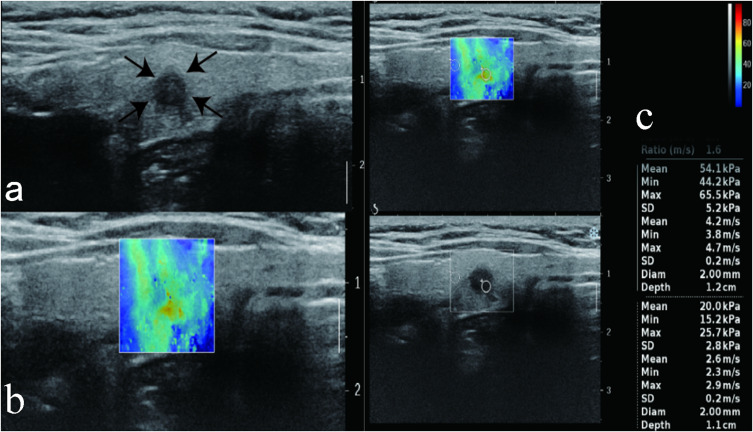
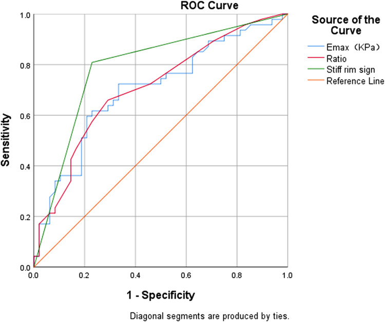
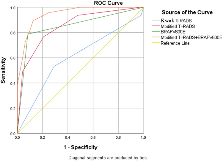
Methods: Ninety five thyroid nodules (48 benign and 47 malignant) from 81 patients were examined using Kwak Thyroid Imaging Reporting and Data System (TI-RADS) were subjected to shear wave elasticity (SWE), BRAFV600E genotyping and fine needle aspiration (FNA) cytology.
Results: The modified TI-RADS exhibited superior diagnostic accuracy compared to TI-RADS in differentiating benign nodules from malignant thyroid nodules. Moreover, the AUC of modified TI-RADS in conjunction with BRAFV600E was the highest at 95% CI (0.898-0.992, p=0.003), surpassing other diagnostic methods in enhanced sensitivity and maintaining high specificity.
Conclusion: The diagnostic efficiency of this combination surpassed that of individual diagnostic methods.
Keywords: BRAFV600E; modified TI-RADS; papillary thyroid cancer; shear wave elasticity; stiff rim sign.
© 2024 Wang et al.
Conflict of interest statement
The authors of this manuscript declare no relationships with any companies, whose products or services may be related to the subject matter of the article.
Figures
References
-
- Mulita F, Anjum F. Thyroid Adenoma. Study Guide from StatPearls Publishing, Treasure Island (FL), 24 Sep 2020. Available from: https://europepmc.org/article/MED/32965923. Accessed 28 June, 2024. - PubMed
-
- Haugen BR, Alexander EK, Bible KC, et al. 2015 American thyroid association management guidelines for adult patients with thyroid nodules and differentiated thyroid cancer: the American thyroid association guidelines task force on thyroid nodules and differentiated thyroid cancer. Thyroid. 2016;26(1):1–133. doi: 10.1089/thy.2015.0020 - DOI - PMC - PubMed
LinkOut - more resources
Full Text Sources
