. 2024 Jun 21:39:101761.
doi: 10.1016/j.bbrep.2024.101761. eCollection 2024 Sep.

Increased levels of thymidine kinase 1 in malignant cell-derived extracellular vesicles

Affiliations

Increased levels of thymidine kinase 1 in malignant cell-derived extracellular vesicles

Ehsan Manouchehri Doulabi et al. Biochem Biophys Rep. .

Abstract

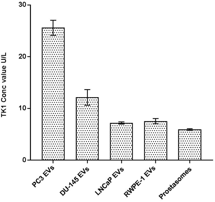
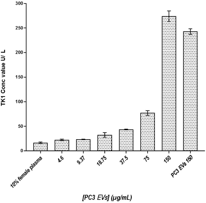
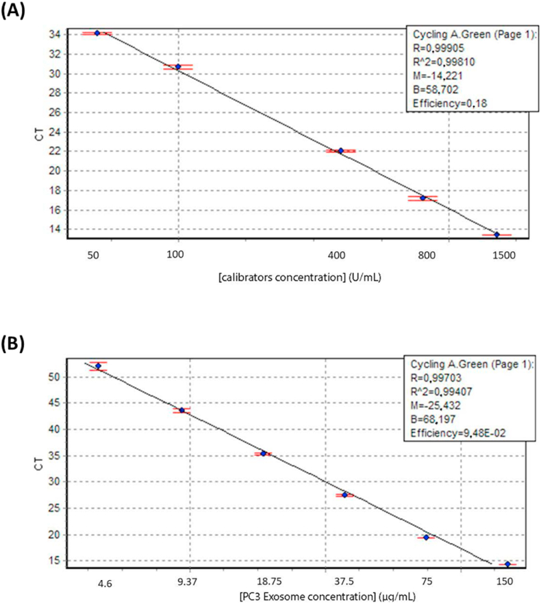
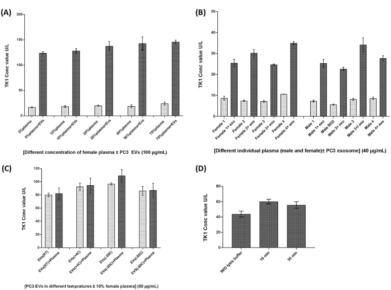
Extracellular vesicles (EVs), whose main subtypes are exosomes, microparticles, and apoptotic bodies, are secreted by all cells and harbor biomolecules such as DNA, RNA, and proteins. They function as intercellular messengers and, depending on their cargo, may have multiple roles in cancer development. Thymidine kinase 1 (TK1) is a cell cycle-dependent enzyme used as a biomarker for cell proliferation. TK1 is usually elevated in cancer patients' serum, making the enzyme a valuable tumor proliferation biomarker that strongly correlates with cancer stage and metastatic capabilities. Here, we investigated the presence of TK1 in EVs derived from three prostate cancer cell lines with various p53 mutation statuses (LNCaP, PC3, and DU145), EVs from the normal prostate epithelial cell line RWPE-1 and EVs isolated from human seminal fluid (prostasomes). We measured the TK1 activity by a real-time assay for these EVs. We demonstrated that the TK1 enzyme activity is higher in EVs derived from the malignant cell lines, with the highest activity from cells deriving from the most aggressive cancer, compared to the prostasomes and RWPE-1 EVs. The measurement of TK1 activity in EVs may be essential in future prostate cancer studies.

Keywords: Cancer; Diagnostics; Extracellular vesicles; Prostasomes; Prostate cancer; Thymidine kinase 1 (TK1); p53.


Conflict of interest statement

Per Stålhandske is employed by Biovica International AB commercializing the TK1 measurement assay. The other authors state that there is no conflict of interest regarding the publication of this article.

Figures

Fig. 1
Fig. 1
Measured TK1 activity for EVs isolated from the three cancer cell lines, PC3 DU145, LNCaP, from the normal epithelial prostate cell line RWPE-1, and prostasomes isolated from seminal fluids from healthy individuals. TK1 activities were normalized by the total protein concentration of 40 μg/mL for each sample. The mean values of TK1 activity for EVs isolated from the cell lines PC3, DU145, LNCaP, RWPE-1, and from prostasomes, 25, 12.1, 7.2, 7.5, and 5.9 U/L, respectively (n = 3, mean ± s.d.).
Fig. 2
Fig. 2
TK1 activity measurement from purified PC3-derived EVs at different protein concentrations, a 2-fold serial dilution with starting concentration at 4.6 μg/mL, spiked in 10 % female plasma (n = 3, mean ± s.d.). 10 % female plasma without spiked EVs and pure PC3-derived EVs at the highest protein concentration (150 μg/mL) were also analyzed.
Fig. 3
Fig. 3
Top graph (A) displays the real-time assay control run of the calibrators A-D. The R2 > 0.998 indicates a usual assay run. In bottom graph (B), the real-time assay control was run with a 2-fold serial dilution of PC3 cell line EVs with an R2 > 0.994. Data are represented as mean ± s.d. (n = 3).
Fig. 4
Fig. 4
(A), TK1 activity measured in 100 μg/mL PC3-derived EVs spiked in pooled female plasma ranging from 5 % to 75 % in PBS. (B) The background levels of the TK1 activity were analyzed in 10 % plasma samples from four men and four women with and without 40 μg/mL spiked PC3-derived EVs. (C) Analysis of the TK1 activity in 80 μg/mL of the PC3-derived EVs in PBS or 10 % plasma after storing at RT, 4 °C, −20 °C and −80 °C for 24 h. (D) The TK1 activity was investigated in 80 μg/mL PC3-derived EVs that were treated with or without lysis and at either 10 or 30 min of lysis incubation. Data are represented as mean ± s.d. (n = 3).

Similar articles

Cited by

References

    1. Colombo M., Raposo G., Théry C. Biogenesis, secretion, and intercellular interactions of exosomes and other extracellular vesicles. Annu. Rev. Cell Dev. Biol. 2014;30 doi: 10.1146/annurev-cellbio-101512-122326. - DOI - PubMed
    1. Doyle L.M., Wang M.Z. Overview of extracellular vesicles, their origin, composition, purpose, and methods for exosome isolation and analysis. Cells. 2019;8(7) doi: 10.3390/cells8070727. - DOI - PMC - PubMed
    1. Wickman G., Julian L., Olson M.F. How apoptotic cells aid in the removal of their own cold dead bodies. Cell Death Differ. 2012;19(5) doi: 10.1038/cdd.2012.25. - DOI - PMC - PubMed
    1. Babiker A.A., Nilsson B., Ronquist G., Carlsson L., Ekdahl K.N. Transfer of functional prostasomal CD59 of metastatic prostatic cancer cell origin protects cells against complement attack. Prostate. 2005;62(2) doi: 10.1002/pros.20102. - DOI - PubMed
    1. Ronquist K.G., Sanchez C., Dubois L., et al. Energy-requiring uptake of prostasomes and PC3 cell-derived exosomes into non-malignant and malignant cells. J. Extracell. Vesicles. 2016;5(1) doi: 10.3402/jev.v5.29877. - DOI - PMC - PubMed

LinkOut - more resources