. 2024 Jun 5;32(3):200823.
doi: 10.1016/j.omton.2024.200823. eCollection 2024 Sep 19.

Oncolytic vaccinia virus harboring CLEC2A gene enhances viral replication and antitumor efficacy

Affiliations

Oncolytic vaccinia virus harboring CLEC2A gene enhances viral replication and antitumor efficacy

Chunqing Gao et al. Mol Ther Oncol. .

Abstract

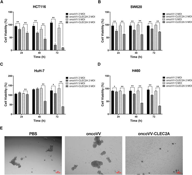
In the field of innovative cancer treatment strategies, oncolytic vaccinia virus (VV)es have gained traction as promising vectors. In the current study, we inserted the human C-type lectin domain family 2 member A (CLEC2A) gene into VV, creating a replicating therapeutic, oncoVV-CLEC2A. The findings reveal that oncoVV-CLEC2A effectively suppresses colorectal proliferation of mouse xenografts and a range of human cancer cell lines by augmenting viral reproduction capabilities, including the lung cancer H460 cell line, colorectal cancer cell lines (HCT116 and SW620), and hepatocellular carcinoma HuH-7 cell line. Moreover, it is evident that oncoVV-CLEC2A can induce antitumor immunity by boosting cytokine production but not antivirus response, and enhancing calreticulin expression. Further investigation indicates that oncoVV-CLEC2A can enhance antitumor capabilities by activating natural killer cells to produce interferon-γ and induce M1-like macrophage polarization. These findings shed light on the antitumor mechanisms of oncoVV-CLEC2A, provide a theoretical basis for oncolytic therapies, and lay the groundwork for novel strategies for modifying VVs.

Keywords: C-type lectin domain family 2 member A; MT: Regular Issue; NK cells; antitumor; oncolytic vaccinia virus; viral replication.


Conflict of interest statement

The authors declare no conflict of interest.

Figures

None
Graphical abstract
Figure 1
Figure 1
The high expression of CLEC2A in oncoVV-CLEC2A treatment (A) Schematic of engineering the oncoVV-CLEC2A recombinant. (B–E) The expression of CLEC2A in HCT116 cells (B), SW620 cells (C), HuH-7 cells (D), and H460 cells (E) at 36 h after infection with oncoVV-CLEC2A or oncoVV. The mRNA level of CLEC2A was identified by qPCR assay. Data are shown as the mean ± SD (∗p < 0.05, ∗∗p < 0.01). (F and G) The expression levels of CLEC2A in HCT116 cells (F) and HuH-7 cells (G) after treatment with oncoVV-CLEC2A in cells. GAPDH was used as the internal control.
Figure 2
Figure 2
The cell cytotoxicity of oncoVV-CLEC2A on tumor cells and tumor spheroids (A–D) Cell viability in HCT116 (A), SW620 (B), HuH-7 (C), and H460 (D) cell lines. Data are shown as the mean ± SD (∗p < 0.05, ∗∗p < 0.01). (E) Morphology of the HCT116 tumor spheroids observed by inverted microscope at day 5 after infection with 5 MOI of oncoVV or oncoVV-CLEC2A.
Figure 3
Figure 3
OncoVV-CLEC2A promoted apoptosis (A–D) The proportion of apoptotic cells was determined using flow cytometry in HCT116 (A), SW620 (B), HuH-7 (C), and H460 (D) cell lines at 36 h after infection with oncoVV-CLEC2A or oncoVV. Data are shown as the mean ± SD (∗p < 0.05, ∗∗p < 0.01). (E) The expression levels of cleaved caspase-9, caspase-9, and caspase-3 after treatment with oncoVV-CLEC2A in cells. GAPDH was used as the internal control.
Figure 4
Figure 4
The promotion of oncoVV-CLEC2A in viral yields (A and B) The replicative capacity of oncoVV and oncoVV-CLEC2A was assessed using TCID50 assay in HCT116 cells (A) and HuH-7 cells (B). Data are shown as the mean ± SD (∗p < 0.05, ∗∗p < 0.01). (C and D) The levels of A27L in HCT116 (C) and HuH-7 (D) cells at 36 h after infection with oncoVV or oncoVV-CLEC2A were determined using Western blot. GAPDH was used as the internal control.
Figure 5
Figure 5
Inflammatory response mechanism induced by oncoVV-CLEC2A (A and B) The transcription of cytokines in HCT116 cells (A) and HuH-7 cells (B) at 36 h after infection with oncoVV or oncoVV-CLEC2A. The mRNA levels of IFN-α, IL-6 IFN-β and TNF-α were measured using qPCR. Data are shown as the mean ± SD (∗p < 0.05, ∗∗p < 0.01).
Figure 6
Figure 6
Inflammatory response mechanism induced by oncoVV-CLEC2A (A–C) Transcriptional activity of AP-1 (A), NF-κB (B), and ISRE (C) were investigated by dual luciferase reporter gene assay in HCT116 and HuH-7 cells. Data are shown as the mean ± SD (∗p < 0.05, ∗∗p < 0.01). (D) The protein expression levels of c-Fos, p-c-Fos, c-Jun, and p-c -Jun in HCT116 and HuH-7 cells were quantified using western blot analysis at the 36-h time point after infection with oncoVV-CLEC2A or oncoVV. GAPDH was used as the internal control for loading normalization.
Figure 7
Figure 7
The antitumor efficacy of oncoVV-CLEC2A on HCT116 tumors (A) The volume of the tumor was evaluated every 3 days after treatment. (B) Proportion of tumor weight in the body weight of mice. Data are shown as the mean ± SD (∗p < 0.05, ∗∗p < 0.01). (C) Photo of tumors taken from sacrificed mice. (D) HE staining of tumor tissue obtained from the mice treated with 0.9% NaCl, oncoVV or oncoVV-CLEC2A. (E and F) IHC detected A27L (E) and CALR (F) expression in the tumor tissue of mice treated with 0.9% NaCl, oncoVV or oncoVV-CLEC2A.
Figure 8
Figure 8
OncoVV-CLEC2A enhanced the cytotoxicity of NK cell (A) The serum levels of IFN-γ and TNF-α in the mice. (B–D) FCM analysis and quantitative analysis of percentage of NK cells (CD49b+ IFN-γ+) (B), M1 macrophagocytes (F4/80+ IFN-γ+) (C) and M2 macrophagocytes (F4/80+ IL-4+) (D) of three groups. Data are shown as the mean ± SD (∗p < 0.05, ∗∗p < 0.01).

Similar articles

References

    1. Fukuhara H., Ino Y., Todo T. Oncolytic virus therapy: A new era of cancer treatment at dawn. Cancer Sci. 2016;107:1373–1379. - PMC - PubMed
    1. Mondal M., Guo J., He P., Zhou D. Recent advances of oncolytic virus in cancer therapy. Hum. Vaccines Immunother. 2020;16:2389–2402. - PMC - PubMed
    1. Jefferson A., Cadet V.E., Hielscher A. The mechanisms of genetically modified vaccinia viruses for the treatment of cancer. Crit. Rev. Oncol. Hematol. 2015;95:407–416. - PubMed
    1. Smith G.L., Moss B. Infectious poxvirus vectors have capacity for at least 25 000 base pairs of foreign DNA. Gene. 1983;25:21–28. - PubMed
    1. Breitbach C.J., Burke J., Jonker D., Stephenson J., Haas A.R., Chow L.Q.M., Nieva J., Hwang T.H., Moon A., Patt R., et al. Intravenous delivery of a multi-mechanistic cancer-targeted oncolytic poxvirus in humans. Nature. 2011;477:99–102. - PubMed

LinkOut - more resources