. 2024;143(1):27.
doi: 10.1186/s13358-024-00323-8. Epub 2024 Jul 12.

New insights into the early morphological evolution of sea turtles by re-investigation of Nichollsemys baieri, a three-dimensionally preserved fossil stem chelonioid from the Campanian of Alberta, Canada

Affiliations

New insights into the early morphological evolution of sea turtles by re-investigation of Nichollsemys baieri, a three-dimensionally preserved fossil stem chelonioid from the Campanian of Alberta, Canada

Juliette C L Menon et al. Swiss J Palaeontol. 2024.

Abstract

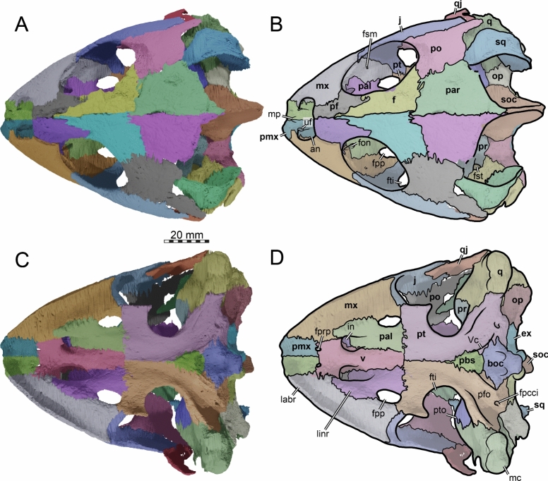
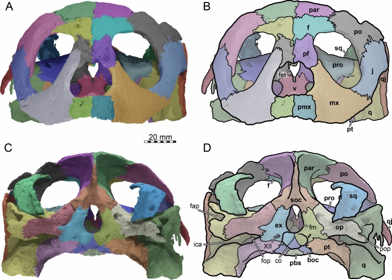
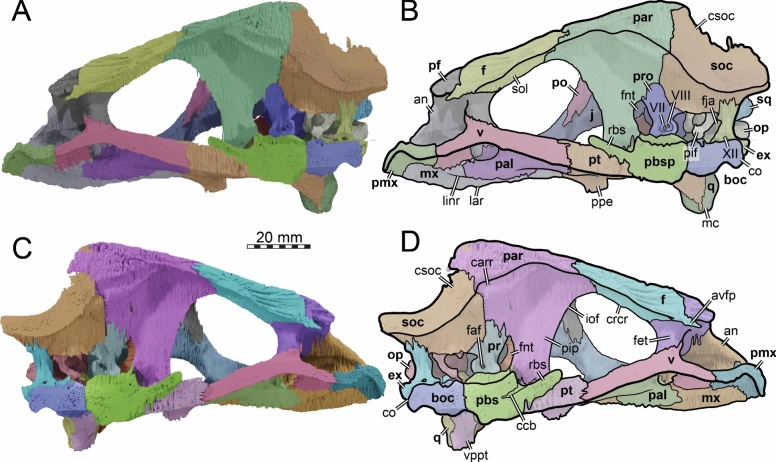
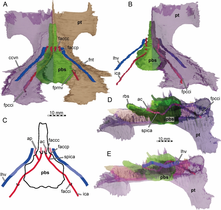
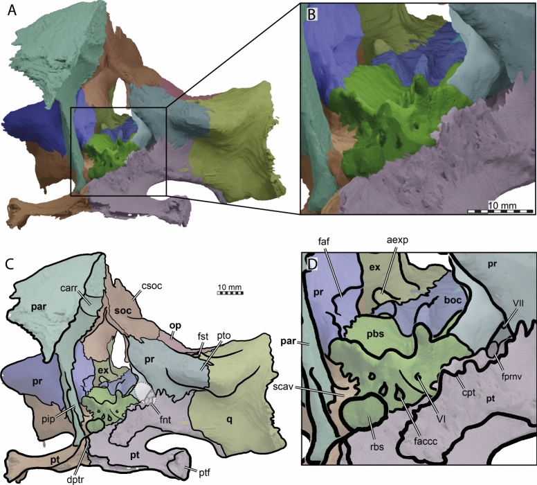
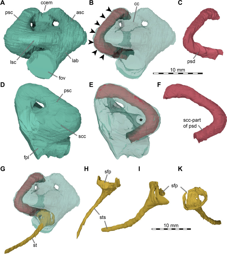
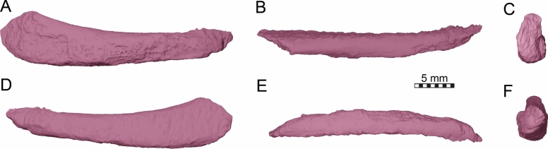
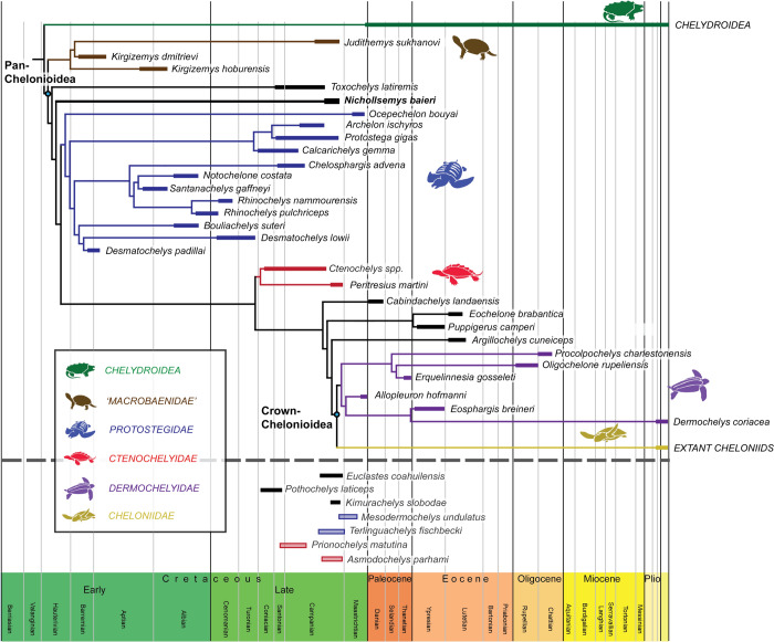
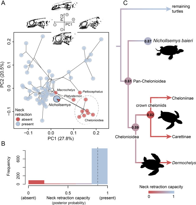
The early evolution of Pan-Chelonioidea (sea turtles) is poorly understood. This is in part due to the rarity of undeformed skulls of definitive early stem chelonioids. In this work, we redescribe the holotype of Nichollsemys baieri using µCT scans and segmentations of the skull. This fossil is the best 3D preserved skull of any Campanian sea turtle, and includes partial "soft tissue" preservation. Nichollsemys is morphologically similar but clearly distinct from Toxochelys spp., and both show a mosaic of plesiomorphic and derived chelonioid features. The internal cranial anatomy documents the presence of derived characters in Nichollsemys baieri that are absent in Toxochelys spp., such as the loss of the epipterygoids and the rod-like shape of the rostrum basisphenoidale. Among the numerous plesiomorphic characters is the presence of a splenial bone, which was unnoticed before. An updated phylogenetic analysis retrieves Nichollsemys baieri as a non-protostegid early stem chelonioid in a slightly more crownward position than Toxochelys latiremis. Our phylogeny includes macrobaenids and protostegids as pan-chelonioids, and we find unorthodox results for dermochelyids. Thus, although Nichollsemys baieri provides important new insights into the early morphological evolution of sea turtles, much work remains to be done. As a completely 3D preserved specimen, we included Nichollsemys baieri into a recent landmark-based skull shape dataset of turtles. Morphospace analysis reveals an intermediate position between cryptodires and crown chelonioids. Based on these data, we also predict that Nichollsemys baieri was still capable of neck retraction, constraining the loss of this trait to more crownward pan-chelonioids.

Supplementary information: The online version contains supplementary material available at 10.1186/s13358-024-00323-8.

Keywords: CT scan; Evolution; Neck retraction; Pan-Chelonioidea; Phylogeny; Sea turtle.


Conflict of interest statement

Competing interestsThe authors declare no competing interests.

Figures

Fig. 1
Fig. 1
Photographs of the holotype skull of Nichollsemys baieri (TMP 1997.99.1). A dorsal view. B ventral view. C left lateral view. D right lateral view. E anterior view. F posterior view
Fig. 2
Fig. 2
Three-dimensional renderings of the cranium of the holotype specimen of Nichollsemys baieri (TMP 1997.99.1). A dorsal view; B interpretative line-drawing; C ventral view; D interpretative line-drawing. Note that bones are labelled in bold and features labelled in regular font. an apertura naris, boc basioccipital, ex exoccipital, f frontal, fon foramen orbito-nasale, fpcci foramen posterius canalis caroticus interni, fpp foramen palatinum posterius, fprp foramen praepalatinum, fsm foramen supramaxillare, fst foramen stapedio-temporale, fti inferior temporal fossa, in internal nares, j jugal, labr labial ridge, linr lingual ridge, mc mandibular condyle, mx maxilla, mp median pocket, op opisthotic, pal palatine, par parietal, pbs parabasisphenoid, pf prefrontal, pfo pterygoid fossa, pmx premaxilla, po postorbital, pr prootic, pt pterygoid, pto processus trochlearis oticum, q quadrate, qj quadratojugal, soc supraoccipital, sq squamosal, uf unnamed foramen, v vomer, Vc “V” shaped ventral crest
Fig. 3
Fig. 3
Three-dimensional renderings of the cranium of the holotype specimen of Nichollsemys baieri (TMP 1997.99.1). A anterior view; B interpretative line-drawing; C posterior view; D interpretative line-drawing. Note that bones are labelled in bold and features labelled in regular font. an apertura naris, boc basioccipital, ex exoccipital, f frontal, fap foramen antrum postoticum, fet fissura ethmoidalis, XII foramen nervi hypoglossi, fm foramen magnum, fop fenestra postotica, ica incisura columella auris, j jugal, mx maxilla, op opisthotic, pal palatine, par parietal, pbs parabasisphenoid, pf prefrontal, pmx premaxilla, po postorbital, pop paraoccipital process, pr prootic, pt pterygoid, qj quadratojugal, q quadrate, soc supraoccipital, sq squamosal, v vomer
Fig. 4
Fig. 4
Three-dimensional renderings of the cranium of the holotype specimen of Nichollsemys baieri (TMP 1997.99.1). A left lateral view; B interpretative line-drawing; C right lateral view; D interpretative line-drawing. Note that bones are labelled in bold and features labelled in regular font. Also note that on the right skull side, the jugal facets on the postorbital and quadratojugal indicate an originally larger extent of the bone, indicated in D by hashed color and a dashed outline. ap antrum postoticum, crcr crista cranii, csoc crista supraoccipitalis, f frontal, fap foramen antrum postoticum, fon foramen orbito-nasale, ica incisura columella auris, j jugal, mc mandibular condyle, mx maxilla, pal palatine, par parietal, pbs parabasisphenoid, pf prefrontal, pmx premaxilla, po postorbital, pt pterygoid, ptf pterygoid flange, q quadrate, qap quadrate articular process, qj quadratojugal, soc supraoccipital, sq squamosal, v vomer, vqjp ventral quadratojugal process
Fig. 5
Fig. 5
Three-dimensional renderings of the cranium of the holotype specimen of Nichollsemys baieri (TMP 1997.99.1). A right internal view; B interpretative line-drawing; C left internal view; D interpretative line-drawing. Note that bones are labelled in bold and features labelled in regular font. an apertura naris, avfp anteroventral process of the frontal, boc basioccipital, carr cartilaginous rider, ccb canalis caroticus cerebralis, co occipital condyle, csoc crista supraoccipitalis, ex exoccipital, f frontal, faf fossa acustico-facialis, fet fissura ethmoidalis, fnt foramen nervi trigemini, fon foramen orbito-nasale, iof interorbital fenestra, j jugal, mc mandibular condyle, mx maxilla, op opisthotic, pal palatine, par parietal, pbs parabasisphenoid, pf prefrontal, pif processus interfenestralis, pip processus inferior parietalis, pmx premaxilla, po postorbital, pr prootic, pt pterygoid, qj quadratojugal, q quadrate, rbs rostrum basisphenoidale, soc supraoccipital, sol sulcus olfactorius, sq squamosal, XII foramen nervi hypoglossi, v vomer, VII foramen nervi facialis, VIII foramen nervi acusticus, vppt ventral process of the pterygoid
Fig. 6
Fig. 6
Three-dimensional renderings of the partial braincase of the holotype specimen of Nichollsemys baieri (TMP 1997.99.1), A anterolateral view onto the trigeminal area of the left skull side; B anterolateral view of the internal basicranium; C close-up on the parabasisphenoid area based on B; D interpretative line-drawing. Note that bones are labelled in bold and features labelled in regular font. avr anterior ventral ridge, boc basioccipital, btb basis tuberculum basalis, cci canalis caroticus internus, clp clinoid process, faccc foramen anterius canalis caroticus cerebralis, faccp foramen anterius canalis carotici palatinum, faf fossa acustico-facialis, fcav foramen cavernosum, fnt foramen nervi trigemini, par parietal, pbs parasphenoid, pip processus inferior parietalis, pr prootic, pt pterygoid, q quadrate, rbs rostrum basisphenoidale, scav sulcus cavernosus, soc supraoccipital, ‘st' vestigial sella turcica, VI foramen nervi abducentis
Fig. 7
Fig. 7
Three-dimensional renderings of the partial left lateral skull side of Nichollsemys baieri (TMP 1997.99.1) with the squamosal rotated into alternative positions. A, C, E squamosal with low inclination. B, D, F squamosal with high inclination. A, B left lateral view; C, D dorsal view; E, F posterior view. Note that bones are labelled in bold and features labelled in regular font. aspq anterior squamosal tapering process, fst foramen stapedio-temporale, j jugal, op opisthotic, plf posterolateral fossa, po postorbital, pop paraoccipital process prootic, q quadrate, qj quadratojugal, sq squamosal, sqms squamosal medial sheet, sqpp squamosal posterior process
Fig. 8
Fig. 8
Three-dimensional reconstruction of the partial basicranium of the holotype specimen of Nichollsemys baieri (TMP 1997.99.1) with inferred cranial circulation based on canal endocasts. A dorsal view, with left side rendered transparent; B ventral view, with left side rendered transparent; C interpretative line-drawing of the dorsal view; D lateral left view without lateral head vein; E lateral left view including lateral head vein. Note that bones are labelled in bold and features labelled in regular font. ac cerebral artery, ap palatine artery, faccc foramen anterius canalis caroticus cerebralis, faccp foramen anterius canalis carotici palatinum, fnt foramen nervi trigemini, fprnv foramen pro ramo nervi vidiani, ica internal carotid artery, lhv lateral head vein, pbs parabasisphenoid, pt pterygoid, rbs rostrum basisphenoidale, spica split of the internal carotid artery
Fig. 9
Fig. 9
Three-dimensional renderings of the partial braincase of the holotype specimen of Nichollsemys baieri (TMP 1997.99.1) in anterior and slight lateral view. A overview of braincase morphology; B close-up onto the parabasisphenoid and anterior canalis cavernosus area; C interpretative line-drawing based on A; D interpretative line-drawing based on B. Note that bones are labelled in bold and features labelled in regular font. aexp anterior (hooked) exoccipital process, boc basioccipital, carr cartilaginous rider, cpt crista pterygoidei, csc crista supraoccipitalis, dptr dorsal interpterygoid ridge, faccc foramen anterius canalis caroticus cerebralis, faf fossa acustico-facialis, fnt foramen nervi trigemini, fprnv foramen pro ramo nervi vidiani, fst foramen stapedio-temporale, par parietal, pbs parabasisphenoid, pip processus inferior parietalis, pr prootic, pt pterygoid, ptf pterygoid flange, pto processus trochlearis oticum, q quadrate, rbs rostrum basisphenoidale, scav sulcus cavernosus, soc supraoccipital, VI foramen nervi abducentis, VII foramen nervi facialis
Fig. 10
Fig. 10
Three-dimensional renderings of right endosseous labyrinth, the stapes, and “soft tissue” preservation of the posterior semicircular duct of the holotype specimen of Nichollsemys baieri (TMP 1997.99.1). A lateral view of the endosseous labyrinth; B lateral view of the endosseous labyrinth rendered transparent with the preserved posterior semicircular duct in solid; C lateral view of the posterior semicircular duct; D posterior view of the endosseous labyrinth; E posterior view of the endosseous labyrinth rendered transparent with the preserved posterior semicircular duct in solid; F posterior view of the posterior semicircular duct; G lateral view of labyrinth structures rendered transparent to see the position of the stapes, which is slightly medially displaced through the fenestra ovalis; H stapes in dorsal view; I stapes in anterior view; K stapes in medial view. Arrows in B show the attachment of the posterior semicircular duct to the outer (dorsal) wall of the semicircular canal. Asterisk in E shows the level of the lateral semicircular canal to highlight how the "soft tissue" bends ventrally around it. Asc anterior semicircular canal, cc common crus, ccem common crus embayment, fov fenestra ovalis, fpl fenestra perilymphatica, lab endosseous labyrinth, lsc lateral semicircular canal, psc posterior semicircular canal, psd posterior semicircular duct, scc secondary common crus, sfp stapedial footplate, st stapes, sts stapedial shaft
Fig. 11
Fig. 11
Three-dimensional renderings of the mandible of the holotype specimen of Nichollsemys baieri (TMP 1997.99.1). A dorsal view; B interpretative line-drawing; C anterior view; D close-up onto the posterior part of the left mandibular ramus in dorsal view; E interpretative line-drawing; F ventral view; G interpretative line-drawing. Note that bones are labelled in bold and features labelled in regular font. An angular, art articular, cor coronoid, den dentary, fM fossa Meckelii, pra prearticular, spl splenial, sur surangular, surl surangular lamina, syn symphyseal notch, tri triturating surface
Fig. 12
Fig. 12
Three-dimensional renderings of the mandible of the holotype specimen of Nichollsemys baieri (TMP 1997.99.1). A left lateral view; B interpretative line-drawing; C right lateral view; D interpretative line-drawing; E medial view onto right mandibular ramus; F interpretative line-drawing; G medial view onto left mandibular ramus; H interpretative line-drawing. Note that the dentary was artificially cut at the symphysis to get undisturbed views onto the medial mandibular sides. Also note that bones are labelled in bold and features labelled in regular font. an angular, art articular, cor coronoid, corp coronoid process, den dentary, fdfm foramen dentofaciale majus, fim foramen intermandibulare medius, fM fossa Meckelii, fnat foramen nervi auriculotemporalis, pra prearticular, sM sulcus Meckelii, spf splenial foramen, spl splenial, sur surangular
Fig. 13
Fig. 13
Three-dimensional renderings of the left cornu branchiale I of the holotype specimen of Nichollsemys baieri (TMP 1997.99.1). A external lateral view; B dorsal view; C anterior view; D medial lateral view; E ventral view; F posterior view
Fig. 14
Fig. 14
Strict consensus tree obtained from 2 MPTs from our implied weighting analysis using ordered characters based on our fully revised matrix plotted through time. Note that only the pan-chelonioid part of the tree is shown (full tree available as Supplementary file S5). Thick bars on terminal branches represent stratigraphic occurrences of focal taxa. Note that some additional taxa not included in our phylogeny are also plotted below the dotted gray line to highlight the diversity of Late Cretaceous sea turtles. Silhouettes by Juliette Menon and Walter Joyce
Fig. 15
Fig. 15
Morphospace position of Nichollsemys baieri and predictions of neck retraction. A Cranial morphospace of turtles, showing first two axes of a PCA. Nichollsemys is highlighted as a pie chart, indicating the probability of being capable of neck retraction predicted by pfda. Constellation of points above the plot indicates the consensus shape (gray) and extremes of the first two PCA axes (black) in left lateral view. B Histogram of posterior probabilities for the presence of neck retraction in Nichollsemys baieri across the set of pfda replicates. Dashed line indicates the mean value (0.86). C Ancestral reconstructions of neck retraction mapped onto a simplified phylogenetic tree of pan-chelonioid turtles. Numbers at nodes represent the ancestral probability of neck retraction capacity for pan-chelonioids (0.61), crown chelonioids (0.59) and crown cheloniids (0.02). Chelonia mydas silhouette by Edwin Price, and Dermochelys coriacea silhouette by Guillaume Dera, both retrieved from phylopic.org (under CC0 1.0)

Similar articles

References

    1. Adams DC. A method for assessing phylogenetic least squares mod- els for shape and other high-dimensional multivariate data. Evolution. 2014;68:2675–2688. doi: 10.1111/evo.12463. - DOI - PubMed
    1. Adams, D.C., Collyer, M.L. & Kaliontzopoulou, A. (2020). Geomorph: software for geometric morphometric analyses. R package version 3.2.1. Available via https://cran.r-project.org/package=geomorph
    1. Albrecht PW. The cranial arteries of turtles and their evolutionary significance. Journal of Morphology. 1976;149(2):159–182. doi: 10.1002/jmor.1051490203. - DOI - PubMed
    1. Anquetin J. Reassessment of the phylogenetic interrelationships of basal turtles (Testudinata) Journal of Systematic Palaeontology. 2012;10(1):3–45. doi: 10.1080/14772019.2011.558928. - DOI
    1. Anquetin J, Püntener C, Billon-Bruyat J-P. Portlandemys gracilis n. sp., a new coastal marine turtle from the Late Jurassic of Porrentruy (Switzerland) and a reconsideration of plesiochelyid cranial anatomy. PLoS ONE. 2015;10(6):e0129193. doi: 10.1371/journal.pone.0129193. - DOI - PMC - PubMed

LinkOut - more resources