. 2024 Jun 28:11:1419093.
doi: 10.3389/fmolb.2024.1419093. eCollection 2024.

Diagnostic model for hepatocellular carcinoma using small extracellular vesicle-propagated miRNA signatures

Affiliations

Diagnostic model for hepatocellular carcinoma using small extracellular vesicle-propagated miRNA signatures

Xinyi Luo et al. Front Mol Biosci. .

Abstract

Background: Hepatocellular carcinoma (HCC) is the most common type of liver cancer. Small extracellular vesicles (sEVs) are bilayer lipid membrane vesicles containing RNA that exhibit promising diagnostic and prognostic potential as cancer biomarkers.

Aims: To establish a miRNA panel from peripheral blood for use as a noninvasive biomarker for the diagnosis of HCC.

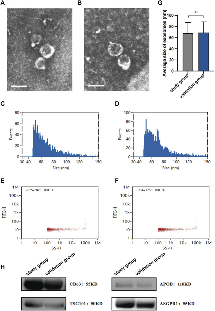
Methods: sEVs obtained from plasma were profiled using high-throughput sequencing. The identified differential miRNA expression patterns were subsequently validated using quantitative real-time polymerase chain reaction analysis.

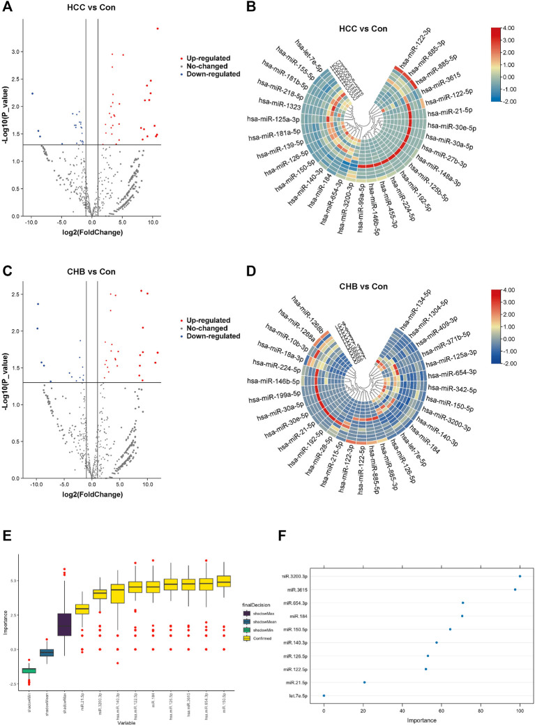
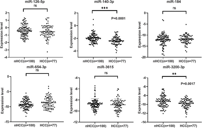
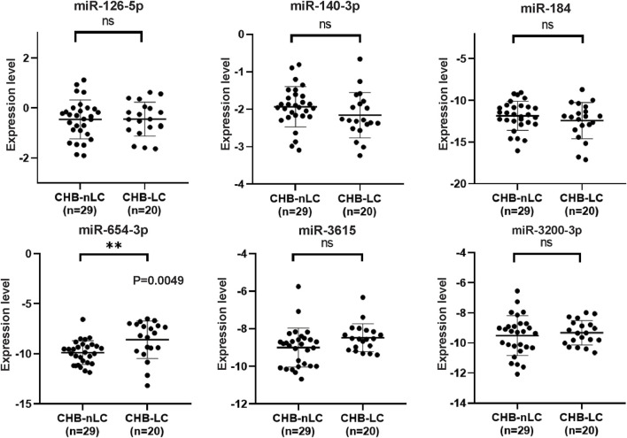
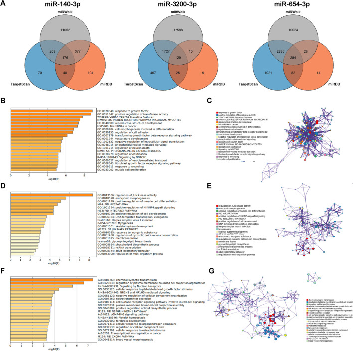
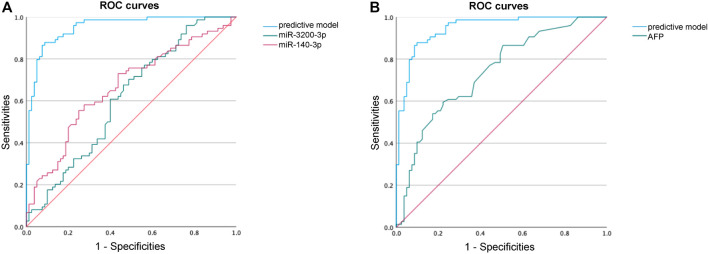
Results: The random forest method identified ten distinct miRNAs distinguishing HCC plasma from non-HCC plasma. During validation, miR-140-3p (p = 0.0001) and miR-3200-3p (p = 0.0017) exhibited significant downregulation. Enrichment analysis uncovered a notable correlation between the target genes of these miRNAs and cancer development. Utilizing logistic regression, we developed a diagnostic model incorporating these validated miRNAs. Receiver operating characteristic (ROC) curve analysis revealed an area under the curve (AUC) of 0.951, with a sensitivity of 90.1% and specificity of 87.8%.

Conclusion: These aberrantly expressed miRNAs delivered by sEVs potentially contribute to HCC pathology and may serve as diagnostic biomarkers for HCC.

Keywords: biomarker; cancer; hepatocellular carcinoma; microRNA; small extracellular vesicle.


Conflict of interest statement

The authors declare that the research was conducted in the absence of any commercial or financial relationships that could be construed as a potential conflict of interest.

Figures

FIGURE 1
FIGURE 1
Characterization of isolated sEVs. The morphology of sEVs from the study group (A) and the validation group (B) was observed by TEM. Scales bars, 100 nm. The size of sEVs from the study group (C) and the validation group (D) was assessed by NTA. The concentration of sEVs from the study group (E) and the validation group (F) was assessed by NTA. (G) Average sizes of sEVs from the study group and the validation group. (H) Expression of protein markers of sEV, as evaluated by western blotting. Data are mean ± standard deviation (SD); ns: p > 0.05.
FIGURE 2
FIGURE 2
Aberrantly expressed miRNAs in HCC and CHB. (A) The volcano plot shows the expression of sEV-delivered miRNAs in HCC patients, with red and blue spots representing significantly upregulated and downregulated miRNAs in HCC, respectively. (B) Hierarchical clustering analysis of the sEV-delivered miRNA expression profile in HCC patients, displaying the top 30 enriched miRNAs. (C) The volcano plot represents the expression of sEV-delivered miRNAs in CHB patients. (D) Hierarchical clustering analysis of the sEV-delivered miRNA expression profile in CHB patients, showing the top 30 enriched miRNAs. (E) The boxplot displays the minimum, average, and maximum shadow scores of all combined features, with yellow variables representing important miRNAs confirmed by random forest analysis. (F) The dot plot visually represents the importance of variables. Each dot corresponds to a miRNA, and its position on the axis reflects its level of importance. Generally, more important miRNAs are positioned higher on the axis.
FIGURE 3
FIGURE 3
The expression levels of six selected miRNAs analyzed by qRT-PCR in HCC and nHCC samples. The expression values were normalized to miR103a-3p, and the experiments were performed three times. The results revealed that miR-140-3p and miR-3200-3p exhibited downregulation in the plasma sEVs of HCC patients. However, there were no significant changes observed in the expression levels of miR-126-5p, miR-184, miR-654-3p, and miR-3615.
FIGURE 4
FIGURE 4
qRT-PCR analysis of six selected miRNAs in CHB-LC patients compared to CHB-nLC patients. The results demonstrate that miR-654-3p showed a significant upregulation in the plasma sEVs of CHB patients with liver cirrhosis. On the other hand, the expression levels of the other five candidate miRNAs remained unchanged.
FIGURE 5
FIGURE 5
Prediction of target genes. (A) Venn diagram of the common predicted genes for the three miRNAs. (B,D, F) GO enrichment analysis for miR-140-3p, miR-3200-3p, and miR-654-3p, respectively. The top 20 clusters are shown in the bar graph based on the list of input genes, with the color representing the p-values. The log10(P) indicates the p-value in logarithmic base 10. (C,E,G) Network visualization of the selected enriched terms. The top 20 informative entries from the obtained GO clusters are depicted. Nodes in the network are colored based on their cluster IDs.
FIGURE 6
FIGURE 6
ROC curve analysis to identify HCC. (A) ROC curve performance of our predictive model compared with miR-140-3p and miR-3200-3p. (B) ROC curve performance of our predictive model compared with AFP.

Similar articles

Cited by

References

    1. Arroyo J. D., Chevillet J. R., Kroh E. M., Ruf I. K., Pritchard C. C., Gibson D. F., et al. (2011). Argonaute2 complexes carry a population of circulating microRNAs independent of vesicles in human plasma. Proc. Natl. Acad. Sci. U. S. A. 108, 5003–5008. 10.1073/pnas.1019055108 - DOI - PMC - PubMed
    1. Bray F., Laversanne M., Sung H., Ferlay J., Siegel R. L., Soerjomataram I., et al. (2024). Global cancer statistics 2022: GLOBOCAN estimates of incidence and mortality worldwide for 36 cancers in 185 countries. CA Cancer J. Clin. 74, 229–263. 10.3322/caac.21834 - DOI - PubMed
    1. Chen Y., Lun A. T. L., Smyth G. K. (2016). From reads to genes to pathways: differential expression analysis of RNA-Seq experiments using Rsubread and the edgeR quasi-likelihood pipeline. F1000Res 5, 1438. 10.12688/f1000research.8987.2 - DOI - PMC - PubMed
    1. Cho O., Kim D.-W., Cheong J.-Y. (2021). Plasma exosomal miRNA levels after radiotherapy are associated with early progression and metastasis of cervical cancer: a pilot study. J. Clin. Med. 10, 2110. 10.3390/jcm10102110 - DOI - PMC - PubMed
    1. Diener C., Keller A., Meese E. (2022). Emerging concepts of miRNA therapeutics: from cells to clinic. Trends Genet. 38, 613–626. 10.1016/j.tig.2022.02.006 - DOI - PubMed

LinkOut - more resources