. 2024 Jun 20:47:87-96.
doi: 10.1016/j.jot.2024.05.005. eCollection 2024 Jul.

Comparative effect of skeletal stem cells versus bone marrow mesenchymal stem cells on rotator cuff tendon-bone healing

Affiliations

Comparative effect of skeletal stem cells versus bone marrow mesenchymal stem cells on rotator cuff tendon-bone healing

Linfeng Wang et al. J Orthop Translat. .

Abstract

Background: Bone marrow mesenchymal stem cells (BMSCs) have immense potential in applications for the enhancement of tendon-bone (T-B) healing. Recently, it has been well-reported that skeletal stem cells (SSCs) could induce bone and cartilage regeneration. Therefore, SSCs represent a promising choice for cell-based therapies to improve T-B healing. In this study, we aimed to compare the therapeutic potential of SSCs and BMSCs for tendon-bone healing.

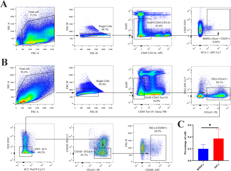
Methods: SSCs and BMSCs were isolated by flow cytometry, and their proliferation ability was measured by CCK-8 assay. The osteogenic, chondrogenic, and adipogenic gene expression in cells was detected by quantitative real-time polymerase chain reaction (qRT-PCR). C57BL/6 mice underwent unilateral supraspinatus tendon detachment and repair, and the mice were then randomly allocated to 4 groups: control group (tendon-bone interface without any treatment), hydrogel group (administration of blank hydrogel into the tendon-bone interface), hydrogel + BMSCs group (administration of hydrogel with BMSCs into the tendon-bone interface), and hydrogel + SSCs group (administration of hydrogel with SSCs into the tendon-bone interface). Histological staining, Micro-computed tomography (Micro-CT) scanning, biomechanical testing, and qRT-PCR were performed to assay T-B healing at 4 and 8 weeks after surgery.

Results: SSCs showed more cell proportion, exhibited stronger multiplication capacity, and expressed higher osteogenic and chondrogenic markers and lower adipogenic markers than BMSCs. In vivo assay, the SSCs group showed a better-maturated interface which was characterized by richer chondrocytes and more proteoglycan deposition, as well as more newly formed bone at the healing site and increased mechanical properties when compared to other there groups. qRT-PCR analysis revealed that the healing interface in the SSCs group expressed more transcription factors essential for osteogenesis and chondrogenesis than the interfaces in the other groups.

Conclusions: Overall, the results demonstrated the superior therapeutic potential of SSCs over BMSCs in tendon-bone healing.

The translational potential of this article: This current study provides valuable insights that SSCs may be a more effective cell therapy for enhancing T-B healing compared to BMSCs.

Keywords: Bone marrow mesenchymal stem cells; Cell-based therapies; Skeletal stem cells; Tendon-bone healing; Therapeutic potential.


Figures

Image 1
Graphical abstract of the overall experimental process.
Fig. 1
Fig. 1
Fluorescence-activated cell sorting for BMSCs and SSCs. (A) Representative flow cytometry plots gating strategy for isolation of BMSCs. (B) Representative flow cytometry plots gating strategy for isolation of SSCs. (C) Quantification of flow cytometry data of the sorted cells. n = 6 per group. Data are represented as mean ± SD, *p < 0.05 represent significant differences between the indicated columns.
Fig. 2
Fig. 2
Cellular characterization of BMSCs and SSCs. (A) Representative morphology of BMSCs and SSCs. Scale bar: 200 μm; (B) Proliferative capacity of BMSCs and SSCs evaluated by Cell Counting Kit-8 (CCK-8) assays from day 1 to day 7. n = 6 per group. (C) qRT-PCR analysis for the expression of adipogenic gene (FABP4, ADPN, and PPAR-γ), osteogenic gene (RUNX2, OCN, and COL1A1) and chondrogenic gene (ACAN, SOX9, and COL2A1) in BMSCs and SSCs. n = 6 per group. Data are represented as mean ± SD, *p < 0.05, **p < 0.01, ***p < 0.001 represent significant differences between the indicated columns.
Fig. 3
Fig. 3
Histologic evaluation of ST enthesis at 4 weeks postoperatively. (A) Representative images of H&E and TB&FG staining at the repaired site. The area selected by the rectangle dashed line is the local magnified area. scale bar = 200 μm; (B) Tendon-to-bone insertion maturing score of regenerated enthesis. n = 6 per group. ST, supraspinatus tendon; H&E, hematoxylin and eosin; TB&FG, toluidine blue and fast green; All data are mean ± SD. *p < 0.05, **p < 0.01, ***p < 0.001 represent significant differences between the indicated columns. (For interpretation of the references to colour in this figure legend, the reader is referred to the Web version of this article.)
Fig. 4
Fig. 4
Histologic evaluation of ST enthesis at 8 weeks postoperatively. (A) Representative images of H&E and TB&FG staining at the repaired site. The area selected by the rectangle dashed line is the local magnified area. scale bar = 200 μm. (B) Tendon-to-Bone insertion maturing score of the regenerated enthesis. n = 6 per group. ST, supraspinatus tendon; H&E, hematoxylin and eosin; TB&FG, toluidine blue and fast green; All data are mean ± SD. *p < 0.05, **p < 0.01, ***p < 0.001 represent significant differences between the indicated columns. (For interpretation of the references to colour in this figure legend, the reader is referred to the Web version of this article.)
Fig. 5
Fig. 5
Radiological analysis by Micro-CT for RC healing site at 4 and 8 weeks postoperatively. (A) three-dimensional reconstruction image of the humeral head, Scale bar = 500 μm. (B) The morphological parameters of new bone at the healing site. n = 6 per group. RC, rotator cuff; BV/TV, bone volume/total volume; Tb.Th, trabecular thickness; Tb.N, trabecular number; Tb.Sp, trabecular separation. All data are mean ± SD. *p < 0.05, **p < 0.01, ***p < 0.001 represent significant differences between the indicated columns.
Fig. 6
Fig. 6
Biomechanical testing for the insertion site of supraspinatus tendon–humerus complexes at postoperative weeks 4 and 8, as expressed by failure load, stiffness, ultimate stress, and cross-sectional area. n = 10 per group. Data are represented as mean ± SD, *p < 0.05, **p < 0.01, ***p < 0.001 represent significant differences between the indicated columns.
Fig. 7
Fig. 7
qRT-PCR analysis for the expression of osteogenic gene (RUNX2, OCN, and COL1A1) and chondrogenic gene (ACAN, SOX9, and COL2A1) in the mouse ST enthesis after injury‐repair surgery. n = 6 per group. The fold change of expression was determined by comparing the expression level of repaired ST enthesis in different treatment groups to that in the normal enthesis. The data are presented as mean ± SD. *p < 0.05, **p < 0.01, ***p < 0.001 represent significant differences between the indicated columns. ST, supraspinatus tendon.

Similar articles

Cited by

References

    1. Fang F., Xiao Y., Zelzer E., Leong K.W., Thomopoulos S. A mineralizing pool of Gli1-expressing progenitors builds the tendon enthesis and demonstrates therapeutic potential. Cell Stem Cell. 2022;29(12) [eng] - PMC - PubMed
    1. Zhang T., Li S., Chen Y., Xiao H., Wang L., Hu J., et al. Characterize the microstructure change after tendon enthesis injury using synchrotron radiation μCT. J Orthop Res. 2022;40(11):2678–2687. [eng] - PubMed
    1. Zhou Y., Xie S., Tang Y., Li X., Cao Y., Hu J., et al. Effect of book-shaped acellular tendon scaffold with bone marrow mesenchymal stem cells sheets on bone-tendon interface healing. J Orthop Translat. 2021;26:162–170. [eng] - PMC - PubMed
    1. Xiao H., Zhang T., Li C.J., Cao Y., Wang L.F., Chen H.B., et al. Mechanical stimulation promotes enthesis injury repair by mobilizing cells via ciliary TGF-β signaling. Elife. 2022;11 [eng] - PMC - PubMed
    1. Yea J.-H., Bae T.S., Kim B.J., Cho Y.W., Jo C.H. Regeneration of the rotator cuff tendon-to-bone interface using umbilical cord-derived mesenchymal stem cells and gradient extracellular matrix scaffolds from adipose tissue in a rat model. Acta Biomater. 2020;114:104–116. [eng] - PubMed

LinkOut - more resources