. 2024 Jun 21:47:105-115.
doi: 10.1016/j.jot.2024.06.007. eCollection 2024 Jul.

Upregulation of FABP4 induced inflammation in the pathogenesis of chronic tendinopathy

Affiliations

Upregulation of FABP4 induced inflammation in the pathogenesis of chronic tendinopathy

Zebin Ma et al. J Orthop Translat. .

Abstract

Objectives: Excessive inflammation contributes to the pathogenesis of tendinopathy. Fatty acid binding protein 4 (FABP4) is a pro-inflammatory adipokine mediating various metabolic and inflammatory diseases. This study aimed to examine the expression of FABP4 and its association with the expressions of inflammatory cytokines in tendinopathy. The effects of a single injection of FABP4 on tendon pathology and inflammation were examined. The effect of FABP4 on the expressions of inflammatory cytokines and the effect of IL-1β on the expression of FABP4 in tendon-derived stem/progenitor cells (TDSCs) were also investigated.

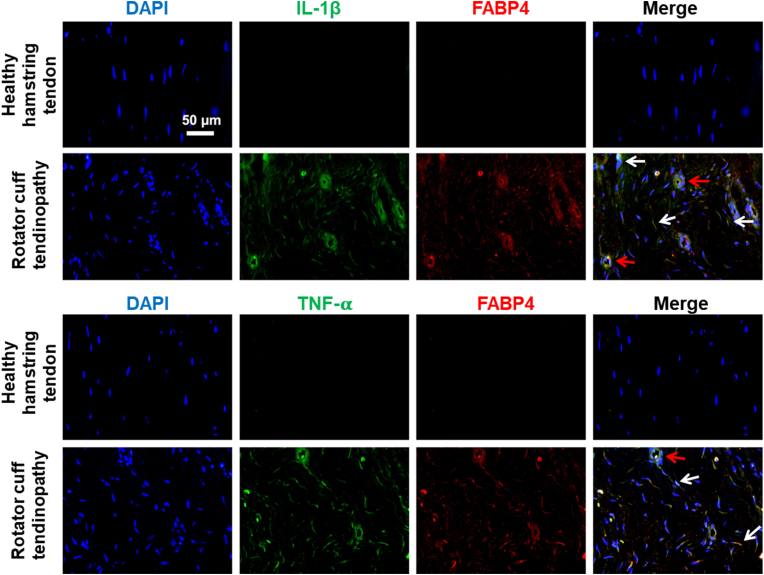
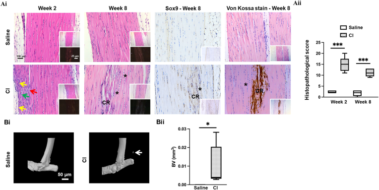
Methods: 1) Clinical patellar tendinopathy samples, healthy hamstring tendon samples, and healthy patellar tendon samples, 2) rotator cuff tendinopathy samples and healthy hamstring tendon samples; and 3) Achilles tendons of mice after saline or collagenase injection (CI) were stained for FABP4, IL-1β, IL-6, TNF-α and IL-10 by immunohistochemistry (IHC). For the rotator cuff tendinopathy samples, co-localization of FABP4 with IL-1β and TNF-α was done by immunofluorescent staining (IF). Mouse Achilles tendons injected with FABP4 or saline were collected for histology and IHC as well as microCT imaging post-injection. TDSCs were isolated from human and mouse tendons. The mRNA expressions of inflammatory cytokines in human and mouse TDSCs after the addition of FABP4 was quantified by qRT-PCR. The expression of FABP4 in TDSCs isolated from rotator cuff tendinopathy samples and healthy hamstring tendon samples was examined by IF. Mouse Achilles TDSCs were treated with IL-1β. The mRNA and protein expressions of FABP4 were examined by qRT-PCR and IF, respectively.

Results: There was significant upregulation of FABP4 in the patellar tendinopathy samples and rotator cuff tendinopathy samples compared to their corresponding controls. FABP4 was mainly expressed in the pathological areas including blood vessels, hypercellular and calcified regions. The expressions of IL-1β and TNF-α increased in human rotator cuff tendinopathy samples and co-localized with the expression of FABP4. Collagenase induced tendinopathic-like histopathological changes and ectopic calcification in the mouse Achilles tendinopathy model. The expressions of inflammatory cytokines (IL-1β, IL-6, TNF-α, IL-10) and FABP4 increased in hypercellular region, round cells chondrocyte-like cells and calcified regions in the mouse Achilles tendons post-collagenase injection. A single injection of FABP4 in mouse Achilles tendons induced histopathological changes resembling tendinopathy, with increased cell rounding, loss of collagen fiber alignment, and additionally presence of chondrocyte-like cells and calcification post-injection. The expressions of IL1-β, IL-6, TNF-α and IL-10 increased in mouse Achilles tendons post-FABP4 injection. FABP4 increased the expressions of IL10, IL6, and TNFa in human TDSCs as well as the expressions of Il1b, Il6, and Il10 in mouse TDSCs. Human tendinopathy TDSCs expressed higher level of FABP4 compared to healthy hamstring TDSCs. Besides, IL-1β increased the expression of FABP4 in mouse TDSCs.

Conclusion: In conclusion, an upregulation of FABP4 is involved in excessive inflammation and pathogenesis of tendinopathy. TDSCs is a potential source of FABP4 during tendon inflammation.

Translation potential of this article: FABP4 can be a potential treatment target of tendinopathy.

Keywords: Collagenase-induced tendon injury; FABP4; Inflammation; Tendinopathy; Tendon-derived stem / progenitor cells.


Conflict of interest statement

We don't any potential conflicts of interest.

Figures

Image 1
Graphical abstract
Fig. 1
Fig. 1
Upregulation of FABP4 in patellar and rotator cuff tendinopathy. (A) Photomicrographs showing the immunohistochemical staining of FABP4 in (i) healthy hamstring tendon samples, (ii) healthy patellar tendon samples, and (ii) patellar tendinopathy samples. Scale bar = 100 μm, yellow arrow: blood vessel; red arrow: hypercellular area; CR: calcified region. (B) Boxplot showing IOD/mm2 of FABP4 signal. n = 6/group; **p < 0.01; ***p < 0.001 (C) Photomicrographs showing the immunohistochemical staining of FABP4 in healthy human hamstring tendon samples and human rotator cuff tendinopathy samples. Scale bar = 100 μm; yellow arrow: blood vessel; red arrow: hypercellular area; CR: calcified region. (D) Boxplot showing IOD/mm2 of FABP4 signal. n = 5/group; **p < 0.01.
Fig. 2
Fig. 2
Co-localization of FABP4 with inflammatory cytokines in rotator cuff tendinopathy. Photomicrographs showing co-localization of FABP4 with IL-1β or TNF-α in human rotator cuff tendinopathy samples. Scale bar = 50 µm; n = 5/group; white arrows: co-localized signal in tendon cells; red arrows: co-localized signal in blood vessels.
Fig. 3
Fig. 3
Collagenase injection induced histopathological and radiological changes in mouse Achilles tendons resembling human tendinopathy. (Ai) Photomicrographs showing H&E staining and corresponding polarized images, IHC of Sox9 and Von Kossa staining of representative samples of mouse Achilles tendons at week 2 and 8 after saline or collagenase injection. (Aii) Histopathological scoring of H&E images. n = 5/group; Scale bar = 100 μm (insert 25 μm); yellow arrow: blood vessels; red arrow: hypercellular area; green arrow: small, nucleated cells; CR: calcified region; *: chondrocyte-like cells; black arrow: immunopositive cells; ***p < 0.001 (Bi) microCT images showing ectopic calcification inside Achilles tendons at week 8 after saline or collagenase injection. (Bii) Boxplot showing the bone volume (BV) inside tendon in different groups. n = 6/group; Scale bar = 50 μm; white arrow: ectopic calcification; *p < 0.05.
Fig. 4
Fig. 4
Upregulation of FABP4 and inflammatory cytokines in mouse CI animal model.
Fig. 5
Fig. 5
FABP4 injection induced damages, ectopic bone and inflammation in mouse Achilles tendons resembling human tendinopathy. (Ai) Photomicrographs showing H&E staining and corresponding polarized images of representative samples of mouse Achilles tendons at week 2 and week 8 after saline or FABP4 (0.5 µg) injection. (Aii) Histopathological scoring of H&E images. n = 5/group; Scale bar = 100 µm (insert 25 µm); yellow arrow: blood vessels; red arrow: hypercellular area; green arrow: small, nucleated cells; CR: calcified region; *: chondrocyte-like cells; ***p < 0.001. (Bi) microCT images showing ectopic calcification inside Achilles tendons at week 8 after saline or FABP4 (0.5 µg) injection. (Bii) Boxplot showing the bone volume (BV) inside tendon in different groups. n = 6/group; Scale bar = 50 µm; white arrow: ectopic bone; *p < 0.05. (Ci) Photomicrographs showing the immunohistochemical staining of IL-1β, IL-6, TNF-α and IL-10 in representative samples of mouse Achilles tendons at week 2 and week 8 after saline or FABP4 (0.5 µg) injection. (Cii) Boxplots showing the IOD/mm2 of L-1β, IL-6, TNF-α and IL-10 at week 2 and week 8 after saline or FABP4 (0.5 µg) injection. n=5/group; Scale bar = 100 µm (insert 25 µm); red arrow: hypercellular area; *: chondrocyte-like cells; black arrow: immunopositive cells; *p < 0.05; **p < 0.01; ***p < 0.001.
Fig. 6
Fig. 6
FABP4 increased the mRNA expression of inflammatory cytokines. Boxplots showing (A) the expressions of IL1b, IlL, and TNFa in human TDSCs and; (B) the expressions of Il1b, Il6, and Il10 in mouse TDSCs after the addition of FABP4 (120 ng/mL) for 24 h *p < 0.05; **p < 0.01; ***p < 0.001; n = 4/group.
Fig. 7
Fig. 7
Expression of FABP4 in human rotator cuff tendinopathy TDSCs and IL-1β-treated mouse TDSCs. (A) Photomicrographs showing protein expression of FABP4 in human rotator cuff tendinopathy TDSCs compared to healthy hamstring tendon TDSCs; Scale bar: 100 μm; n = 4. The cell nucleus was stained by DAPI. (B) Photomicrographs showing protein expression of FABP4 in mouse TDSCs treated with IL-1β (10 ng/mL) for 48 h; Scale bar: 100 μm; n = 6. The cell nucleus was stained by DAPI. (C) Boxplot showing mRNA expression of Fabp4. *p < 0.05; n = 4–5/group.
None
figs1

References

    1. Fu S.C., Rolf C., Cheuk Y.C., Lui P.P., Chan K.M. Deciphering the pathogenesis of tendinopathy: a three-stages process. Sports Med Arthrosc Rehabil Ther Technol. 2010;2:30. - PMC - PubMed
    1. Rees J.D., Stride M., Scott A. Tendons--time to revisit inflammation. Br J Sports Med. 2014;48(21):1553–1557. - PMC - PubMed
    1. Challoumas D., Biddle M., Millar N.L. Recent advances in tendinopathy. Fac Rev. 2020;9:16. - PMC - PubMed
    1. Hu J.J., Yin Z., Shen W.L., Xie Y.B., Zhu T., Lu P., et al. Pharmacological regulation of in situ tissue stem cells differentiation for soft tissue calcification treatment. Stem Cell. 2016;34(4):1083–1096. - PubMed
    1. Rui Y.F., Lui P.P., Wong Y.M., Tan Q., Chan K.M. Altered fate of tendon-derived stem cells isolated from a failed tendon-healing animal model of tendinopathy. Stem Cell Dev. 2013;22(7):1076–1085. - PMC - PubMed

LinkOut - more resources