

A noval noninvasive targeted therapy for osteosarcoma: the combination of LIFU and ultrasound-magnetic-mediated SPIO/TP53/PLGA nanobubble
- PMID: 39007051
- PMCID: PMC11239426
- DOI: 10.3389/fbioe.2024.1418903
A noval noninvasive targeted therapy for osteosarcoma: the combination of LIFU and ultrasound-magnetic-mediated SPIO/TP53/PLGA nanobubble
Abstract
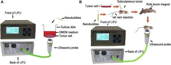
Purpose: Osteosarcoma (OS) is the most common type of primary malignant bone tumor. Transducing a functional TP53 gene can effectively inhibit OS cell activity. Poly lactic acid-glycolic acid (PLGA) nanobubbles (NBs) mediated by focused ultrasound (US) can introduce exogenous genes into target cells in animal models, but this technique relies on the passive free diffusion of agents across the body. The inclusion of superparamagnetic iron oxide (SPIO) in microbubbles allows for magnetic-based tissue localization. A low-intensity-focused ultrasound (LIFU) instrument was developed at our institute, and different intensities of LIFU can either disrupt the NBs (RLI-LIFU) or exert cytocidal effects on the target tissues (RHI-LIFU). Based on these data, we performed US-magnetic-mediated TP53-NB destruction and investigated its ability to inhibit OS growth when combined with LIFU both in vitro and in vivo.
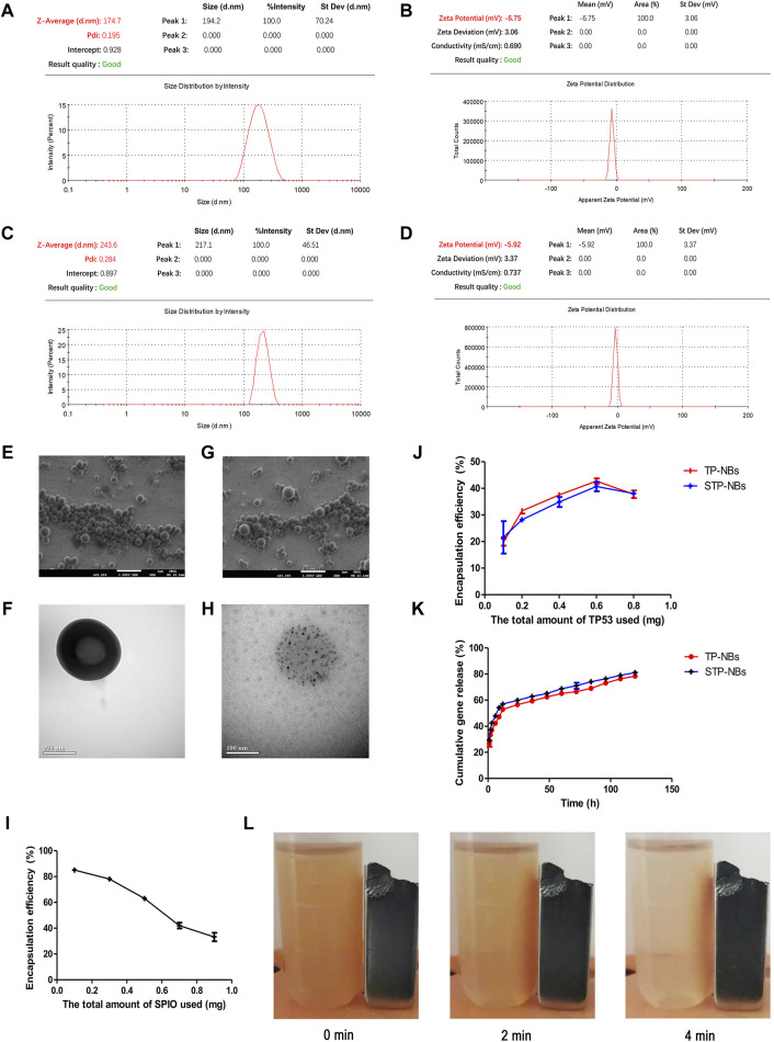
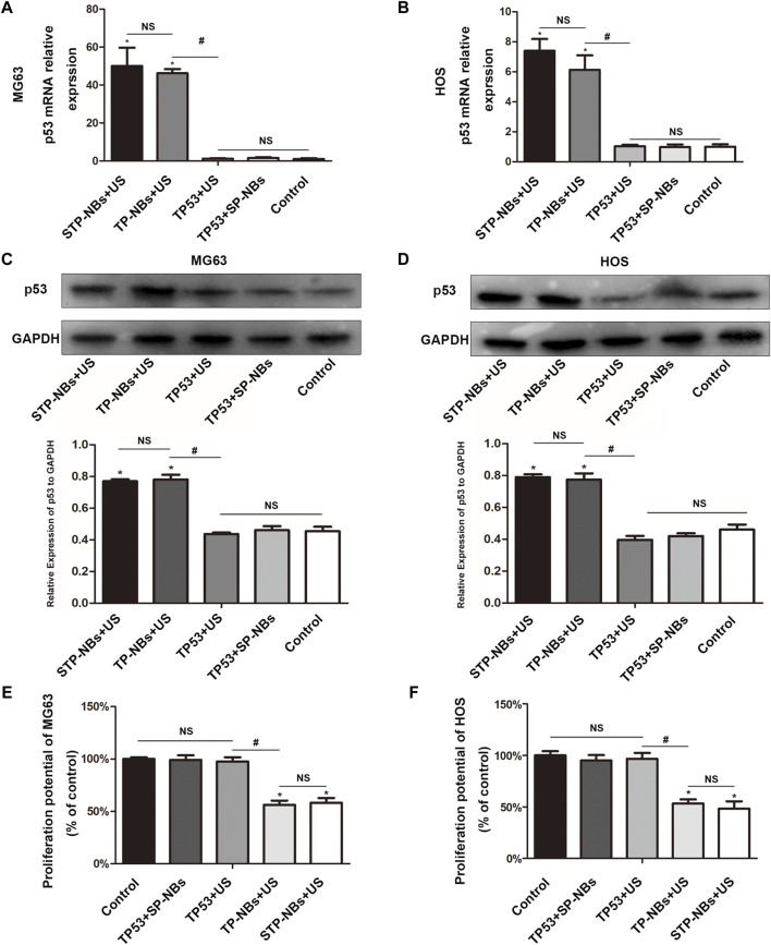
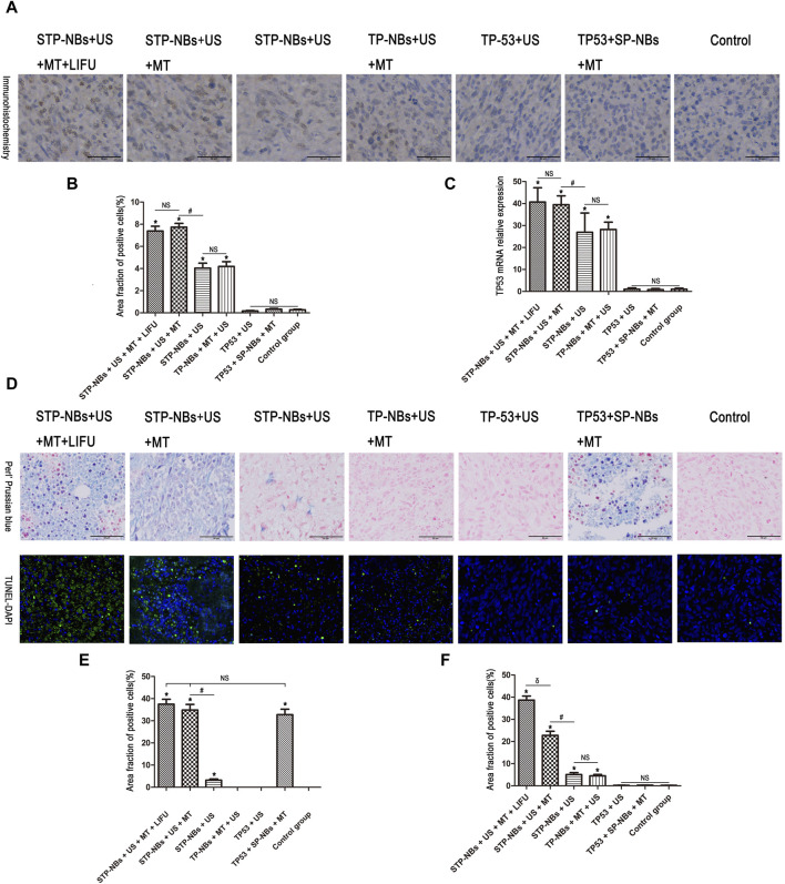
Methods: Several SPIO/TP53/PLGA (STP) NB variants were prepared and characterized. For the in vitro experiments, HOS and MG63 cells were randomly assigned into five treatment groups. Cell proliferation and the expression of TP53 were detected by CCK8, qRT-PCR and Western blotting, respectively. In vivo, tumor-bearing nude mice were randomly assigned into seven treatment groups. The iron distribution of Perls' Prussian blue-stained tissue sections was determined by optical microscopy. TUNEL-DAPI was performed to examine apoptosis. TP53 expression was detected by qRT-PCR and immunohistochemistry.
Results: SPIO/TP53/PLGA NBs with a particle size of approximately 200 nm were prepared successfully. For in vitro experiments, ultrasound-targeted transfection of TP53 overexpression in OS cells and efficient inhibition of OS proliferation have been demonstrated. Furthermore, in a tumor-bearing nude mouse model, RLI-LIFU-magnetic-mediated SPIO/TP53/PLGA NBs increased the transfection efficiency of the TP53 plasmid, resulting in apoptosis. Adding RHI-LIFU to the treatment regimen significantly increased the apoptosis of OS cells in vivo.
Conclusion: Combining LIFU and US-magnetic-mediated SPIO/TP53/PLGA NB destruction is potentially a novel noninvasive and targeted therapy for OS.
Keywords: PLGA; low-intensity-focused ultrasound; nanobubbles; osteosarcoma; superparamagnetic iron oxide.
Copyright © 2024 Ren, Xiang, Liu, Wang, Zhou and Hu.
Conflict of interest statement
The authors declare that the research was conducted in the absence of any commercial or financial relationships that could be construed as a potential conflict of interest.
Figures
Similar articles
-
Ultrasound-mediated delivery of RGD-conjugated nanobubbles loaded with fingolimod and superparamagnetic iron oxide nanoparticles: targeting hepatocellular carcinoma and enhancing magnetic resonance imaging.RSC Adv. 2020 Oct 27;10(64):39348-39358. doi: 10.1039/d0ra06415g. eCollection 2020 Oct 21. RSC Adv. 2020. PMID: 35518389 Free PMC article.
-
Brain Delivery of Curcumin Through Low-Intensity Ultrasound-Induced Blood-Brain Barrier Opening via Lipid-PLGA Nanobubbles.Int J Nanomedicine. 2021 Nov 4;16:7433-7447. doi: 10.2147/IJN.S327737. eCollection 2021. Int J Nanomedicine. 2021. PMID: 34764649 Free PMC article.
-
Nerve growth factor delivery by ultrasound-mediated nanobubble destruction as a treatment for acute spinal cord injury in rats.Int J Nanomedicine. 2017 Mar 2;12:1717-1729. doi: 10.2147/IJN.S128848. eCollection 2017. Int J Nanomedicine. 2017. PMID: 28280337 Free PMC article.
-
A Comprehensive Review of Low-Intensity Focused Ultrasound Parameters and Applications in Neurologic and Psychiatric Disorders.Neuromodulation. 2025 Jan;28(1):1-15. doi: 10.1016/j.neurom.2024.07.008. Epub 2024 Sep 4. Neuromodulation. 2025. PMID: 39230530 Free PMC article. Review.
-
The therapeutic potential of low-intensity focused ultrasound for treating substance use disorder.Front Psychiatry. 2024 Nov 19;15:1466506. doi: 10.3389/fpsyt.2024.1466506. eCollection 2024. Front Psychiatry. 2024. PMID: 39628494 Free PMC article. Review.
References
-
- Asano Y., Meguro R., Odagiri S., Li C., Iwatsuki H., Shoumura K. (2006). Visualization of non-heme ferric and ferrous iron by highly sensitive non-heme iron histochemistry in the stress-induced acute gastric lesions in the rat. Histochem Cell Biol. 125, 515–525. 10.1007/s00418-005-0097-6 - DOI - PubMed
-
- Chen Z. Y., Liang K., Qiu R. X. (2010). Targeted gene delivery in tumor xenografts by the combination of ultrasound-targeted microbubble destruction and polyethylenimine to inhibit survivin gene expression and induce apoptosis. J. Exp. Clin. Cancer Res. 29, 152. 10.1186/1756-9966-29-152 - DOI - PMC - PubMed
LinkOut - more resources
Full Text Sources
Research Materials
Miscellaneous