. 2024 May 30;134(14):e176230.
doi: 10.1172/JCI176230.

Molecular analysis of primary and metastatic sites in patients with renal cell carcinoma

Affiliations

Molecular analysis of primary and metastatic sites in patients with renal cell carcinoma

Shuchi Gulati et al. J Clin Invest. .

Abstract

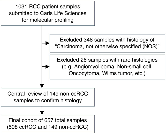
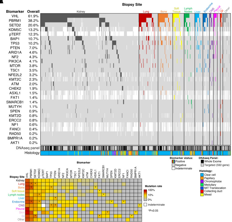
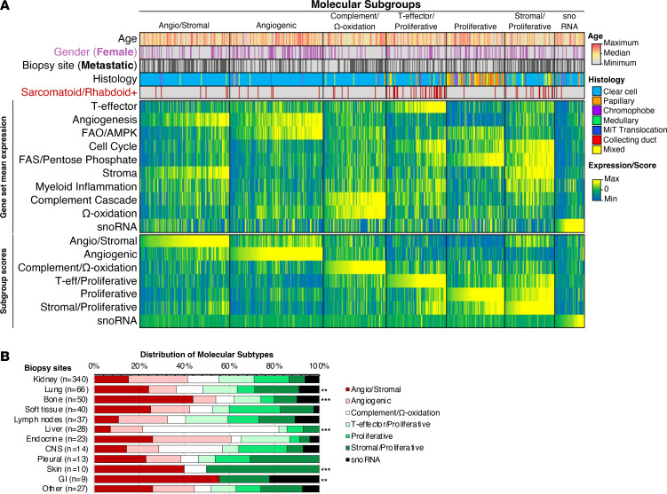
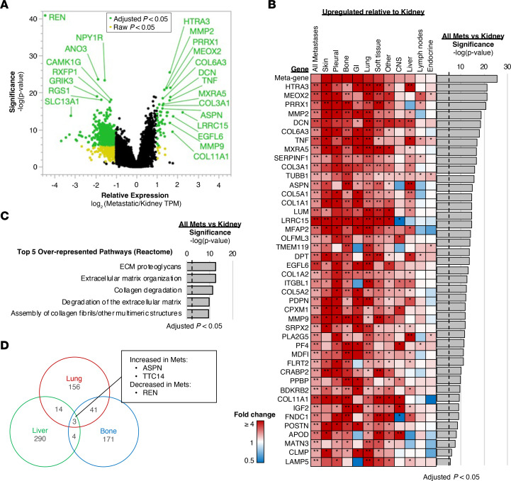
BACKGROUNDMetastases are the hallmark of lethal cancer, though underlying mechanisms that drive metastatic spread to specific organs remain poorly understood. Renal cell carcinoma (RCC) is known to have distinct sites of metastases, with lung, bone, liver, and lymph nodes being more common than brain, gastrointestinal tract, and endocrine glands. Previous studies have shown varying clinical behavior and prognosis associated with the site of metastatic spread; however, little is known about the molecular underpinnings that contribute to the differential outcomes observed by the site of metastasis.METHODSWe analyzed primary renal tumors and tumors derived from metastatic sites to comprehensively characterize genomic and transcriptomic features of tumor cells as well as to evaluate the tumor microenvironment at both sites.RESULTSWe included a total of 657 tumor samples (340 from the primary site [kidney] and 317 from various sites of metastasis). We show distinct genomic alterations, transcriptomic signatures, and immune and stromal tumor microenvironments across metastatic sites in a large cohort of patients with RCC.CONCLUSIONWe demonstrate significant heterogeneity among primary tumors and metastatic sites and elucidate the complex interplay between tumor cells and the extrinsic tumor microenvironment that is vital for developing effective anticancer therapies.

Keywords: Cancer; Oncology.


Conflict of interest statement

Conflict of interest: Provided in the Supplemental Materials.

Figures

Figure 1
Figure 1. Consort diagram of study inclusion process.
Figure 2
Figure 2. Distribution of histological subtypes by sites.
Clear cell RCC is the most common histological subtype among all sites (primary as well as metastatic tumors). No difference was noted in histological subtype distribution by metastatic site relative to the kidney. GI, gastrointestinal; MiT, Microphthalmia transcription factor.
Figure 3
Figure 3. Somatic DNA alterations by site of patient tumor biopsy.
(A) Oncoprint of the most common alterations (≥ 2% overall mutation rate), along with alterations present in the top 10 most frequently altered genes for any individual biopsy site. (B) Heatmap of mutation rates by metastatic site for genes shown in panel 2A. *P < 0.05. Abbreviations: CNS: central nervous system; GI, gastrointestinal; MiT, Microphthalmia transcription factor; pTERT, TERT promoter.
Figure 4
Figure 4. Sites of metastases have distinct distributions of molecular subtypes.
(A) Heatmap of patient age, gender, tumor histology, average gene set expression levels, and molecular subgroup composite scores, with patient samples sorted in ascending order (left to right) of composite scores for each subgroup. (B) Distribution of molecular subtypes by organ site. **P < 0.01 and ***P < 0.001 when compared with kidney. FAO, fatty acid oxidation; AMPK, AMP-activated protein kinase; FAS, fatty acid synthesis; GI, gastrointestinal; MiT, Microphthalmia transcription factor.
Figure 5
Figure 5. Gene expression profiling of metastatic and kidney biopsy sites.
(A) Volcano plot of relative gene expression of all metastatic samples compared with kidney. Colored data points indicate log2 (fold changes) ≥ 1 or ≤ –1 and raw (yellow) or adjusted (green) P value < 0.05. (B) Expression of genes significantly upregulated in panel A for each metastatic site relative to kidney. *Raw P < 0.05, **Adjusted P < 0.05. (C) Pathway analysis of the significantly upregulated genes in panel A, with the top 5 over-represented pathways from Reactome database shown. (D) Venn diagram representing differentially expressed genes for lung, bone, and liver sites compared with kidney. met, metastases; GI, gastrointestinal.
Figure 6
Figure 6. TME and immunotherapy-associated predictive biomarkers by site of metastases.
(A) Heatmap of median cell abundance and gene expression by biopsy site. *P < 0.05, **P < 0.01, ***P < 0.001, and ****P < 0.0001 when compared with kidney. Frequency of biomarker-positive samples for (B) PDL1 IHC (SP142 antibody), (C) TMB-High (≥ 10 mut/Mb), and (D) dMMR/MSI-High. NK, natural killer; CTLA4, The cytotoxic T-lymphocyte-associated antigen-4; TIM3, T cell immunoglobulin domain and mucin domain3; LAG3, Lymphocyte-Activation Gene3; PD-1, programmed death-1; PDL1, programmed death ligand-1; PDL2, programmed death ligand-2; GI, gastrointestinal.

References

    1. SEER Cancer Stat Facts: Cancer of Any Site. National Cancer Institute. https://seer.cancer.gov/statfacts/html/all.html.
    1. Gong J, et al. Metastasis in renal cell carcinoma: biology and implications for therapy. Asian J Urol. 2016;3(4):286–292. doi: 10.1016/j.ajur.2016.08.006. - DOI - PMC - PubMed
    1. Wood E, et al. Adjuvant therapy for localized high-risk renal cell carcinoma. Urol Clin North Am. 2020;47(3):345–358. doi: 10.1016/j.ucl.2020.04.007. - DOI - PubMed
    1. Comen E, et al. Clinical implications of cancer self-seeding. Nat Rev Clin Oncol. 2011;8(6):369–377. doi: 10.1038/nrclinonc.2011.64. - DOI - PubMed
    1. Paget S. The distribution of secondary growths in cancer of the breast. 1889. Cancer Metastasis Rev. 1989;8(2):98–101. - PubMed