. 2024 Aug 1;4(8):1963-1977.
doi: 10.1158/2767-9764.CRC-24-0196.

Estrogen Receptor Blockade Potentiates Immunotherapy for Liver Metastases by Altering the Liver Immunosuppressive Microenvironment

Affiliations

Estrogen Receptor Blockade Potentiates Immunotherapy for Liver Metastases by Altering the Liver Immunosuppressive Microenvironment

Yasmine Benslimane et al. Cancer Res Commun. .

Abstract

Liver metastases (LM) remain a major cause of cancer-related death and are a major clinical challenge. LM and the female sex are predictors of a poorer response to immunotherapy but the underlying mechanisms remain unclear. We previously reported on a sexual dimorphism in the control of the tumor microenvironment (TME) of colorectal carcinoma liver metastases (CRCLM) and identified estrogen as a regulator of an immunosuppressive TME in the liver. Here we aimed to assess the effect of estrogen deprivation on the cytokine/chemokine profile associated with CRCLM, using a multiplex cytokine array and the RNAscope technology, and its effects on the innate and adaptive immune responses in the liver. We also evaluated the benefit of combining the selective estrogen-receptor degrader Fulvestrant with immune checkpoint blockade for the treatment of CRCLM. We show that estrogen depletion altered the cytokine/chemokine repertoire of the liver, decreased macrophage polarization, as reflected in reduced accumulation of tumor infiltrating M2 macrophages and increased the accumulation of CCL5+/CCR5+ CD8+ T and NKT cells in the liver TME. Similar results were obtained in a murine pancreatic ductal adenocarcinoma model. Importantly, treatment with Fulvestrant also increased the accumulation of CD8+CCL5+, CD8+CCR5+ T and NK cells in the liver TME and enhanced the therapeutic benefit of anti-PD1 immunotherapy, resulting in a significant reduction in the outgrowth of LM. Taken together, our results show that estrogen regulates immune cell recruitment to the liver and suggest that inhibition of estrogen action could potentiate the tumor-inhibitory effect of immunotherapy in hormone-independent and immunotherapy-resistant metastatic cancer.

Significance: The immune microenvironment of the liver plays a major role in controlling the expansion of hepatic metastases and is regulated by estrogen. We show that treatment of tumor-bearing mice with an estrogen receptor degrader potentiated an anti-metastatic effect of immunotherapy. Our results provide mechanistic insight into clinical findings and a rationale for evaluating the efficacy of combination anti-estrogen and immunotherapy for prevention and/or treatment of hepatic metastases in female patients.


Conflict of interest statement

P. Brodt reports grants from Canadian Institute for Health Research during the conduct of the study. No disclosures were reported by the other authors.

Figures

Figure 1
Figure 1
Estrogen depletion increases NKT cytotoxicity in the liver TME. FC was performed on hepatic immune cells isolated following injection of 5 × 10⁵ MC-38 cells and immunolabeled with the indicated antibodies. Shown in A are flow cytometric contour plots and the respective bar graphs (n = 4). Shown in B (top) is the workflow of the NKT cytotoxicity assay, in B (bottom-left) representative images of green fluorescent (dead) MC-38 cells after co-culture with NKT cells for 30 hours and in B (bottom right) quantification of dead MC-38 cells as measured over time, normalized to time 0 hour (n = 2–3). ****, P ≤ 0.0001; Two-way ANOVA. (Created with BioRender.com.)
Figure 2
Figure 2
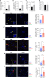
Estrogen depletion alters the cytokines/chemokines repertoire of the liver and increases expression of CCR5 and CCL5 in NKT and CD8+ T cells. A, Cytokine array analysis was conducted on whole liver protein lysates (four livers/group) derived from MC-38 tumor-bearing mice. Data were normalized to SHAM-operated values. In B–E are FC results for the indicated cell populations (n = 4). In F (left)—representative RNAscope images of frozen liver sections probed with CCL5 and NK1.1, followed by CD3 staining, and in F (right) image analysis results based on 15 fields per group (n = 3). Yellow arrows denote NK1.1+CCL5+CD3+ cells, and green arrows denote NK1.1+CD3+ cells with high CCL5 density. Experimental LM were generated by intrasplenic/portal injection of 105 MC-38 cells. Intraperitoneal Mvc or vehicle injections began the following day and continued for 6 days. Mice were sacrificed and LM enumerated on day 18 post-inoculation. Results shown are of a representative experiment of two performed. The number of metastases per liver are shown in G (n = 4–7; bars indicate medians) and representative livers are shown in H. *, P ≤ 0.05; **, P ≤ 0.01; ***, P ≤ 0.001, Student t test (FC) and Mann–Whitney test (LM).
Figure 3
Figure 3
Increased CXCR3 expression in hepatic macrophages associated with early stages of the metastatic process. FC was conducted on hepatic immune cells from mice injected with 5 × 10⁵ MC-38 cells 8 days earlier. Shown in A–D are FC results for the indicated cell populations (n = 3–4). Confocal images (E, F, H) were acquired for 10 μm liver cryostat sections from these mice that were immunostained with the indicated antibodies and counterstained with 4′,6-diamidino-2-phenylindole. Cell counts per field (means ± SD, n = 10) are shown on the right. Representative RNAscope images were obtained from cryostat sections probed for CXCL10 expression in NKT (G) or macrophages (I), as indicated. Image analysis (G, I, right) was based on 15 fields per group (n = 2–3). Yellow arrows denote NK1.1+ or CD68+ cells expressing CXCL10, and green arrows denote cells with high CXCL10 expression. *, P ≤ 0.05; **, P ≤ 0.01; ***, P ≤ 0.001; ****, P ≤ 0.0001, Student t test.
Figure 4
Figure 4
Estrogen depletion increases the M1/M2 macrophage ratio in both the recruited and liver-resident macrophage populations. FC was performed on macrophages isolated 12 days post injection of 5 × 10⁵ MC-38 cells and stained with the indicated antibodies. Shown in A and B are flow cytometric contour plots (left) and M1/M2 macrophage ratios (right, n = 4). Representative confocal images obtained from 15 μm liver sections labeled as indicated are shown in C (left) and cell counts per field (means ± SD, n = 10) are shown in C (right). *, P ≤ 0.05; ****, P ≤ 0.0001, Student t test.
Figure 5
Figure 5
Fulv reprograms the liver TME and inhibits CRCLM outgrowth. Experimental LM were generated by intrasplenic/portal injection of 105 MC-38 cells. Mice received eight subcutaneous injections of Fulv or vehicle on alternate days and were sacrificed 21 days post tumor inoculation. Results shown are of a representative experiment of two performed. In A are metastases counted per liver (n = 5–6), in B mean nodule size (mm; n = 5–6), and in C representative liver images. Hepatic immune cells were isolated in a separate experiment 7 days post-injection, stained with the indicated antibodies, and analyzed by FC. Shown in D (left) are representative FC contour plots (n = 3–4) and in D (right) cell counts (means ± SD). *, P ≤ 0.05, Mann–Whitney test (metastases) or Student t test (FC).
Figure 6
Figure 6
Fulv enhances the inhibitory effect of anti-PD-1 immunotherapy against CRCLM. Experimental LM were generated by intrasplenic/portal injection of 105 MC-38 cells. Treatment consisted of subcutaneous injection of 20 mg/kg Fulv or vehicle on day 1 post-injection, followed by intraperitoneal administration of 10 mg/kg anti-PD-1 or control IgG on day 2, with no treatment on day 3, a protocol that was repeated until day 15 [as shown in A]. Mice were sacrificed and metastases enumerated on day 18. Shown in B are metastases counts per liver (n = 4–9, pooled data of two separate experiments), in C nodule size range (n = 4–9), in D representative liver images, and in E representative H&E-stained formalin-fixed, paraffin-embedded sections. *, P ≤ 0.05; **, P ≤ 0.01; ***, P ≤ 0.001, Mann–Whitney test.
Figure 7
Figure 7
The combinatorial Fulv and anti-PD-1 immunotherapy reprograms the IME of CRCLM. Hepatic immune cells were isolated following the treatment regimen depicted in Fig. 6A. Shown in A–J are results of FC performed on cells from the indicated treatment groups (n = 4). Shown in K and L are results of RT-qPCR performed on whole liver RNA (n = 3), normalized to GAPDH, and expressed as means (± SD) fold change relative to vehicle (assigned a value of 1). *, P ≤ 0.05; **, P ≤ 0.01; ***, P ≤ 0.001, Student t test.
Figure 8
Figure 8
A proposed model for the role of estrogen in the regulation of the TME of CRCLM and the effect of ER blockade by Fulv. The diagram depicts postulated mechanisms for the immunosuppressive role of estrogen in the liver and the effects of its depletion or blockade. ER/E2 signaling can promote CRCLM by regulating the recruitment and activity of innate and adaptive immune cells. Ovariectomy or Fulv treatment result in increased accumulation of NK/NKT cells and M1-like macrophages, increasing the antitumor immune response. Fulv treatment can thereby potentiate the response to immunotherapy and augment the eradication of metastatic tumor cells.

References

    1. Zhou H, Liu Z, Wang Y, Wen X, Amador EH, Yuan L, et al. . Colorectal liver metastasis: molecular mechanism and interventional therapy. Signal Transduct Target Ther 2022;7:70. - PMC - PubMed
    1. Siegel RL, Jakubowski CD, Fedewa SA, Davis A, Azad NS. Colorectal cancer in the young: epidemiology, prevention, management. Am Soc Clin Oncol Educ Book 2020;40:1–14. - PubMed
    1. Wu Y, Xu W, Wang L. IDDF2021-ABS-0191 gender matters: sex disparities in colorectal cancer liver metastasis survival: a population-based study. Gut 2021;70:A142–3.
    1. Miller KD, Nogueira L, Mariotto AB, Rowland JH, Yabroff KR, Alfano CM, et al. . Cancer treatment and survivorship statistics, 2019. CA Cancer J Clin 2019;69:363–85. - PubMed
    1. Siegel R, Naishadham D, Jemal A. Cancer statistics, 2012. CA Cancer J Clin 2012;62:10–29. - PubMed

Publication types

MeSH terms