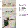
Methods for Tattooing Xenopus laevis with a Rotary Tattoo Machine
- PMID: 39007614
- PMCID: PMC11292787
- DOI: 10.3791/67086
Methods for Tattooing Xenopus laevis with a Rotary Tattoo Machine
Abstract
Animal models expand the scope of biomedical research, furthering our understanding of developmental, molecular, and cellular biology and enabling researchers to model human disease. Recording and tracking individual animals allows researchers to reduce the number of animals required for study and refine practices to improve animal wellbeing. Several well-documented methods exist for marking and tracking mammals, including ear punching and ear tags. However, methods for marking aquatic amphibian species are limited, with the existing resources being outdated, ineffective, or prohibitively costly. In this manuscript, we outline methods and best practices for marking Xenopus laevis with a rotary tattoo machine. Proper tattooing results in high-quality tattoos, making individuals easily distinguishable for researchers and posing minimal risk to animals' health. We also highlight the causes of poor-quality tattoos, which can result in tattoos that fade quickly and cause unnecessary harm to animals. This approach allows researchers and veterinarians to mark amphibians, enabling them to track biological replicates and transgenic lines and to keep accurate records of animal health.
Conflict of interest statement
DISCLOSURES:
The authors declare no competing interests.
Figures


Similar articles
-
Integrating computer vision algorithms and RFID system for identification and tracking of group-housed animals: an example with pigs.J Anim Sci. 2024 Jan 3;102:skae174. doi: 10.1093/jas/skae174. J Anim Sci. 2024. PMID: 38908015 Free PMC article.
-
Systemic pharmacological treatments for chronic plaque psoriasis: a network meta-analysis.Cochrane Database Syst Rev. 2021 Apr 19;4(4):CD011535. doi: 10.1002/14651858.CD011535.pub4. Cochrane Database Syst Rev. 2021. Update in: Cochrane Database Syst Rev. 2022 May 23;5:CD011535. doi: 10.1002/14651858.CD011535.pub5. PMID: 33871055 Free PMC article. Updated.
-
Perceptions and experiences of the prevention, detection, and management of postpartum haemorrhage: a qualitative evidence synthesis.Cochrane Database Syst Rev. 2023 Nov 27;11(11):CD013795. doi: 10.1002/14651858.CD013795.pub2. Cochrane Database Syst Rev. 2023. PMID: 38009552 Free PMC article.
-
Behavioral interventions to reduce risk for sexual transmission of HIV among men who have sex with men.Cochrane Database Syst Rev. 2008 Jul 16;(3):CD001230. doi: 10.1002/14651858.CD001230.pub2. Cochrane Database Syst Rev. 2008. PMID: 18646068
-
Dressings and topical agents for treating venous leg ulcers.Cochrane Database Syst Rev. 2018 Jun 15;6(6):CD012583. doi: 10.1002/14651858.CD012583.pub2. Cochrane Database Syst Rev. 2018. PMID: 29906322 Free PMC article.
References
-
- Sive H. Xenopus: A Laboratory Manual. Cold Spring Harbor Press; (2023).
Publication types
MeSH terms
Grants and funding
LinkOut - more resources
Full Text Sources
