Review
. 2024 Jul 15;20(14):e845-e864.
doi: 10.4244/EIJ-D-23-01050.

Durability of transcatheter aortic valve implantation

Affiliations
Review

Durability of transcatheter aortic valve implantation

Julien Ternacle et al. EuroIntervention. .

Abstract

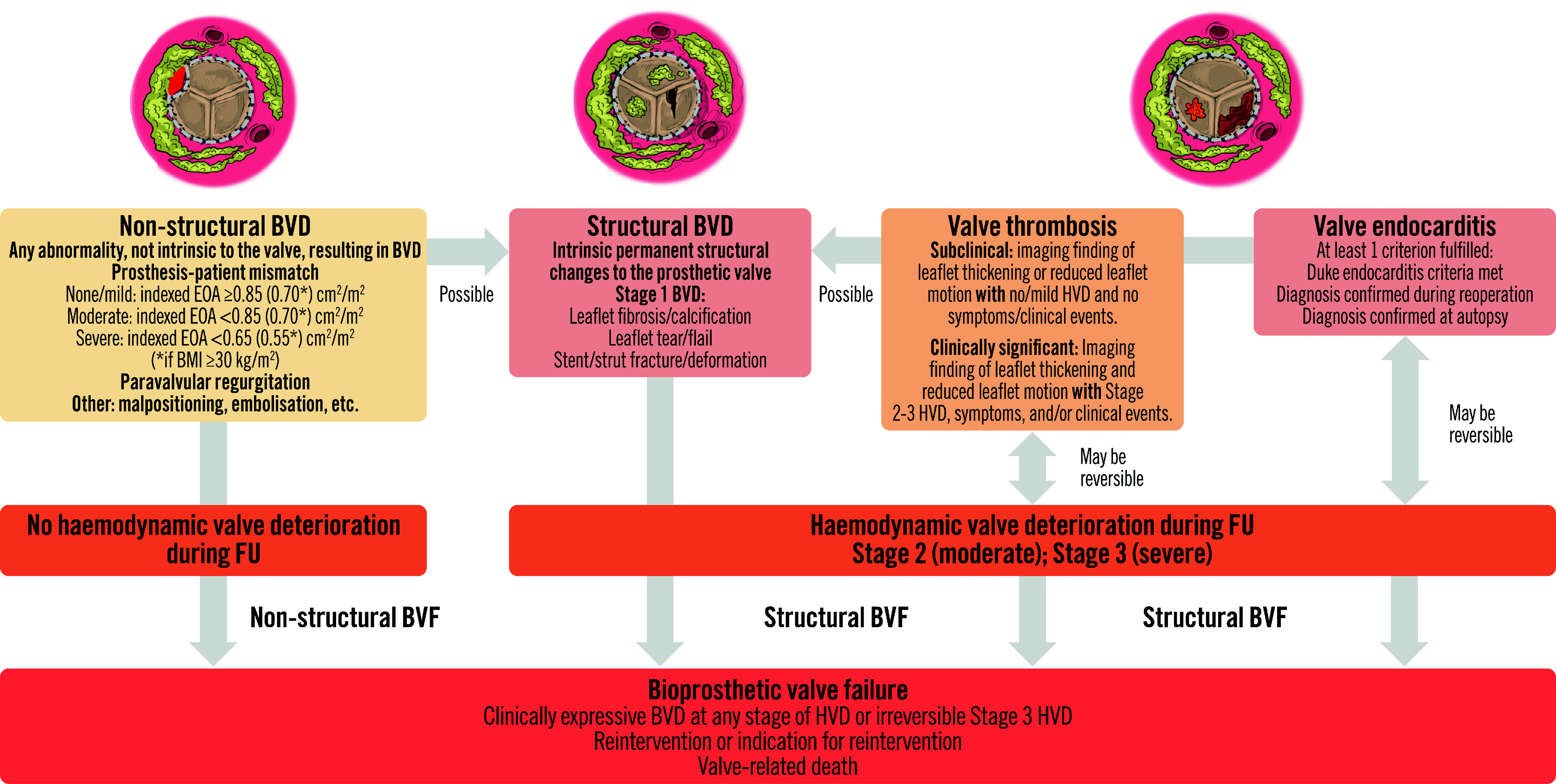
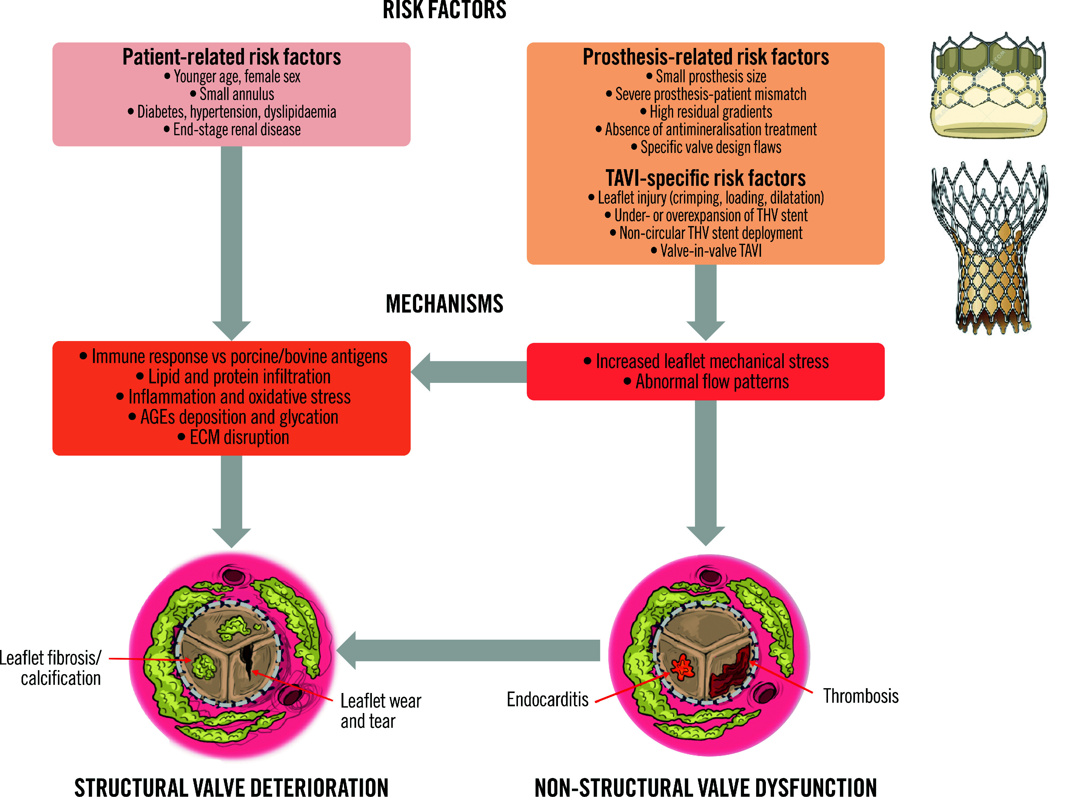
Transcatheter aortic valve implantation (TAVI) is now utilised as a less invasive alternative to surgical aortic valve replacement (SAVR) across the whole spectrum of surgical risk. Long-term durability of the bioprosthetic valves has become a key goal of TAVI as this procedure is now considered for younger and lower-risk populations. The purpose of this article is to present a state-of-the-art overview on the definition, aetiology, risk factors, mechanisms, diagnosis, clinical impact, and management of bioprosthetic valve dysfunction (BVD) and failure (BVF) following TAVI with a comparative perspective versus SAVR. Structural valve deterioration (SVD) is the main factor limiting the durability of the bioprosthetic valves used for TAVI or SAVR, but non-structural BVD, such as prosthesis-patient mismatch and paravalvular regurgitation, as well as valve thrombosis or endocarditis may also lead to BVF. The incidence of BVF related to SVD or other causes is low (<5%) at midterm (5- to 8-year) follow-up and compares favourably with that of SAVR. The long-term follow-up data of randomised trials conducted with the first generations of transcatheter heart valves also suggest similar valve durability in TAVI versus SAVR at 10 years, but these trials suffer from major survivorship bias, and the long-term durability of TAVI will need to be confirmed by the analysis of the low-risk TAVI versus SAVR trials at 10 years.


Conflict of interest statement

J. Ternacle has been a consultant for Abbott, Edwards Lifesciences, Pi-Cardia, Philips Healthcare, and GE HealthCare. M-A. Clavel has received funds from Edwards Lifesciences for computed tomography core laboratory analyses in the field of surgical bioprostheses; and research grants from Edwards Lifesciences, Medtronic, and Pi-Cardia, with no personal compensation. P. Pibarot has received funding from Edwards Lifesciences, Pi-Cardia, and Cardiac Success for echocardiography core laboratory analyses in the field of transcatheter valve therapies, and Medtronic for in vitro analyses, with no personal compensation. The other authors have no conflicts of interest to declare.

Figures

Figure 1
Figure 1. Category, aetiology, and definition of bioprosthetic valve dysfunction and failure.
BMI: body mass index; BVD: bioprosthetic valve dysfunction; BVF: bioprosthetic valve failure; EOA: effective orifice area; FU: follow-up; HVD: haemodynamic valve deterioration
Figure 2
Figure 2. Four-step algorithm for the determination of the presence, aetiology, stage, and clinical consequences of bioprosthetic valve dysfunction.
CT: computed tomography; CECT: contrast-enhanced CT; FU: follow-up; PET: positron emission tomography; TOE: transoesophageal echocardiography; TTE: transthoracic echocardiography
Figure 3
Figure 3. Representative examples of the different stages of bioprosthetic valve dysfunction and proposed management.
Yellow arrows indicate valve thickening and reduced motion; red arrow: colour Doppler flow restriction and turbulence; white arrow: colour Doppler flow paucity. AR: aortic regurgitation; HVD: haemodynamic valve deterioration; MG: mean gradient; TTE: transthoracic echocardiography
Figure 4
Figure 4. Risk factors and mechanisms of structural valve deterioration and failure.
AGEs: advanced glycation end products; ECM: extracellular matrix; TAVI; transcatheter aortic valve implantation; THV: transcatheter heart valve
Figure 5
Figure 5. A case of early valve thrombosis followed by accelerated structural valve deterioration.
A female patient aged 73 years underwent TAVI with a SAPIEN XT 23 mm. At 30 days, she presented with clinical valve thrombosis resulting in severe (Stage 3 HVD) bioprosthetic valve stenosis (A). There was a quasi-complete resolution of the valve thrombosis and stenosis following 2 weeks of VKA treatment (B). However, 3 years later there was evidence of structural valve deterioration resulting in moderate (Stage 2 HVD) bioprosthetic valve stenosis (C). Yellow arrows indicate valve thickening and reduced motion; red arrows: colour Doppler flow restriction/paucity; white arrow: intraprosthetic aortic regurgitation (AR). EOA: effective orifice area; HVD: haemodynamic valve deterioration; SVD: structural valve deterioration; TAVI: transcatheter aortic valve implantation; TOE: transoesophageal echocardiography; TTE: transthoracic echocardiography; VKA: vitamin K antagonist
Figure 6
Figure 6. Incidence of Stage 2 or 3 SVD or of all-cause BVF according to VARC-3 in the TAVI versus SAVR RCTs and registries.
Incidence of Stage 2 (moderate) or 3 (severe) SVD (A) and of all-cause BVF (B) according to VARC-3 definitions in the PARTNER 2A Trial and S3IR, PARTNER 3 Trial, CoreValve HR trial and SURTAVI Trial at 5 years and in the NOTION trial at 10 years. BVF: bioprosthetic valve failure; CoreValve HR: Medtronic CoreValve U.S. Pivotal Trial; RCT: randomised controlled trial; SAVR: surgical aortic valve replacement; SVD: structural valve deterioration; TAVI: transcatheter aortic valve implantation; VARC-3: Valve Academic Research Consortium 3
Figure 7
Figure 7. Management of structural THV failure.
In a patient with transcatheter heart valve failure related to SVD and low-to-intermediate surgical risk, the first step is to discuss the case among the Heart Team in order to select standalone TAVI-in-TAVI, complex TAVI-in-TAVI, or SAVR with THV explantation. Arguments in favour of SAVR are if the patient requires ascending aorta replacement, aortic root enlargement, a repair or replacement of another valve, and/or coronary artery bypass grafting. If TAVI-in-TAVI is considered the preferred option, the next step is to assess the arterial access, the failed THV, the aortic root and the coronary ostia using CT imaging and planning. If there is limited or impossible femoral arterial access, non-transfemoral or surgical transfemoral access should be considered. If there is a risk of coronary obstruction or of impaired coronary access for future percutaneous coronary intervention as identified by a VTC <4 mm and/or a short VTSTJ, leaflet modification or the chimney technique should be considered, and a BEV may be preferable for the TAVI-in-TAVI. If femoral access is feasible and there is no risk of coronary obstruction, the next step is to assess the presence and aetiology (primary vs secondary) of significant MR or TR. If ≥moderate primary MR or TR is present, concomitant transcatheter interventions may be considered. If the MR or TR is secondary, it is preferable to consider a staged approach with standalone TAVI-in-TAVI first, and then the mitral and/or tricuspid transcatheter intervention 3 months later if the MR and/or TR and heart failure symptoms still persist. If pre-existing severe PPM of the failed THV and/or if severe THV stenosis are present, a SEV with supra-annular design should be considered for the TAVI-in-TAVI. In the presence of complex CAD requiring revascularisation, complex percutaneous angioplasty should be considered prior to the TAVI-in-TAVI procedure. The application ‘Redo TAVI’ (KRUTSCH Associates) can be used to select the optimal model and size of THV for the TAVI-in-TAVI. If a high residual gradient (mean gradient >20 mmHg), confirmed by invasive assessment, is present following the TAVI-in-TAVI procedure, a balloon post-dilatation may be considered. *VTSTJ <2.5 mm is considered high risk and 2.5-3.5 mm intermediate risk for coronary artery obstruction. The schematic representation of the VTC and VTSTJ measurements in a scenario of SEV-in-BEV is reproduced with permission. BEV: balloon-expandable valve; CAD: coronary artery disease; CT: computed tomography; MR: mitral regurgitation; PPM: prosthesis-patient mismatch; SAVR: surgical aortic valve replacement; SEV: self-expanding valve; SVD: structural valve deterioration; TAVI: transcatheter aortic valve implantation; THV: transcatheter heart valve; TR: tricuspid regurgitation; VTC: virtual THV to coronary artery distance; VTSTJ: virtual THV to sinotubular junction distance
Figure 8
Figure 8. Balancing the patient’s life expectancy, surgical risk, and expected valve durability in the decision between TAVI versus SAVR.
(A) Schematic of the ratio between life expectancy and valve durability. Adapted with permission. (B) Type of AVR according to life expectancy and surgical risk. SAVR: surgical aortic valve replacement; TAVI: transcatheter aortic valve implantation
Figure 9
Figure 9. Future directions to optimise THV durability.
Lp(a): lipoprotein(a); SGLT2: sodium-glucose cotransporter 2; PCSK9: proprotein convertase subtilisin/hexin type 9; THV: transcatheter heart valve
Central illustration
Central illustration. Durability of TAVI.
The risk factors and mechanisms of valve failure are represented in (A), while (B) presents the data concerning mid- and long-term TAVI durability. BVD: bioprosthetic valve dysfunction; BVF: bioprosthetic valve failure; PPM: prosthesis-patient mismatch; PVL: paravalvular leak; SAVR: surgical aortic valve replacement; TAVI: transcatheter aortic valve implantation; THV: transcatheter heart valve; ViV: valve-in-valve

Similar articles

Cited by

References

    1. Mack MJ, Leon MB, Thourani VH, Pibarot P, Hahn RT, Genereux P, Kodali SK, Kapadia SR, Cohen DJ, Pocock SJ, Lu M, White R, Szerlip M, Ternacle J, Malaisrie SC, Herrmann HC, Szeto WY, Russo MJ, Babaliaros V, Smith CR, Blanke P, Webb JG, Makkar R PARTNER 3 Investigators. Transcatheter Aortic-Valve Replacement in Low-Risk Patients at Five Years. N Engl J Med. 2023;389:1949–60. - PubMed
    1. Forrest JK, Deeb GM, Yakubov SJ, Gada H, Mumtaz MA, Ramlawi B, Bajwa T, Teirstein PS, Tchétché D, Huang J, Reardon MJ Evolut Low Risk Trial Investigators. 4-Year Outcomes of Patients With Aortic Stenosis in the Evolut Low Risk Trial. J Am Coll Cardiol. 2023;82:2163–5. - PubMed
    1. Capodanno D, Petronio AS, Prendergast B, Eltchaninoff H, Vahanian A, Modine T, Lancellotti P, Sondergaard L, Ludman PF, Tamburino C, Piazza N, Hancock J, Mehilli J, Byrne RA, Baumbach A, Kappetein AP, Windecker S, Bax J, Haude M. Standardized definitions of structural deterioration and valve failure in assessing long-term durability of transcatheter and surgical aortic bioprosthetic valves: a consensus statement from the European Association of Percutaneous Cardiovascular Interventions (EAPCI) endorsed by the European Society of Cardiology (ESC) and the European Association for Cardio-Thoracic Surgery (EACTS). Eur Heart J. 2017;38:3382–90. - PubMed
    1. VARC-3 WRITING Généreux P, Piazza N, Alu MC, Nazif T, Hahn RT, Pibarot P, Bax JJ, Leipsic JA, Blanke P, Blackstone EH, Finn MT, Kapadia S, Linke A, Mack MJ, Makkar R, Mehran R, Popma JJ, Reardon M, Rodes-Cabau J, Van Mieghem NM, Webb JG, Cohen DJ, Leon MB. Valve Academic Research Consortium 3: updated endpoint definitions for aortic valve clinical research. Eur Heart J. 2021;42:1825–57. - PubMed
    1. Pibarot P, Herrmann HC, Wu C, Hahn RT, Otto CM, Abbas AE, Chambers J, Dweck MR, Leipsic JA, Simonato M, Rogers T, Sathananthan J, Guerrero M, Ternacle J, Wijeysundera HC, Sondergaard L, Barbanti M, Salaun E, Généreux P, Kaneko T, Landes U, Wood DA, Deeb GM, Sellers SL, Lewis J, Madhavan M, Gillam L, Reardon M, Bleiziffer S, O’Gara PT, Rodés-Cabau J, Grayburn PA, Lancellotti P, Thourani VH, Bax JJ, Mack MJ, Leon MB Heart Valve Collaboratory. Standardized Definitions for Bioprosthetic Valve Dysfunction Following Aortic or Mitral Valve Replacement: JACC State-of-the-Art Review. J Am Coll Cardiol. 2022;80:545–61. - PubMed

MeSH terms

LinkOut - more resources