. 2024 Sep 1:185:126-143.
doi: 10.1016/j.actbio.2024.07.011. Epub 2024 Jul 14.

3D printed O2-generating scaffolds enhance osteoprogenitor- and type H vessel recruitment during bone healing

Affiliations

3D printed O2-generating scaffolds enhance osteoprogenitor- and type H vessel recruitment during bone healing

Naboneeta Sarkar et al. Acta Biomater. .

Abstract

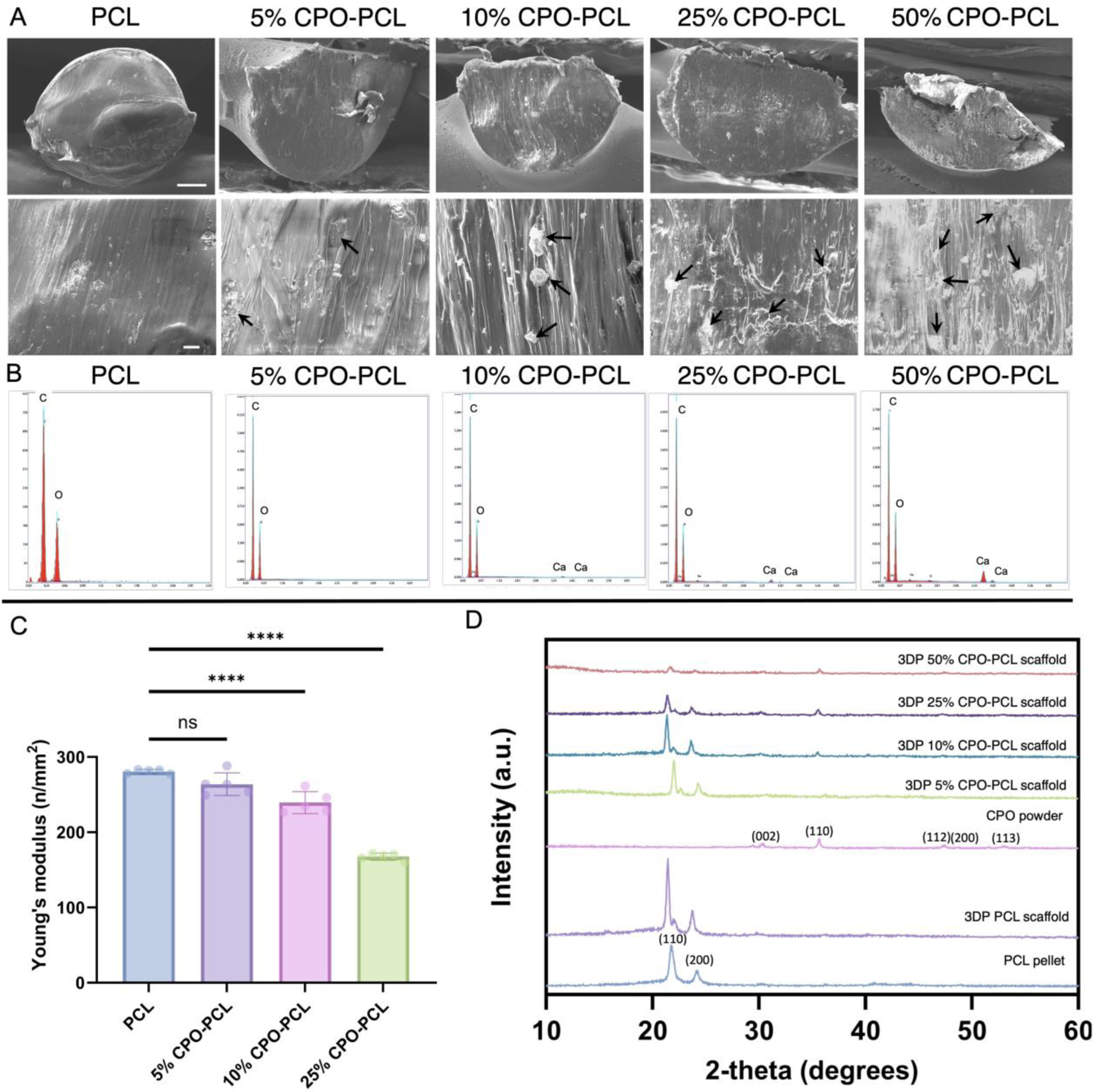
Oxygen (O2)-delivering tissue substitutes have shown tremendous potential for enhancing tissue regeneration, maturation, and healing. As O2 is both a metabolite and powerful signaling molecule, providing controlled delivery is crucial for optimizing its beneficial effects in the treatment of critical-sized injuries. Here, we report the design and fabrication of 3D-printed, biodegradable, O2-generating bone scaffold comprising calcium peroxide (CPO) that once hydrolytically activated, provides long-term generation of oxygen at a controlled, concentration-dependent manner, and polycaprolactone (PCL), a hydrophobic polymer that regulate the interaction of CPO with water, preventing burst release of O2 at early time points. When anoxic conditions were simulated in vitro, CPO-PCL scaffolds maintained the survival and proliferation of human adipose-derived stem/stromal cells (hASCs) relative to PCL-only controls. We assessed the in vivo osteogenic efficacy of hASC-seeded CPO-PCL scaffolds implanted in a non-healing critical-sized 4-mm calvarial defects in nude mice for 8 weeks. Even without exogenous osteoinductive factors, CPO-PCL scaffolds demonstrated increased new bone volume compared to PCL-only scaffolds as verified by both microcomputed tomography analysis and histological assessments. Lastly, we employed a quantitative 3D lightsheet microscopy platform to determine that O2-generating scaffolds had similar vascular volumes with slightly higher presence of CD31hiEmcnhi pro-osteogenic, type H vessels and increased number of Osterix+ skeletal progenitor cells relative to PCL-only scaffolds. In summary, 3D-printed O2 generating CPO-PCL scaffolds with tunable O2 release rates provide a facile, customizable strategy for effectively treating, craniofacial bone defects. STATEMENT OF SIGNIFICANCE: Oxygen(O2)-delivering bone substitutes show promise in defect repair applications by supplying O2 to the cells within or around the graft, improving cell survivability and enhancing bone matrix mineralization. A novel O2-generating bone scaffold has been 3D printed for the first-time which ensures patient and defect specificity. 3D printed calcium peroxide-polycaprolactone (CPO-PCL) bone scaffold provides uninterrupted O2 supply for 22 days allowing cell survival in deprived O2 and nutrient conditions. For the first time, O2-driven bone regenerative environment in mice calvaria has been captured by light-sheet imaging which illuminates abundance of Osterix+ cells, angiogenesis at a single cell resolution indicating active site of bone remodeling and growth in the presence of O2.

Keywords: Angiogenesis; Bone regeneration; Bone tissue engineering; Calcium peroxide; Oxygen.


Conflict of interest statement

Declaration of competing interest The authors declare that they have no known competing financial interests or personal relationships that could have appeared to influence the work reported in this paper.

Figures

Figure 1:
Figure 1:. Fabricating 3D-printed biodegradable CPO-PCL scaffolds.
(A) Schematic depicting design and fabrication of CPO-PCL scaffold. CPO was homogeneously mixed with PCL at varying concentration of CPO-PCL. The powder mixture was fed into a filament extruder to make filaments which were used to print bilayer 3D printed parts using an extrusion-based 3D printer following rigorous optimization. (B) Scanning electron micrograph of CPO-PCL scaffolds with varying CPO content exhibiting increased surface roughness with increased CPO content. (C) Stereomicroscope image of scaffold lattice structure of 5–50% CPO-PCL scaffold were taken to assess the “print quality”, scale 10 mm. Custom MATLAB code was applied to assess (D) average strut thickness measuring 432, 608, 426 and 489 μm for 5%, 10%, 25%, and 50% CPO-PCL scaffold, respectively (n=10) and (E) average pore width measuring 640, 469, 523, 450 μm for 5%, 10%, 25%, and 50% CPO-PCL scaffold, respectively (n=10). [ns P > 0.05, * P ⩽ 0.05, ** P ⩽ 0.01, *** P ⩽ 0.001, **** P ⩽ 0.0001]
Figure 2:
Figure 2:. Characterization of 3D-printed biodegradable CPO-PCL scaffolds.
(A) SEM images show the strut intersection of the 3D-printed PCL, 5% CPO-PCL, 10% CPO-PCL, 25% CPO-PCL and 50% CPO-PCL scaffolds at low and high magnification. The CPO particles (black arrows) appear uniformly distributed within the PCL strut. Scale bars: Top row (100 μm); Bottom row (10 μm). (B) EDAX mapping of the elemental composition at the scaffold strut intersection of 3D-printed PCL, 5% CPO-PCL, 10% CPO-PCL, 25% CPO-PCL and 50% CPO-PCL scaffolds confirming the presence of Ca in composite scaffolds. (C) Compressive strength analysis of the 3D-printed PCL, 5% CPO-PCL, 10% CPO-PCL, and 25% CPO-PCL scaffold suggest that Young’s modulus decreased with increasing CPO concentration. (D) XRD analysis of as-received pre-printed PCL-pellet and CPO powder, alongside post 3D-printed PCL, 5% CPO-PCL, 10% CPO-PCL, 25% CPO-PCL and 50% CPO-PCL scaffolds, verifying that the printing temperature does not alter the physical properties of PCL or CPO. [ns P > 0.05, * P ⩽ 0.05, ** P ⩽ 0.01, *** P ⩽ 0.001, **** P ⩽ 0.0001]
Figure 3:
Figure 3:. Prolonged and controlled release of oxygen by CPO-PCL scaffold.
(A) Schematic showing mechanism of O2 release from CPO-PCL scaffolds and measurement using Agilent Seahorse XF analyzer. Control PCL, and O2-generating 5% CPO-PCL, 10% CPO-PCL, 25% CPO-PCL scaffolds were immersed in aqueous media. CPO releases O2 as soon as it is hydrolytically activated and O2 sensitive fluorophore probe in the Seahorse analyzer measures the dissolved O2 in media. CPO present on the surface of the scaffolds react with H2O and starts releasing O2 as early as 2 hours. (B) In vitro release study showing CPO-PCL scaffolds exhibiting O2 release at a constant rate for 22 days, as well as higher O2 release by PCL-25% CPO compared to PCL with 5 and 10% CPO (inset showing controlled O2 release within the first 24 hours, with no signs of burst release profile). The **** denotes p values based on the statistical differences between adjacent data and control PCL data at each timepoint. (C) pH measurement with Seahorse analyzer indicates the change in pH value remained between pH 7.0–8.0 for all scaffolds during the course of O2 release. The differences in pH values between different composition are not statistically significant. (D) Accelerated degradation study in 5.0 M NaOH showing percentage weight loss rate for scaffolds with varying amount of CPO. 25% CPO-PCL with the lowest amount of hydrophobic PCL polymer degraded earliest, followed by 10% and 5% CPO-PCL scaffolds. [ns P > 0.05, * P ⩽ 0.05, ** P ⩽ 0.01, *** P ⩽ 0.001, **** P ⩽ 0.0001]
Figure 4:
Figure 4:. Controlled oxygen delivery from CPO-PCL scaffolds maintained hASC viability at normoxic condition and enhanced hASC viability and proliferation at anoxic condition.
hASCs were seeded on control PCL and O2-generating 5%, 10%, and 25% CPO-PCL scaffolds and cultured for 7 days in expansion media at normoxic (A-B) and anoxic (C-D) conditions. (A) DNA content (n=4–6) and (B) live-dead staining (n=2) were performed to assess hASCs viability in presence of CPO at normoxic condition after 3 and 7 days of culture, scale bar 100 μm. Controlled release of O2 from CPO-PCL scaffolds allowed cellular attachment and did not exhibit cytotoxicity to the hASCs. (C) DNA content (n=4–6) and (D) live-dead staining (n=2) were performed to assess ASCs viability at anoxic condition after 3 and 7 days of culture, scale bar 100 μm. CPO-PCL scaffolds showed significantly higher DNA content and live cell attachment at anoxia compared to the control PCL scaffold suggesting the constant generation of O2 could replenish the inadequate O2 in the surrounding environment. [ns P > 0.05, * P ⩽ 0.05, ** P ⩽ 0.01, *** P ⩽ 0.001, **** P ⩽ 0.0001]
Figure 5:
Figure 5:. Controlled oxygen delivery from CPO-PCL scaffolds enhanced osteogenic differentiation of hASCs:
hASCs were seeded on control PCL, and O2-generating 5%, 10%, and 25% CPO-PCL scaffolds and cultured for 21 days in osteogenic differentiation media. (A) Calcium content per scaffold (n=4–6) showing presence of CPO significantly enhanced calcium deposition compared to the control PCL at 7, 14 and 21 days of osteogenic culture with the highest calcium deposition by 25% CPO-PCL scaffolds ; (B) von Kossa staining of control PCL, 5%, 10%, and 25% CPO-PCL scaffolds at 7, and 14 days of hASC culture using osteogenic media demonstrating calcium deposition (n = 2; scale bar 50 μm) in dark brown color. Compared to control PCL scaffolds, 25% CPO-PCL showed enhanced mineral deposition after 7 and 14 days of osteogenic culture. (C) Controlled oxygen delivery from CPO-PCL scaffolds enhances inorganic bone mineral deposition shown by alizarin red S content at day 7, and day 14 and day 21 (n=2). (D) Alizarin red S-stained scaffolds after 7, and 14 days of osteogenic induction (n = 2; scale bar 50 μm) showing enhanced inorganic mineral deposition by CPO-PCL scaffolds, compared to the control. (E) CPO-PCL scaffolds upregulate osteogenic differentiation markers. Early (ALPL, RUNX2 and OSN) as well as mid to late-stage osteogenic differentiation markers (COL1A1, OCN) were upregulated in presence of CPO during 7, 16 and 21 days of induction, compared to control PCL scaffolds. [ns P > 0.05, * P ⩽ 0.05, ** P ⩽ 0.01, *** P ⩽ 0.001, **** P ⩽ 0.0001]
Figure 6:
Figure 6:. Without exogenous osteoinductive factors CPO-PCL scaffolds facilitate angiogenic ingrowth and bone regeneration in critical-sized non-healing murine calvarial bone injuries.
(A) In vivo studies in nude athymic mice were carried out to study the effects of PCL only and O2-generating 10% and 25% CPO-PCL scaffolds on mice calvarial bone regeneration. (B) In vivo calvarial defect and scaffold implantation. (C) 3D reconstruction of micro-CT images of whole mice calvaria in control PCL, 10% CPO-PCL, and 25% CPO-PCL groups after 8 weeks post-implantation. Cropped region of interest showing new bone regeneration (green) in control PCL, 10% CPO-PCL, and 25% CPO-PCL groups at 4 mm mice calvarial defect. 25% CPO-PCL group showed the newest bone formation as it can be visualized from the coronal and sagittal view. (D-E) Quantitative analysis determining new bone formation after 8 weeks post-implantation 4 mm calvarial defect in adult mice. 10% and 25% CPO-PCL scaffolds exhibited 2.2 and 2.4-fold higher new bone tissue volume, respectively, compared to the control PCL group. Scale bar 1 mm. [ns P > 0.05, * P ⩽ 0.05, ** P ⩽ 0.01, *** P ⩽ 0.001, **** P ⩽ 0.0001]
Figure 7.
Figure 7.. Regenerating microenvironment in 10% and 25% CPO-PCL scaffolds demonstrates higher total vessel volume and Osterix+ cells compared to the control PCL scaffolds.
(A-B) Whole mount immunostain and 3D lightsheet imaging showing bone regenerating micro-environment at single cell: (A) Maximum intensity projection of (Ai) CD31+ and (Aii) Emcn+ vessels; and (Aiii) Osterix+ skeletal progenitors in mice calvaria 8 weeks post-implantation of 25% CPO-PCL scaffolds. (Aiv) Merged CD31+, Emcn+, Osterix+. Dotted lines indicate 4 mm critical-sized defect region. Scale bar 1000 μm (D) Zoomed-in images showing spatial correlation between (Bi) CD31+ vessels with Osterix+ skeletal progenitors (Bii) Emcn+ vessels with Osterix+ skeletal progenitors. Spatial location of (Biii) Osterix+ skeletal progenitors on 10% CPO-PCL scaffold struts within the 4 mm critical-sized defect area. (Biv) CD31hi and Emcnhi type H vessels wrapping around the 3D printed CPO-PCL scaffold strut depicting vascular ingrowth at 8 weeks post implantation. (C) 5 × 5 mm volumes of interest (VOIs) containing the scaffolds were used to quantify vascular and osteoprogenitor recruitment within the 4 mm critical-sized calvarial defect site, 8 weeks after scaffold implantation. (D) Presence of CD31hi and Emcnhi type H vessels within the ROI. Green = CD31; Red = Emcn; Cyan = Osterix. Scale bar 500 μm (C-G) Quantification of vessel phenotypes from the VOI showing (E) total vessel volume (F) CD31hiEmcnhi (G) CD31hiEmcn (H) CD31loEmcnhi and (I) Osterix+ osteoprogenitor cells. [ns P > 0.05, * P ⩽ 0.05, ** P ⩽ 0.01, *** P ⩽ 0.001, **** P ⩽ 0.0001]
Figure 8:
Figure 8:. CPO-PCL scaffolds promote osteocalcin and type I collagen deposition and lower inflammatory cell recruitment within regenerating microenvironment.
Immunohistochemical staining for (A) osteocalcin (orange) and cell nuclei (blue) in critical-sized calvarial defect reveals organized and compact deposition of bone matrix protein in both 10% CPO-PCL and 25% CPO-PCL groups whereas comparatively irregular pattern of osteocalcin is observed in control PCL group after 8 weeks post-implantation. (B) Quantification of osteocalcin (% covered area) from the immunofluorescence stained images showing significantly higher osteocalcin deposition by 25% CPO-PCL group. (C) Collagen type I staining (red) and cell nuclei (blue) shows enhanced collagen deposition within the critical sized defect area in 25% CPO-PCL groups compared to the control PCL group. (scale bar: left panel= 500 μm, right panel= 100 μm). Histological analysis using (D) Quantification of % covered area of collagen1A1 from the immunofluorescence stained sections showing significantly higher collagen formation in both 10% CPO-PCL and 25% CPO-PCL groups, compared to the control. (E) H&E staining shows presence of inflammatory cells (dark pink) in control PCL, as well as higher osteoid or new bone formation (light pink) in 10% and 25% CPO-PCL defect microenvironment compared to the control PCL. (F) Masson’s Trichrome staining of the decalcified tissue-material sections further supports enhanced collagen deposition in 10% CPO-PCL, and 25% CPO-PCL groups compared to control PCL after 8 weeks post-implantation (scale bar: top panel= 1000 μm, middle panel = 200 μm, bottom panel= 100 μm) [ns P > 0.05, * P ⩽ 0.05, ** P ⩽ 0.01, *** P ⩽ 0.001, **** P ⩽ 0.0001]

Similar articles

Cited by

References

    1. Novosel EC, Kleinhans C, Kluger PJ, Vascularization is the key challenge in tissue engineering, Adv. Drug Deliv. Rev 63 (2011) 300–311. 10.1016/J.ADDR.2011.03.004. - DOI - PubMed
    1. Rouwkema J, Khademhosseini A, Vascularization and Angiogenesis in Tissue Engineering: Beyond Creating Static Networks, Trends Biotechnol 34 (2016) 733–745. 10.1016/J.TIBTECH.2016.03.002. - DOI - PubMed
    1. Suvarnapathaki S, Nguyen A, Goulopoulos A, Camci-Unal G, Oxygen-Generating Scaffolds for Cardiac Tissue Engineering Applications, ACS Biomater. Sci. Eng 9 (2023) 409–426. 10.1021/ACSBIOMATERIALS.2C00853/ASSET/IMAGES/LARGE/AB2C00853_0008.JPEG. - DOI - PMC - PubMed
    1. Coronel MM, Liang JP, Li Y, Stabler CL, Oxygen Generating Biomaterial Improves the Function and Efficacy of Beta Cells within a Macroencapsulation Device, Biomaterials. 210 (2019) 1. 10.1016/J.BIOMATERIALS.2019.04.017. - DOI - PMC - PubMed
    1. Pedraza E, Coronel MM, Fraker CA, Ricordi C, Stabler CL, Preventing hypoxia-induced cell death in beta cells and islets via hydrolytically activated, oxygen-generating biomaterials, Proc. Natl. Acad. Sci. U. S. A 109 (2012) 4245–4250. 10.1073/PNAS.1113560109/-/DCSUPPLEMENTAL. - DOI - PMC - PubMed

Publication types

LinkOut - more resources