. 2024 Aug;300(8):107576.
doi: 10.1016/j.jbc.2024.107576. Epub 2024 Jul 14.

Two-metal ion mechanism of DNA cleavage by activated, filamentous SgrAI

Affiliations

Two-metal ion mechanism of DNA cleavage by activated, filamentous SgrAI

Zelin Shan et al. J Biol Chem. 2024 Aug.

Abstract

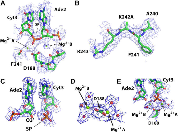
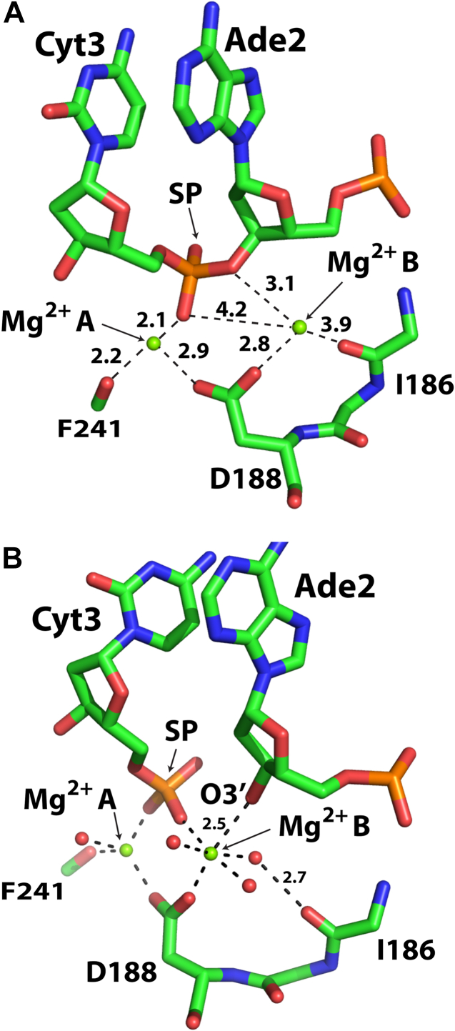
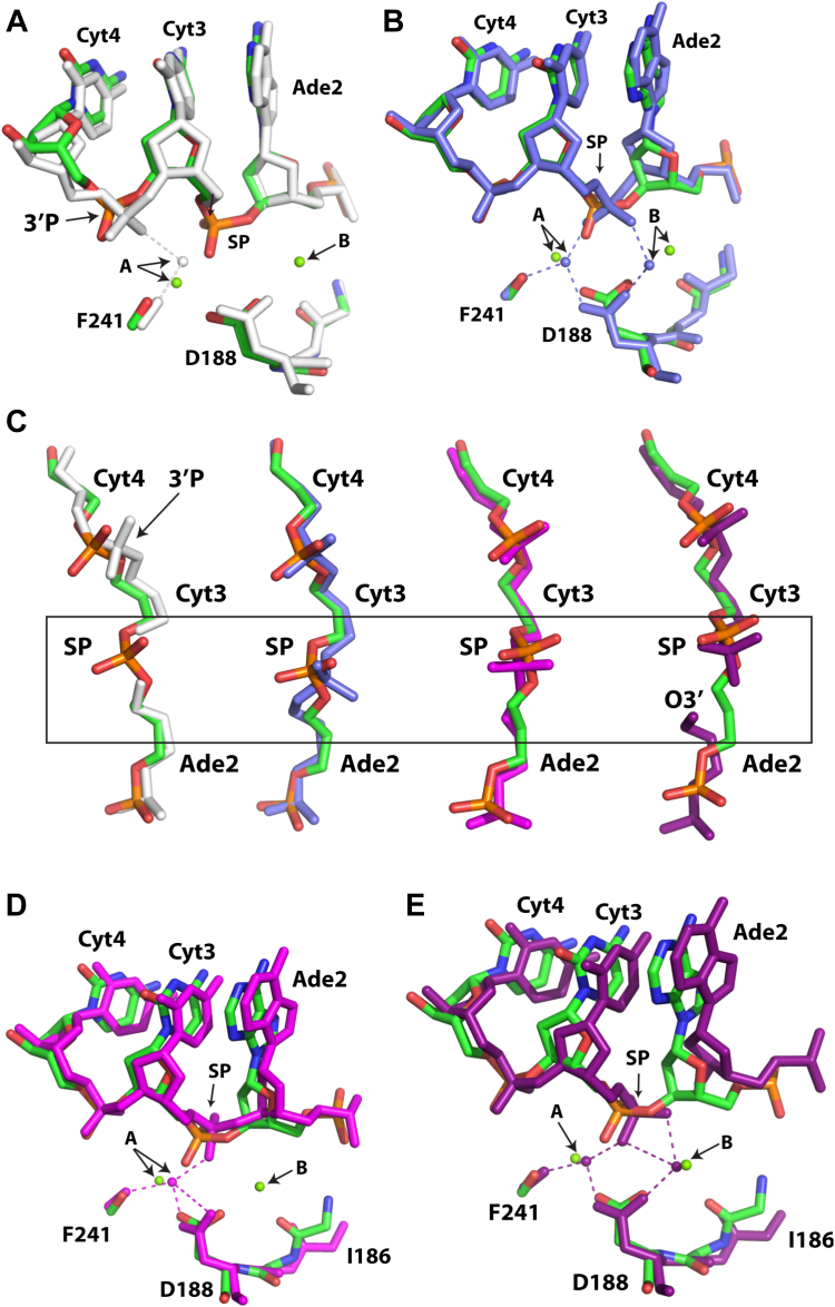
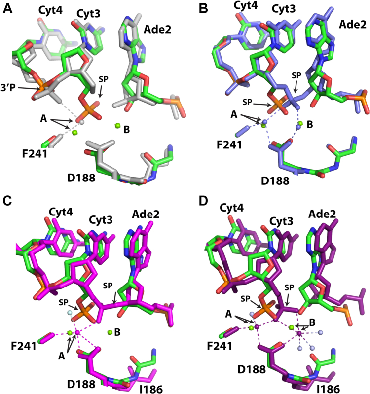
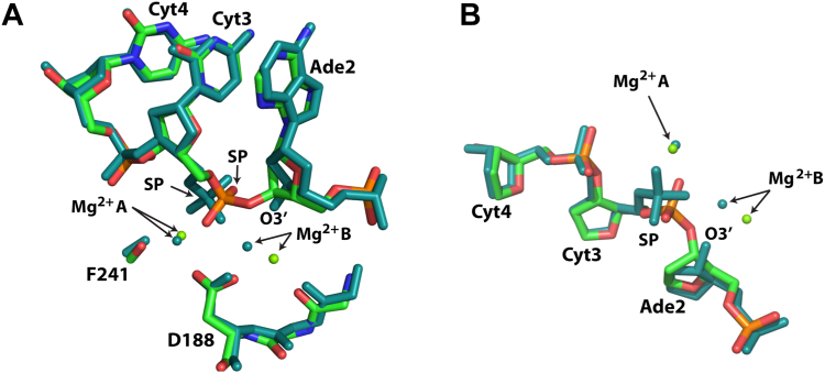
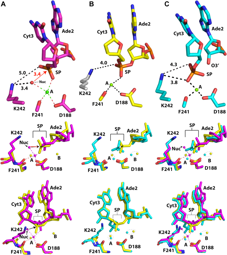
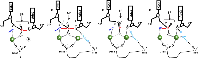
Enzymes that form filamentous assemblies with modulated enzymatic activities have gained increasing attention in recent years. SgrAI is a sequence specific type II restriction endonuclease that forms polymeric filaments with accelerated DNA cleavage activity and expanded DNA sequence specificity. Prior studies have suggested a mechanistic model linking the structural changes accompanying SgrAI filamentation to its accelerated DNA cleavage activity. In this model, the conformational changes that are specific to filamentous SgrAI maximize contacts between different copies of the enzyme within the filament and create a second divalent cation binding site in each subunit, which in turn facilitates the DNA cleavage reaction. However, our understanding of the atomic mechanism of catalysis is incomplete. Herein, we present two new structures of filamentous SgrAI solved using cryo-EM. The first structure, resolved to 3.3 Å, is of filamentous SgrAI containing an active site mutation that is designed to stall the DNA cleavage reaction, which reveals the enzymatic configuration prior to DNA cleavage. The second structure, resolved to 3.1 Å, is of WT filamentous SgrAI containing cleaved substrate DNA, which reveals the enzymatic configuration at the end of the enzymatic cleavage reaction. Both structures contain the phosphate moiety at the cleavage site and the biologically relevant divalent cation cofactor Mg2+ and define how the Mg2+ cation reconfigures during enzymatic catalysis. The data support a model for the activation mechanism that involves binding of a second Mg2+ in the SgrAI active site as a direct result of filamentation induced conformational changes.

Keywords: DNA nuclease; allostery; antiviral strategies; enzyme filaments; enzyme mechanism; enzyme regulation; polymeric enzymes; protein oligomerization.


Conflict of interest statement

Conflict of interest The authors declare that they have no conflicts of interest with the contents of this article.

Figures

Figure 1
Figure 1
Structures of filamentous and nonfilamentous SgrAI and the two metal ion mechanism.A, structure of the SgrAI filament showing approximately four DNA bound SgrAI dimers (DBD, each colored individually in red, green, yellow, or purple) per turn in a left-handed helix. B, superposition of filamentous (cyan) and nonfilamentous (magenta) SgrAI structures using one chain of a DBD showing an 11° rotation of the other subunit. DNA from the nonfilamentous structure shown in gray in surface rendering. C, view of the active site in the superimposed chains from filamentous (cyan, from PDB ID 7SS5) and nonfilamentous (magenta, from PDB ID 3DVO) DBD showing a shift of residues 184 to 187, which creates a second divalent cation binding site in the filamentous structure. D, schematic of the two-metal-ion mechanism. The nucleophile of the reaction is a water or hydroxide (dark blue) which is positioned by its coordination to metal ion A for in-line attack on the phosphorus atom of the scissile phosphate (SP). Both metal ions A and B coordinate a nonbridging oxygen of the SP, and metal ion B coordinates the leaving group as well as a water molecule positioned to donate a proton to the leaving group following bond cleavage (the bond to be cleaved is shown in red). An active site lysine found in many restriction endonucleases is shown interacting with the nucleophile and may serve in its positioning and activation.
Figure 2
Figure 2
Atomic models and experimental cryo-EM density maps in the active site.A, SgrAIK242A/40-1/Mg2+ around active site. Map contoured at 3σ. B, as in A, showing area around the K242A mutation. Map contoured at 4σ. C, SgrAIWT/40-1/Mg2+ at scissile phosphate. Map contoured at 4.5σ. D and E, as in C, but around Mg2+ in the active site. Map contoured at 5σ.
Figure 3
Figure 3
Active site geometry and Mg2+positioning in SgrAIK242A/40-1/Mg2+and SgrAIWT/40-1/Mg2+.A, the active site of SgrAIK242A/40-1/Mg2+. All distances between Mg2+ and oxygen ligands are 1.9 to 2.2 Å unless otherwise indicated. B, as in A, but for SgrAIWT/40-1/Mg2+. Distances shown in Å. SP, scissile phosphate.
Figure 4
Figure 4
Active site superpositions of SgrAIK242A/40-1/Mg with other SgrAI/DNA structures.A, superposition using all atoms of chain A of SgrAIK242A/40-1/Mg (green, blue, red, and orange) with those of 6OBJ (white). SP, scissile phosphate. B, as in (A), but with 7SS5 (slate). C, as in (A), but with view for SP conformation. 3DVO in magenta, 3MQY in dark purple. Boxed region indicates cleavage site. D, as in (A) but with 3DVO (magenta). E, as in A but 3MQY (dark purple).
Figure 5
Figure 5
Comparison of SgrAIWT/40-1/Mg2+with other SgrAI structures.A, all atoms of one chain were used in alignments. SgrAIWT/40-1/Mg2+ is shown in green, red, blue, orange, and SgrAIWT/PC/Mg2+ (PDB ID 6OBJ) is shown in white. PC is a precleaved version of 40-1 missing the phosphate at the cleavage site (i.e. the scissile phosphate or SP). B, superposition of SgrAIWT/40-1/Mg2+ (green, red, blue, and orange) with SgrAIWT/40-1/Ca2+ (PDB ID 7SS5, slate). C, superposition of SgrAIWT/40-1/Mg2+ (green, red, blue, and orange) with SgrAIWT/18-1/Ca2+ (PDB ID 3DVO, magenta, water molecule in light blue). D, superposition of SgrAIWT/40-1/Mg2+ (green, red, blue, and orange) with SgrAIWT/18-1/Mg2+ (PDB ID 3MQY, magenta, water molecules in light blue). PDB, Protein Data Bank.
Figure 6
Figure 6
Comparison of SgrAIK242A/40-1/Mg2+and SgrAIWT/40-1/Mg2+structures.A, view of the active site after superposition using all chain A atoms. WT SgrAI shown in teal, K242A mutant shown in green, red, blue, and orange. SP, scissile phosphate. B, as in (A), view from above showing the DNA backbone at the SP and positions of Mg2+ ions.
Figure 7
Figure 7
Position of K242 side chain in pre- and post-cleavage structures.A, top panel, active site arrangements in 3DVO. Middle and lower panels, superposition using all atoms of one chain of 3DVO (pink) and SgrAIK242A/40-1/Mg2+ (yellow). B, top panel, active site arrangement in SgrAIK242A/40-1/Mg2+. Side chain of K242 (white) is from 3DVO to show its possible position. Middle and lower panels, superposition using all atoms of a single chain of SgrAIK242A/40-1/Mg2+ (yellow) and SgrAIWT/40-1/Mg2+ (cyan). C, top panel, active site arrangement in SgrAIWT/40-1/Mg2+, middle and lower panels, superposition using all atoms of a single chain of 3DVO (pink), SgrAIK242A/40-1/Mg2+ (yellow) and SgrAIWT/40-1/Mg2+ (cyan).
Figure 8
Figure 8
Updatedmodel of active DNA cleavage by SgrAI.

Similar articles

References

    1. Park C.K., Horton N.C. Structures, functions, and mechanisms of filament forming enzymes: a Renaissance of enzyme filamentation. Biophys. Rev. 2019;11:927–994. - PMC - PubMed
    1. Park C.K., Horton N.C. Novel insights into filament-Forming enzymes. Nat. Rev. Mol. Cell Biol. 2020;21:1–2. - PubMed
    1. Lynch E.M., Kollman J.M., Webb B.A. Filament Formation by metabolic enzymes-a new twist on regulation. Curr. Opin. Cell Biol. 2020;66:28–33. - PMC - PubMed
    1. Simonet J.C., Burrell A.L., Kollman J.M., Peterson J.R. Freedom of assembly: metabolic enzymes Come Together. Mol. Biol. Cell. 2020;31:1201–1205. - PMC - PubMed
    1. Prouteau M., Loewith R. Regulation of cellular metabolism through phase separation of enzymes. Biomolecules. 2018;8:160–173. - PMC - PubMed

MeSH terms

LinkOut - more resources