. 2024 Jul 30;11(7):ENEURO.0165-24.2024.
doi: 10.1523/ENEURO.0165-24.2024. Print 2024 Jul.

Assembling a Coculture System to Prepare Highly Pure Induced Pluripotent Stem Cell-Derived Neurons at Late Maturation Stages

Affiliations

Assembling a Coculture System to Prepare Highly Pure Induced Pluripotent Stem Cell-Derived Neurons at Late Maturation Stages

Masuma Akter et al. eNeuro. .

Abstract

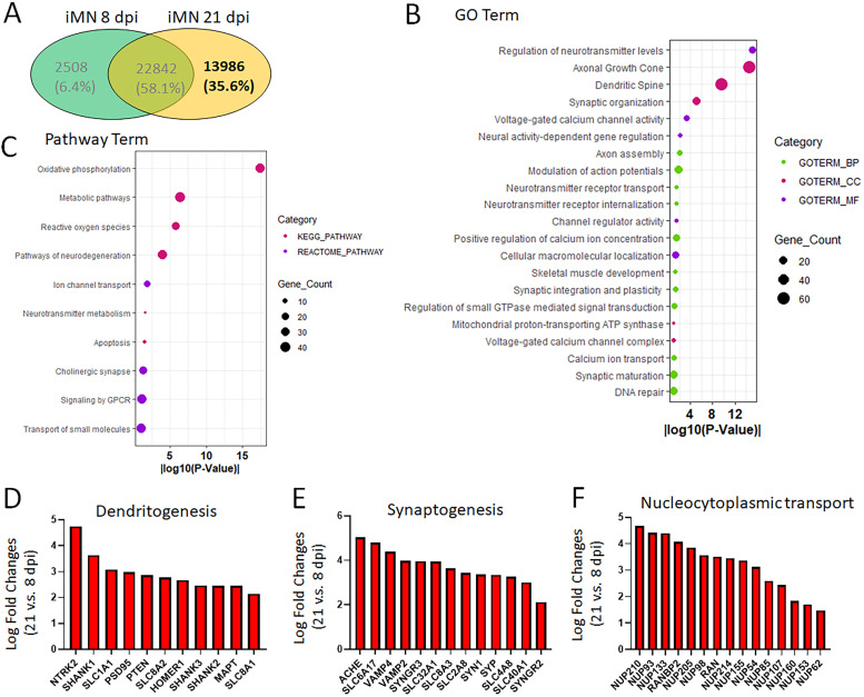
Generation of human induced pluripotent stem cell (hiPSC)-derived motor neurons (MNs) offers an unprecedented approach to modeling movement disorders such as dystonia and amyotrophic lateral sclerosis. However, achieving survival poses a significant challenge when culturing induced MNs, especially when aiming to reach late maturation stages. Utilizing hiPSC-derived motor neurons and primary mouse astrocytes, we assembled two types of coculture systems: direct coculturing of neurons with astrocytes and indirect coculture using culture inserts that physically separate neurons and astrocytes. Both systems significantly enhance neuron survival. Compared with these two systems, no significant differences in neurodevelopment, maturation, and survival within 3 weeks, allowing to prepare neurons at maturation stages. Using the indirect coculture system, we obtained highly pure MNs at the late mature stage from hiPSCs. Transcriptomic studies of hiPSC-derived MNs showed a typical neurodevelopmental switch in gene expression from the early immature stage to late maturation stages. Mature genes associated with neurodevelopment and synaptogenesis are highly enriched in MNs at late stages, demonstrating that these neurons achieve maturation. This study introduces a novel tool for the preparation of highly pure hiPSC-derived neurons, enabling the determination of neurological disease pathogenesis in neurons at late disease onset stages through biochemical approaches, which typically necessitate highly pure neurons. This advancement is particularly significant in modeling age-related neurodegeneration.

Keywords: coculture; human induced pluripotent stem cells (hiPSCs); motor neurons (MNs); neurodevelopment; synaptogenesis; transcriptomics.


Conflict of interest statement

The authors declare no competing financial interests.

Figures

None
Visual Abstract
Figure 1.
Figure 1.
Limited survival when culturing iPSC-derived MNs alone. A, Schematic shows the process of generation iPSC-MNs. iPSC, induced pluripotent stem cells; NPCs, neuronal progenitor cells; MNs, motor neurons. B, The timeline, treatment, and culture conditions during the process of generation of iPSC-MNs. The time at lentiviral transduction was set as Day 0. C, Representative confocal images of iPSC-MNs directly cocultured with astrocytes at 14 d postviral infection (dpi; for HB9 and ISL1) and 21 dpi (for ChAT). TUBB3 (tubulin beta 3 class III) served as a neuronal marker and Hoechst 33342 (HST) stained nuclei. Nuclear HB9 (motor neurons and pancreas homeobox 1) and ISL1 (ISL LIM homeobox 1) were used as early-stage MN markers, and ChAT (choline acetyltransferase) was used as a late-stage MN marker. Scale bar, 25 μm. D, Representative micrograph of iPSC-MNs cultured alone at 4 dpi shows typical healthy outgrowth with complex neurites that are well attached on the culture plate. Scale bar, 100 μm. E, Representative micrograph of iPSC-MNs cultured alone at 10 dpi shows most neurons are degenerated and start floating. Scale bar, 100 μm. F, Percentage of neurons showed degeneration at different time points. Results represent three independent experiments. Data presented as mean ± SD.
Figure 2.
Figure 2.
Coculturing with astrocytes enables full maturation of iPSC-MNs. A, A representative micrograph of mouse primary astrocytes. Scale bar, 50 μm. B, Confocal micrographs of primary astrocytes immunoassayed with astrocyte marker, glial fibrillary acidic protein (GFAP), and the nuclear dye Hoechst 33342 (HST). Scale bar, 25 μm. C, Confocal micrograph of iPSC-MNs directly cocultured with astrocytes at 3 weeks postviral infection (wpi). Scale bar, 25 μm. D, Repetitive AP waveforms recorded under current-clamp mode of iPSC-MNs at 3 wpi. E, Degenerated neurons at different time points. Data presented as mean ± SD.
Figure 3.
Figure 3.
Assembling a coculture system using culture inserts to physically separate astrocytes and MNs. A, Schematic shows the process of assembling the coculture system with culture inserts that physically separate neurons and cocultured astrocytes. B, Representative confocal micrographs of MNs at 14 dpi with direct coculture with astrocytes and coculture with astrocytes using culture insert. Neuronal marker TUBB3 shows the soma and neuron processes, Hoechst 33342 (HST) stained nuclei, and glial fibrillary acidic protein (GFAP) was used as an astrocyte marker. Scale bar, 25 μm. C, RT-PCR assay shows the relative gene expression levels of HB9 and MAP2 in indicated conditions. Samples of iPSC-MNs at 7 dpi were set up as an early-stage control, and samples of cocultured iPSC-MNs with culture inserts were collected at 21 dpi. Target gene expression was normalized with TUBB3. Data presented as mean ± SD. ns, not significant; ***p < 0.001; ****p < 0.0001. ANOVA. D, The time course of survived neurons under indicated conditions. The number of neurons at 7 dpi was set as a starting number (100%) and the numbers of surviving neurons at late time points were normalized by the starting number. Results represent three independent experiments. Data presented as mean ± SD. Direct coculture versus coculture with inserts: ns, not significant; *p < 0.05. Student's t test.
Figure 4.
Figure 4.
Transcriptomic studies characterized iPSC-MNs prepared using culture inserts. A, Venn diagram shows the RNAseq results of iPSC-MNs at different development stages. The early-stage samples of iPSC-MNs (8 dpi) were cultured alone and the late-stage samples of iPSC-MNs (21 dpi) were cultured using culture inserts. B, A volcano plot shows the DEGs (21 vs 8 dpi) based on the cut off log2FC >1 and p value <0.05. Blue and red dots represent down- and upregulated genes, respectively. Gray dots represent the remaining genes with no significant difference. C, Heat map of RNAseq results of all DEGs in iPSC-MNs (21 vs 8 dpi), including 5,020 downregulated genes and 7,009 upregulated genes. D, The developmental switch of some temporally regulated genes in iPSC-MNs at 21 dpi. Genes highly expressed at early developmental stages were downregulated, and genes implicated in neuron maturation are upregulated. E, GO analysis of significantly upregulated genes in iPSC-MNs at 21 dpi with the enrichment of terms related to neuron maturation and synaptogenesis. F, Pathway enrichment analysis with KEGG and Reactome of significantly upregulated genes in iPSC-MNs at 21 dpi. G, GO analysis of significantly downregulated genes in iPSC-MNs at 21 dpi with the enrichment of GO terms related to early neuron differentiation. H, Pathway enrichment analysis with KEGG and Reactome of significantly downregulated genes in iPSC-MNs at 21 dpi.
Figure 5.
Figure 5.
Characterization of gene expression in hiPSC-MNs at late mature stages. A, Venn diagram shows the RNAseq results of the number of genes that were only identified in late-stage iPSC-MNs at 21 dpi prepared with culture inserts. B, GO analysis of genes only expressed in iPSC-MNs at 21 dpi with the enrichment of terms related to neuron functions. C, Pathway enrichment analysis with KEGG and Reactome of genes only expressed in iPSC-MNs at 21 dpi. D, Representative genes involved in dendritogenesis are upregulated in iPSC-MNs at 21 dpi. E, Representative genes involved in synaptogenesis are upregulated in iPSC-MNs at 21 dpi. F, Representative genes involved in nucleocytoplasmic transport are upregulated in iPSC-MNs at 21 dpi.

References

    1. Akter M, Cui H, Chen Y-H, Ding B (2021) Generation of two induced pluripotent stem cell lines with heterozygous and homozygous GAG deletion in TOR1A gene from a healthy hiPSC line. Stem Cell Res 56:102536. 10.1016/j.scr.2021.102536 - DOI - PMC - PubMed
    1. Akter M, Cui H, Hosain MA, Liu J, Duan Y, Ding B (2024) RANBP17 overexpression restores nucleocytoplasmic transport and ameliorates neurodevelopment in induced DYT1 dystonia motor neurons. J Neurosci 44:e1728232024. 10.1523/JNEUROSCI.1728-23.2024 - DOI - PMC - PubMed
    1. Akter M, Cui H, Sepehrimanesh M, Hosain MA, Ding B (2022) Generation of highly pure motor neurons from human induced pluripotent stem cells. STAR Protoc 3:101223. 10.1016/j.xpro.2022.101223 - DOI - PMC - PubMed
    1. Akter M, Ding B (2022) Modeling movement disorders via generation of hiPSC-derived motor neurons. Cells 11:3796. 10.3390/cells11233796 - DOI - PMC - PubMed
    1. Alves CJ, Dariolli R, Jorge FM, Monteiro MR, Maximino JR, Martins RS, Strauss BE, Krieger JE, Callegaro D, Chadi G (2015) Gene expression profiling for human iPS-derived motor neurons from sporadic ALS patients reveals a strong association between mitochondrial functions and neurodegeneration. Front Cell Neurosci 9:289. 10.3389/fncel.2015.00289 - DOI - PMC - PubMed

LinkOut - more resources