. 2024 Jul;30(7):e14859.
doi: 10.1111/cns.14859.

Prediction of disease progression in individuals with subjective cognitive decline using brain network analysis

Affiliations

Prediction of disease progression in individuals with subjective cognitive decline using brain network analysis

Simin Deng et al. CNS Neurosci Ther. 2024 Jul.

Abstract

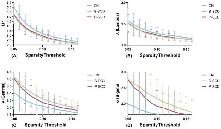
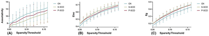
Objective: The objective of this study is to explore potential differences in brain functional networks at baseline between individuals with progressive subjective cognitive decline (P-SCD) and stable subjective cognitive decline (S-SCD), as well as to identify potential indicators that can effectively distinguish between P-SCD and S-SCD.

Methods: Alzheimer's Disease Neuroimaging Initiative (ADNI) database was utilized to enroll SCD individuals with a follow-up period of over 3 years. This study included 39 individuals with S-SCD, 15 individuals with P-SCD, and 45 cognitively normal (CN) individuals. Brain functional networks were constructed based on the AAL template, and graph theory analysis was performed to determine the topological properties.

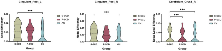
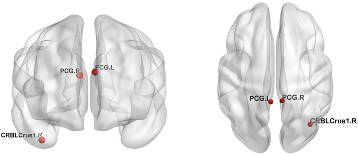
Results: For global metric, the S-SCD group exhibited stronger small-worldness with reduced connectivity among nearby nodes and accelerated compensatory information transfer capacity. For nodal efficiency, the S-SCD group showed increased connectivity in bilateral posterior cingulate gyri (PCG). However, for nodal local efficiency, the P-SCD group exhibited significantly reduced connectivity in the right cerebellar Crus I compared with the S-SCD group.

Conclusion: There are differences in brain functional networks at baseline between P-SCD and S-SCD groups. Furthermore, the right cerebellar Crus I region may be a potentially useful brain area to distinguish between P-SCD and S-SCD.

Keywords: SCD; brain functional networks; early identification; graph theory.


Conflict of interest statement

The author reports no conflicts of interest in this work.

Figures

FIGURE 1
FIGURE 1
Small‐worldness of the whole‐brain resting‐state functional network among the three groups at a sparsity rating of 0.05–0.16. (A) LP indicates the shortest path length. The AUCs of LP were significantly lower in the S‐SCD group compared with the CN group. The P‐SCD group exhibited AUC values intermediate between the S‐SCD and CN groups, but there were no statistically significant differences among the groups. (B) λ indicates the normalized characteristic path length. The AUC values of λ were significantly lower in the S‐SCD group compared with the CN group. The P‐SCD group exhibited values intermediate between the S‐SCD and CN groups, but there was no statistically significant difference between the groups. (C) γ indicates the normalized clustering coefficient. The AUC values of γ were significantly higher in the S‐SCD group compared with the CN group. Similarly, the P‐SCD group had values intermediate to those of the S‐SCD and CN groups, with no statistically significant differences from the other groups. (D) σ indicates the small‐worldness coefficient. The AUC values of σ were significantly higher in the S‐SCD group compared with the CN group. Similarly, the AUC values of the P‐SCD group were intermediate between the S‐SCD and CN groups, without statistically significant differences between the groups. CN, cognitively normal; LP, shortest path length; P‐SCD, progressive subjective cognitive decline; S‐SCD, stable subjective cognitive decline; γ, normalized clustering coefficient; λ, normalized characteristic path length; σ, small‐worldness coefficient.
FIGURE 2
FIGURE 2
Whole‐brain resting‐state functional network of the three groups at a sparsity rating of 0.05–0.16. (A) The AUC value of assortativity was lower in the S‐SCD group compared with the P‐SCD and CN groups. (B) Eloc indicates the local efficiency. The AUC values of Eloc were higher in the S‐SCD group compared with the CN group. The P‐SCD group showed AUC values intermediate between the S‐SCD and CN groups, but without statistically significant differences among the groups. (C) Eg indicates the global efficiency. The AUC values of Eg were higher in the S‐SCD group compared with the CN group. The P‐SCD group showed AUC values intermediate between the S‐SCD and CN groups, but without statistically significant differences among the groups. CN, cognitively normal; Eg, global efficiency; Eloc, local efficiency; P‐SCD, progressive subjective cognitive decline; S‐SCD, stable subjective cognitive decline.
FIGURE 3
FIGURE 3
Differences in nodal local efficiency among the three groups. Compared with the CN group, both the S‐SCD and P‐SCD groups exhibited changes in the Eloc of nodes in bilateral PCG. In particular, the S‐SCD group demonstrated a significant increase in the Eloc in this area. The P‐SCD group exhibited significantly lower connectivity in the right cerebellum Crus I compared with the S‐SCD group. CN, cognitively normal; Eloc, local efficiency; PCG, posterior cingulate gyrus; P‐SCD, progressive subjective cognitive decline; S‐SCD, stable subjective cognitive decline. *** indicates p < 0.05, indicating statistically significant differences between groups.
FIGURE 4
FIGURE 4
Characteristics of nodal properties. CRBLCrus 1.R, right cerebellar Crus I; PCG.L, posterior cingulate gyrus, left; PCG.R, posterior cingulate gyrus, right.

Similar articles

References

    1. Abbott A. Conquering Alzheimer's: a look at the therapies of the future. Nature. 2023;616(7955):26‐28. - PubMed
    1. 2023 Alzheimer's disease facts and figures. Alzheimers Dement. 2023;19(4):1598‐1695. - PubMed
    1. Self WK, Holtzman DM. Emerging diagnostics and therapeutics for Alzheimer disease. Nat Med. 2023;29(9):2187‐2199. - PubMed
    1. Jessen F, Wolfsgruber S, Kleineindam L, et al. Subjective cognitive decline and stage 2 of Alzheimer disease in patients from memory centers. Alzheimers Dement. 2023;19(2):487‐497. - PubMed
    1. Jessen F, Amariglio RE, van Boxtel M, et al. A conceptual framework for research on subjective cognitive decline in preclinical Alzheimer's disease. Alzheimers Dement. 2014;10(6):844‐852. - PMC - PubMed

Publication types

LinkOut - more resources