. 2024 Jul 15;15(1):5528.
doi: 10.1038/s41467-024-49600-7.

Beta activity in human anterior cingulate cortex mediates reward biases

Affiliations

Beta activity in human anterior cingulate cortex mediates reward biases

Jiayang Xiao et al. Nat Commun. .

Erratum in

  • Author Correction: Beta activity in human anterior cingulate cortex mediates reward biases.
    Xiao J, Adkinson JA, Myers J, Allawala AB, Mathura RK, Pirtle V, Najera R, Provenza NR, Bartoli E, Watrous AJ, Oswalt D, Gadot R, Anand A, Shofty B, Mathew SJ, Goodman WK, Pouratian N, Pitkow X, Bijanki KR, Hayden B, Sheth SA. Xiao J, et al. Nat Commun. 2025 Jun 25;16(1):5397. doi: 10.1038/s41467-025-61277-0. Nat Commun. 2025. PMID: 40562769 Free PMC article. No abstract available.

Abstract

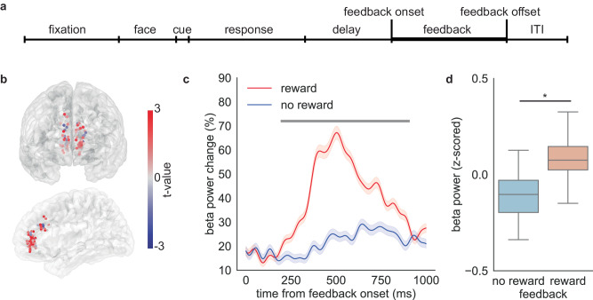
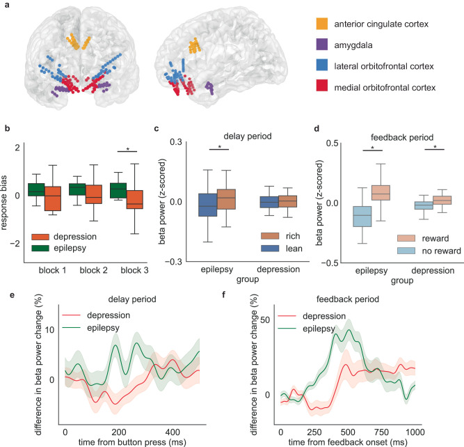
The rewards that we get from our choices and actions can have a major influence on our future behavior. Understanding how reward biasing of behavior is implemented in the brain is important for many reasons, including the fact that diminution in reward biasing is a hallmark of clinical depression. We hypothesized that reward biasing is mediated by the anterior cingulate cortex (ACC), a cortical hub region associated with the integration of reward and executive control and with the etiology of depression. To test this hypothesis, we recorded neural activity during a biased judgment task in patients undergoing intracranial monitoring for either epilepsy or major depressive disorder. We found that beta (12-30 Hz) oscillations in the ACC predicted both associated reward and the size of the choice bias, and also tracked reward receipt, thereby predicting bias on future trials. We found reduced magnitude of bias in depressed patients, in whom the beta-specific effects were correspondingly reduced. Our findings suggest that ACC beta oscillations may orchestrate the learning of reward information to guide adaptive choice, and, more broadly, suggest a potential biomarker for anhedonia and point to future development of interventions to enhance reward impact for therapeutic benefit.


Conflict of interest statement

S.A.S. has consulting agreements with Boston Scientific, NeuroPace, Abbott, and Zimmer Biomet. W.K.G. receives royalties from Nview, LLC and OCDscales, LLC. S.J.M. has served as a consultant to the following companies: Almatica Pharma, Biohaven, BioXcel Therapeutics, Boehringer-Ingelheim, Brii Biosciences, Clexio Biosciences, COMPASS Pathways, Delix Therapeutics, Douglas Pharmaceuticals, Eleusis, Engrail Therapeutics, Freedom Biosciences, Janssen, Liva Nova, Levo Therapeutics, Merck, Neumora, Neurocrine, Perception Neurosciences, Praxis Precision Medicines, Relmada Therapeutics, Sage Therapeutics, Seelos Therapeutics, Signant Health, Sunovion, Xenon Pharmaceuticals, and XW Pharma. S.J.M. has received research support from Boehringer-Ingelheim, Engrail Therapeutics, Merck, Neurocrine, and Sage Therapeutics. N.P. is a consultant for Abbott Laboratories and Sensoria Therapeutics. The remaining authors declare no competing interests.

Figures

Fig. 1
Fig. 1. Task description, behavioral performance, and recording locations.
a Timeline of the probabilistic reward task. b Response bias averaged across all Epilepsy Cohort patients. A positive response bias value indicates there is a preference for choosing the more frequently rewarded stimulus. Boxplots illustrate quartiles at 25% and 75%, with horizontal lines denoting medians, and whiskers extending to 1.5 times the interquartile ranges. n = 16 runs of task. One-sample t test compared with zero, block1: p = 0.036, block2: p = 0.0029, block3: p = 0.0061. ‘*’ represents p < 0.05. c Accuracy for rich and lean stimuli averaged across all epilepsy patients. Boxplots illustrate quartiles at 25% and 75%, with horizontal lines denoting medians, and whiskers extending to 1.5 times the interquartile ranges. n = 16 runs of task. Paired-sample t test, block1: p = 0.026, block2: p = 0.0034, block3: p = 0.0086. ‘*’ represents p < 0.05. d Proportion of choosing the same response when there is a reward or no reward following the response in the previous trial. n = 48 blocks of task. Paired-sample t test, p = 3.9*10-7. ‘*’ represents p < 0.05. e Intracranial recording electrodes sample reward-relevant regions in Epilepsy Cohort patients. Source data are provided as a Source Data file.
Fig. 2
Fig. 2. Increase in ACC beta power in response to the rich stimulus during the delay period.
a Delay period starts from the button press and ends when feedback appears on the screen. b Differences in beta (12–30 Hz) power between the rich and lean trials are plotted at each sEEG contact. The color represents the t value comparing rich with lean trials. Red indicates greater power during rich trials. c Beta power change during the delay period. Red indicates rich trials while blue indicates lean trials. Trials are time-locked to the button press. The horizontal bar indicates time points in the cluster that are statistically significant (p < 0.05), as determined by two-sided cluster-based permutation test. Data are presented as mean values ± SEM. Source data are provided as a Source Data file.
Fig. 3
Fig. 3. Relationship between response bias and neural activity during the delay period.
a Differences in beta power between the rich and lean trials are plotted at each sEEG contact for high response bias blocks or low response bias blocks. b Beta power change during the delay period for high response bias blocks or low response bias blocks. Trials are time-locked to the button press. Horizontal bar depicts time points for which neural activity between rich trials and lean trials is significantly different (two-sided cluster-based permutation test, p < 0.05). Data are presented as mean values ± SEM. c Difference in beta power in high or low response bias blocks. Boxplots illustrate quartiles at 25% and 75%, with horizontal lines denoting medians, and whiskers extending to 1.5 times the interquartile ranges. n = 56 channels for low response bias blocks and n = 52 channels for high response bias blocks. Linear mixed model, p = 0.66 for the low response bias blocks and p = 0.0013 for the high response bias blocks. ‘*’ represents p < 0.05. d Relationship between response bias and the difference in reward response towards rich or lean stimulus in all frequency bands. The statistical test for the Pearson correlation coefficient is two-sided, and no adjustments were made for multiple comparisons. e Correlation between neural activity and response bias in all frequency bands. Center for the error bars represent the correlation coefficient. Error bars represent a 95% confidence interval. p = 0.013 for beta band. ‘*’ represents p < 0.05. Source data are provided as a Source Data file.
Fig. 4
Fig. 4. Increase in ACC beta power in response to reward during the feedback period.
a The feedback period starts when the dollar bill or empty rectangle appears on the screen and ends when the feedback image disappears. b Differences in beta power between the reward and no-reward trials are plotted at each sEEG contact. The color represents the t value comparing reward feedback with neutral feedback. Red indicates greater power during the reward feedback. Only trials with correct response are included in the analysis. c Feedback-aligned beta power change averaged over trials and participants. Red indicates reward trials while blue indicates correct trials with no reward. Trials are time-locked to feedback onset. Horizontal bar depicts time points for which neural activity between reward trials and no-reward trials is significantly different (two-sided cluster-based permutation test, p < 0.05). Data are presented as mean values ± SEM. d Average beta power change across the feedback period. Boxplots illustrate quartiles at 25% and 75%, with horizontal lines denoting medians, and whiskers extending to 1.5 times the interquartile ranges. n = 65 channels. Linear mixed model, p = 1.8*10−36. ‘*’ represents p < 0.05. Source data are provided as a Source Data file.
Fig. 5
Fig. 5. Difference in reward response between Depression Cohort and Epilepsy Cohort.
a Intracranial recording electrodes sample reward-relevant regions in depression patients. b Response bias in Depression Cohort and Epilepsy Cohort. Boxplots illustrate quartiles at 25% and 75%, with horizontal lines denoting medians, and whiskers extending to 1.5 times the interquartile ranges. n = 16 runs for Epilepsy Cohort and n = 12 runs for Depression Cohort. Two-sample t test, block1: p = 0.24, block2: p = 0.24, block3: p = 0.030. ‘*’ represents p < 0.05. c Average beta power change across the delay period. Boxplots illustrate quartiles at 25% and 75%, with horizontal lines denoting medians, and whiskers extending to 1.5 times the interquartile ranges. n = 65 channels for Epilepsy Cohort and n = 36 channels for Depression Cohort. Linear mixed model, p = 0.039 for Epilepsy Cohort, p = 0.77 for Depression Cohort. ‘*’ represents p < 0.05. d Average beta power change across the feedback period. Boxplots illustrate quartiles at 25% and 75%, with horizontal lines denoting medians, and whiskers extending to 1.5 times the interquartile ranges. n = 65 channels for Epilepsy Cohort and n = 36 channels for Depression Cohort. Linear mixed model, p = 1.8*10−36 for Epilepsy Cohort, p = 1.6*10−11 for Depression Cohort. ‘*’ represents p < 0.05. e Difference towards rich or lean stimulus in the beta activity across the delay period in Depression Cohort and Epilepsy Cohort. Data are presented as mean values ± SEM. f Difference towards reward or neutral feedback in the beta activity across the feedback period. Data are presented as mean values ± SEM. Source data are provided as a Source Data file.

References

    1. Averbeck, B. B. & Costa, V. D. Motivational neural circuits underlying reinforcement learning. Nat. Neurosci.20, 505–512 (2017). - PubMed
    1. Howard-Jones, P. A. & Jay, T. Reward, learning and games. Curr. Opin. Behav. Sci.10, 65–72 (2016).
    1. Rangel, A. & Hare, T. Neural computations associated with goal-directed choice. Curr. Opin. Neurobiol.20, 262–270 (2010). - PubMed
    1. Kangas, B. D., Wooldridge, L. M., Luc, O. T., Bergman, J. & Pizzagalli, D. A. Empirical validation of a touchscreen probabilistic reward task in rats. Transl. Psychiatry10, 1–11 (2020). - PMC - PubMed
    1. Pizzagalli, D. A., Jahn, A. L. & O’Shea, J. P. Toward an objective characterization of an anhedonic phenotype: a signal-detection approach. Biol. Psychiatry57, 319–327 (2005). - PMC - PubMed

LinkOut - more resources