. 2024 Oct;16(10):1612-1620.
doi: 10.1038/s41557-024-01584-z. Epub 2024 Jul 15.

Engineering a nanoscale liposome-in-liposome for in situ biochemical synthesis and multi-stage release

Affiliations

Engineering a nanoscale liposome-in-liposome for in situ biochemical synthesis and multi-stage release

Colin P Pilkington et al. Nat Chem. 2024 Oct.

Abstract

Soft-matter nanoscale assemblies such as liposomes and lipid nanoparticles have the potential to deliver and release multiple cargos in an externally stimulated and site-specific manner. Such assemblies are currently structurally simplistic, comprising spherical capsules or lipid clusters. Given that form and function are intertwined, this lack of architectural complexity restricts the development of more sophisticated properties. To address this, we have devised an engineering strategy combining microfluidics and conjugation chemistry to synthesize nanosized liposomes with two discrete compartments, one within another, which we term concentrisomes. We can control the composition of each bilayer and tune both particle size and the dimensions between inner and outer membranes. We can specify the identity of encapsulated cargo within each compartment, and the biophysical features of inner and outer bilayers, allowing us to imbue each bilayer with different stimuli-responsive properties. We use these particles for multi-stage release of two payloads at defined time points, and as attolitre reactors for triggered in situ biochemical synthesis.


Conflict of interest statement

The authors declare no competing interests.

Figures

Fig. 1
Fig. 1. Engineering nanoscale liposomes-in-liposomes.
a, A graphic illustrating the steps involved in the click-chemistry-assisted microfluidic synthesis of concentrisomes. A liposome suspension with alkyne moieties is fed into a MHF chip as a sheathing buffer stream, which is co-flowed alongside a secondary lipid composition that contains azide-functionalized lipids dissolved in ethanol. Micellar/bicellar aggregates tether to the external leaflet of pre-formed vesicles and eventually close to generate a second bilayer. The DBCO moiety, azide and triazole product are represented by a yellow triangle, green star and pink pentagon, respectively. The corresponding chemical structures are shown to the right of each graphic. A representative cryo-TEM micrograph of the concentrisome (double-bilayer liposome) is given on the right. Scale bar, 200 nm. b, A graphical illustration of possible concentrisome functionality: multi-cargo encapsulation in different compartments, triggered sequential multi-stage release of two different payloads, and in situ biochemical synthesis within the attolitre volume of the particle itself, followed by release.
Fig. 2
Fig. 2. Characterization of concentrisomes via DLS and cryo-TEM.
a, Dynamic light scattering data shows a slight shift of the hydrodynamic diameter of input liposomes, functionalized with DBCO, from approximately 100 nm (PDI = 0.16) to 120 nm (PDI = 0.26), after exposure to a new lipid composition via an MHF chip containing azide-functionalized lipids. Correlograms (correlation coefficient (g2−1) versus time) for both Gaussians are shown in the inset. b, Representative cryo-TEM micrograph for input liposomes (yellow), which are unilamellar. c, Representative cryo-TEM micrograph for concentrisomes (blue), showing inner and outer compartments. df, A graphic illustrating the potential stages of concentrisome growth with corresponding cryo-TEM examples. Left to right: pre-formed liposomes (d) act as nucleation points in the MHF chip, around which bicelle/micelles with azide functionality begin to assemble (see central micrograph in e). We propose that these covalently tethered assemblies eventually close over to generate an external spherical bilayer, generating the concentrisome morphology (f). Source data
Fig. 3
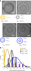
Fig. 3. Relationship between PEGylated-lipid length and intermembrane space.
a, The variation of dinter between successive bilayers, using PEG chains of different molecular weights, and in various combinations, is shown. Micrographs correspond to combinations, with average dinter values of 10, 23, 34 and 44 nm, for no-linker, PEG2KDBCO:PEG2KN3, PEG2KDBCO:PEG5KN3 and PEG5KDBCO:PEG5KN3, respectively. Scale bars, 25 nm. b, A histogram with measured dinter values for each linker combination. Gaussian plots are added for clarity. A gradual increase in dinter is seen with increasing combined PEG length; >25 concentrisomes were analysed to generate each distribution. Source data
Fig. 4
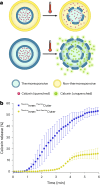
Fig. 4. Validation of layering and compositional control.
a, A graphic illustrating the main events of the assay, showing the ability to control the composition of each bilayer (in this case making them thermoresponsive and non-thermoresponsive). Initially the dye is quenched (non-fluorescent). For concentrisomes with a non-thermoresponsive outer bilayer, calcein was not expected to undergo sufficient dilution via efflux to unquench and produce a fluorescent signal. b, Percent calcein release profiles at 42 °C for each population. When both membranes were thermoresponsive, the fluorescence increase was much higher than when only the inner one was, because, in the latter, calcein remained encapsulated in a small enough volume to prevent unquenching. The release profiles for the additional controls mentioned in the main text can be found in Supplementary Fig. 6, whereas compositional details can be found in Supplementary Fig. 7. Values were calculated from fluorescence intensities after addition of surfactant (Triton X-100, 5 wt%, 2.5 µl min–1). Error bars indicate the s.d. of the average intensities for n = 3. The <20% calcein release for ThermoInner:Non-thermoOuter concentrisomes was thought to originate from unilamellar vesicles that had not undergone click chemistry. Source data
Fig. 5
Fig. 5. Multi-stage payload release.
a, A graphic illustrating multi-stage sequential release of two different cargos from two concentrisomes compartments. The two cargos are self-quenching dyes and thus release leads to an increase in fluorescence. b, Percent release of methylene blue over time from the concentrisome system with outer and inner bilayers composed of DPPC and DSPC, respectively. As methylene blue was isolated to the intermembrane space, the dye was released at 42 °C (above the transition temperature of the outer membrane DPPC composition). Data are represented as mean values ± 1 s.d. for n = 3. c, Percent calcein release over time for the same concentrisome system. In this case calcein was isolated to the inner liposome, thus release was observed only at 52 °C (which is above the transition temperature of this DPSC composition, the main component of the inner bilayer). Data are represented as mean values ± 1 s.d. for n = 3. d, A graphic illustrating the events of a multi-stage sequential release of the same cargo (calcein dye) from the two compartments of a concentrisome. Calcein was encapsulated in both the inner liposome and intermembrane space of a concentrisome system, with the inner bilayer composed of our DSPC composition (Tm ≈ 52 °C), and the outer bilayer of our DPPC composition (Tm ≈ 42 °C). e, Percent release of calcein dye over time, showing two discrete bursts when the samples are heated to the phase transition temperatures of the inner and outer membranes. Data are represented as mean values ± 1 s.d. for n = 3. Further controls can be found in Supplementary Fig. 8. Source data
Fig. 6
Fig. 6. Triggered biochemical synthesis within the nanoparticle.
a, Graphic illustrating in situ enzymatic synthesis in attolitre concentrisome reactors. Thermoresponsive liposomes containing the FDG substrate were recirculated with non-thermoresponsive lipids and β-Gal using an MHF flow regime identical to the flow conditions as before. In the control, liposomes were recirculated with ethanol and β-Gal only. b, Fluorometric data tracking the hydrolysis of FDG to fluorescein (λex = 498 nm; λem = 517 nm) at room temperature. No fluorescence increase was observed indicating enzyme and substrate remained compartmentalized. c, The same populations at 42 °C, showing an increase in fluorescence in the concentrisome sample. This indicates the thermoresponsive inner compartment permeabilizes, leading to the content mixing and subsequent enzymatic reaction occurring within the concentrisome. Error bars indicate the s.d. of the mean between three separate experiments, for both control (blue) and test (yellow) results. Further control experiments can be found in Supplementary Figs. 10–12. Source data

Similar articles

Cited by

References

    1. Gispert, I. et al. Stimuli-responsive vesicles as distributed artificial organelles for bacterial activation. Proc. Natl Acad. Sci. USA119, e2206563119 (2022). - PMC - PubMed
    1. Hindley, J. W. et al. Light-triggered enzymatic reactions in nested vesicle reactors. Nat. Commun.9, 1093 (2018). - PMC - PubMed
    1. Cardoso, B. D., Cardoso, V. F., Lanceros-Méndez, S. & Castanheira, E. M. S. Solid magnetoliposomes as multi-stimuli-responsive systems for controlled release of doxorubicin: assessment of lipid formulations. Biomedicines10, 1207 (2022). - PMC - PubMed
    1. Lee, Y. & Thompson, D. H. Stimuli-responsive liposomes for drug delivery. WIREs Nanomed. Nanobiotechnol.9, e1450 (2017). - PMC - PubMed
    1. Lou, J., Schuster, J. A., Barrera, F. N. & Best, M. D. ATP-responsive liposomes via screening of lipid switches designed to undergo conformational changes upon binding phosphorylated metabolites. J. Am. Chem. Soc.144, 3746–3756 (2022). - PubMed

Publication types

LinkOut - more resources