Review
. 2024 Sep;17(9):373-385.
doi: 10.1007/s12178-024-09911-0. Epub 2024 Jul 16.

Management of Proximal Hamstring Injuries: Non-operative and Operative Treatment

Affiliations
Review

Management of Proximal Hamstring Injuries: Non-operative and Operative Treatment

Thomas R Yetter et al. Curr Rev Musculoskelet Med. 2024 Sep.

Abstract

Purpose of review: To evaluate the current evidence and literature on treatment options for proximal hamstring injuries.

Recent findings: Patients with 3-tendon complete tears with greater than 2 cm of retraction have worse outcomes and higher complication rates compared to those with less severe injuries. Endoscopic and open proximal hamstring repair both have favorable patient reported outcomes at 5-year follow up. Proximal hamstring repair in patients who are male, with isolated semimembranosus injury, and have proximal hamstring free tendon rupture are more likely to have earlier return to sports. The Parisian Hamstring Avulsion Score (PHAS) is a validated patient-reported outcome measure to predict return to sports. Proximal hamstring injuries may occur in both elite and recreational athletes and may present with varying degrees of chronicity and severity. Injuries occur most commonly during forceful eccentric contraction of the hamstrings and often present with ischial tuberosity tenderness, ecchymosis, and hamstring weakness. Treatment decision-making is dictated by the tendons involved and chronicity. Many proximal hamstring injuries can be successfully treated with non-surgical measures. However, operative treatment of appropriately indicated proximal hamstring tendon injuries can result in significantly better functional outcomes and faster and more reliable return to sports compared to nonoperative treatment. Both endoscopic and open surgical repair techniques show high satisfaction levels and excellent patient-reported outcomes at short- and mid-term follow-up. Postoperative rehabilitation protocols vary across the literature and ongoing study is needed to clarify the optimal program, though emphasis on eccentric hamstring strengthening may be beneficial.

Keywords: Endoscopic repair; Hamstring; Open repair; Tendon injury; Treatment outcomes.


Conflict of interest statement

The authors declare no competing interests.

Figures

Fig. 1
Fig. 1
Anatomic cadaveric dissection demonstrating the proximity of the sciatic nerve (SC) lateral to the ischial tuberosity (red line) and proximal insertions of the long head of the biceps femoris (Bl), semitendinosus (ST), and semimembranosus (SM). Permission was obtained for re-use of this figure from the publisher [8]
Fig. 2
Fig. 2
Depiction of Nordic Hamstring Exercise (NHE) otherwise known as Nordic Hamstring Curls. Using an assistant to stabilize the feet, the patient starts in a kneeled position (A), eccentrically loads the hamstrings during lowering phase (B), and then contracts the hamstrings to return to the upright kneeling position (C). Clinicians instructing patients to complete this exercise must adequately pad the knee to avoid instigating or worsening anterior knee pain. Alternatively, if anterior knee pain precludes proper form or participation in part of in full, a GHD (glute-hamstring developer) machine is an excellent tool to perform eccentric hamstring strengthening
Fig. 3
Fig. 3
Endoscopic proximal hamstring repair pre-operative markings for portal placement. A Prone position, left proximal hamstring endoscopic repair portals. The first portal is the medial portal in the gluteal crease; the second portal is the central portal, between the medial and lateral (third) portals. The central portal may be placed further distal depending on the degree of tendon retraction. Distal accessory portals further distal in the thigh may be needed to retrieve significantly retracted torn tendons (greater than 5–7 cm). A proximal trans-gluteal portal is an excellent portal for suture management, distal and lateral to the inferior gluteal nerve. B Same photo as A, with the sciatic nerve roughly outlined with the lateral dotted line and the ischial tuberosity roughly outlined with the medial dotted line
Fig. 4
Fig. 4
A Prone position, oblique view, left proximal hamstring endoscopic repair portals. The arthroscope is in the medial portal; an 8.5 mm diameter cannula is in the central portal (used for suture passage, suture tying, instrumentation); the lateral portal (*) is often used for anchor drilling and suture management; the proximal trans-gluteal portal (#) is a proximal suture management portal. B Same photo as A, direct top-down view
Fig. 5
Fig. 5
A Left proximal hamstring endoscopic repair with two tape double-loaded all-suture suture-anchors; viewing from medial gluteal crease portal; sutures exiting from the trans-gluteal suture management portal. B Completed left proximal hamstring endoscopic repair after side-to-side configuration and standard arthroscopic knot-tying techniques
Fig. 6
Fig. 6
A Right proximal hamstring endoscopic repair with three tape double-loaded all-suture suture-anchors for 5 cm retracted complete full-thickness three tendon tear; viewing from medial gluteal crease portal. B Completed right proximal hamstring endoscopic repair after modified Mason-Allen pulley technique configuration and standard arthroscopic knot-tying techniques
Fig. 7
Fig. 7
Right proximal hamstring endoscopic repair with fluoroscopic visualization of greater trochanter-ischium ischiofemoral impingement. The endoscope is the inferomedial instrument, while a curved radiofrequency device is used as a radiographic marker indicating the proximal margin of the hamstring origin, where the posterior aspect of the greater trochanter makes contact
Fig. 8
Fig. 8
Open proximal hamstring repair demonstrating (A) tendon stump identification, (B) running, locking stitches in the tendon, and (C) final repair construct to the ischium

References

    1. Laszlo S, Jonsson KB. Increasing incidence of surgically treated hamstring injuries: a nationwide registry study in Sweden between 2001 and 2020. Acta Orthop. 2023;94:336–341. Published 2023 Jul 4. 10.2340/17453674.2023.13650. - PMC - PubMed
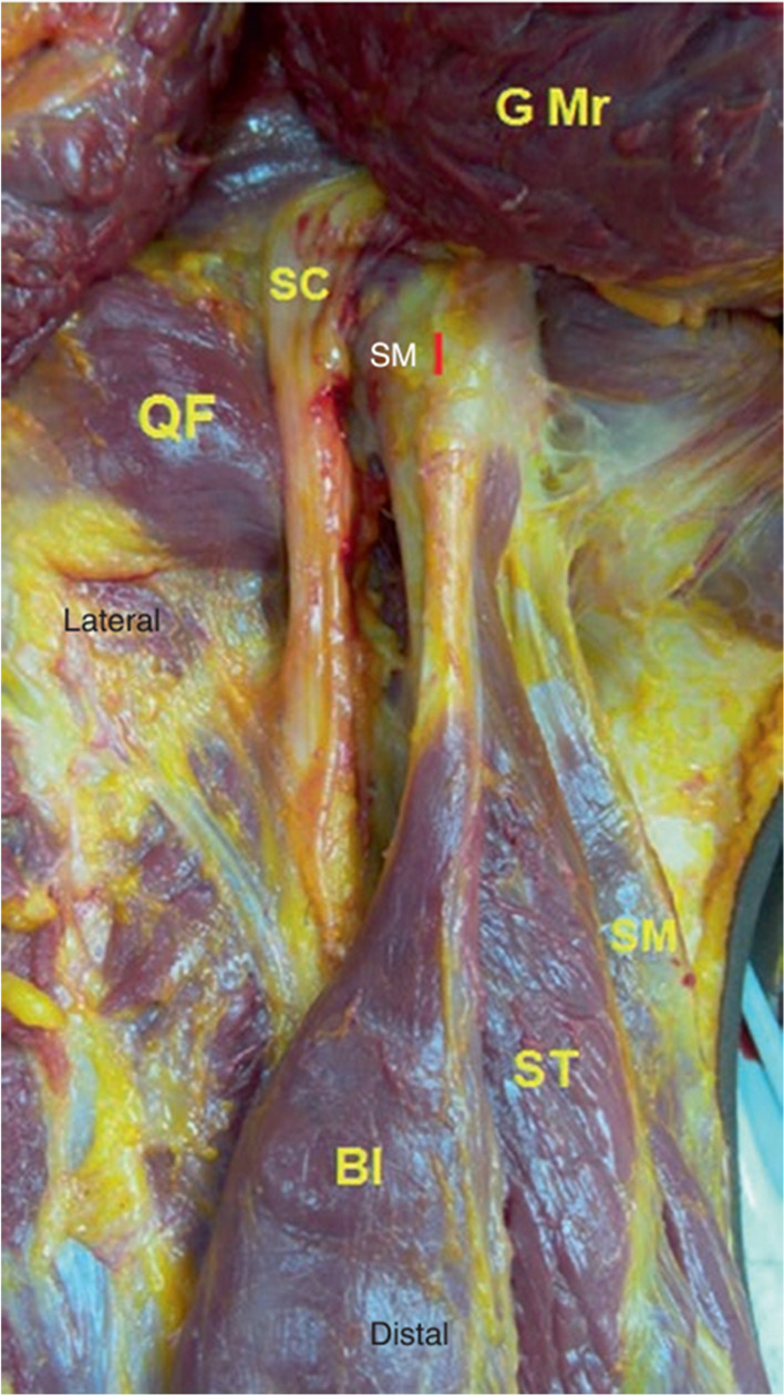
    1. Miller SL, Gill J, Webb GR. The proximal origin of the hamstrings and surrounding anatomy encountered during repair. A cadaveric study [published correction appears in J Bone Joint Surg Am. 2007 Mar;89(3):637]. J Bone Joint Surg Am. 2007;89(1):44–8. 10.2106/JBJS.F.00094. 10.2106/JBJS.F.00094 - DOI - PubMed
    1. Beltran L, Ghazikhanian V, Padron M, Beltran J. The proximal hamstring muscle-tendon-bone unit: a review of the normal anatomy, biomechanics, and pathophysiology. Eur J Radiol. 2012;81(12):3772–9. 10.1016/j.ejrad.2011.03.099. 10.1016/j.ejrad.2011.03.099 - DOI - PubMed
    1. Koulouris G, Connell D. Hamstring muscle complex: an imaging review [published correction appears in Radiographics. 2005 Sep-Oct;25(5):1436]. Radiographics. 2005;25(3):571–86. 10.1148/rg.253045711. 10.1148/rg.253045711 - DOI - PubMed
    1. Kumazaki T, Ehara Y, Sakai T. Anatomy and physiology of hamstring injury [published correction appears in Int J Sports Med. 2012 Dec;33(12):954]. Int J Sports Med. 2012;33(12):950–4. 10.1055/s-0032-1311593. 10.1055/s-0032-1311593 - DOI - PubMed