

Endoplasmic reticulum stress promotes hepatocellular carcinoma by modulating immunity: a study based on artificial neural networks and single-cell sequencing
- PMID: 39010084
- PMCID: PMC11247783
- DOI: 10.1186/s12967-024-05460-9
Endoplasmic reticulum stress promotes hepatocellular carcinoma by modulating immunity: a study based on artificial neural networks and single-cell sequencing
Abstract
Introduction: Hepatocellular carcinoma (HCC) is characterized by the complex pathogenesis, limited therapeutic methods, and poor prognosis. Endoplasmic reticulum stress (ERS) plays an important role in the development of HCC, therefore, we still need further study of molecular mechanism of HCC and ERS for early diagnosis and promising treatment targets.
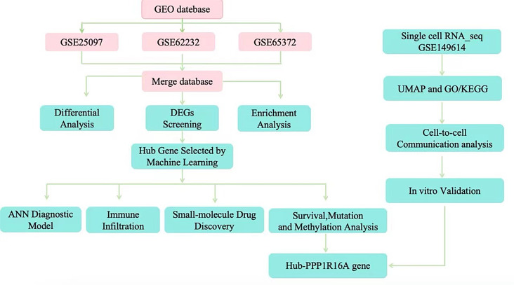
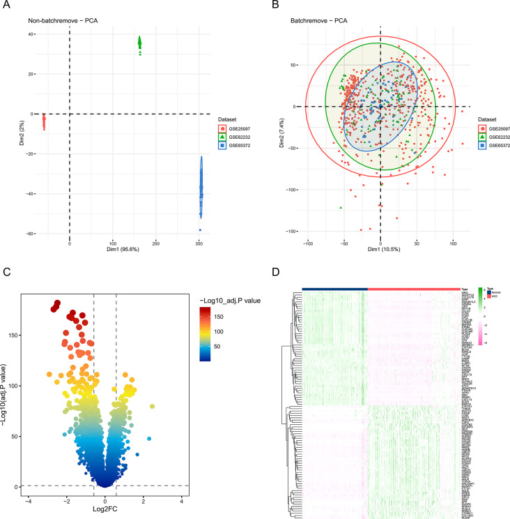
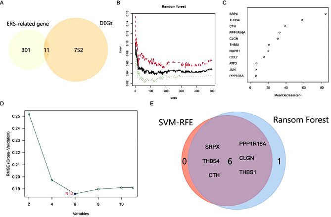
Method: The GEO datasets (GSE25097, GSE62232, and GSE65372) were integrated to identify differentially expressed genes related to HCC (ERSRGs). Random Forest (RF) and Support Vector Machine (SVM) machine learning techniques were applied to screen ERSRGs associated with endoplasmic reticulum stress, and an artificial neural network (ANN) diagnostic prediction model was constructed. The ESTIMATE algorithm was utilized to analyze the correlation between ERSRGs and the immune microenvironment. The potential therapeutic agents for ERSRGs were explored using the Drug Signature Database (DSigDB). The immunological landscape of the ERSRGs central gene PPP1R16A was assessed through single-cell sequencing and cell communication, and its biological function was validated using cytological experiments.
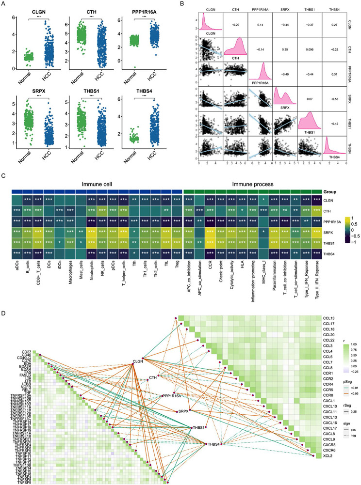
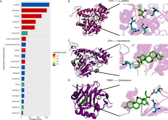
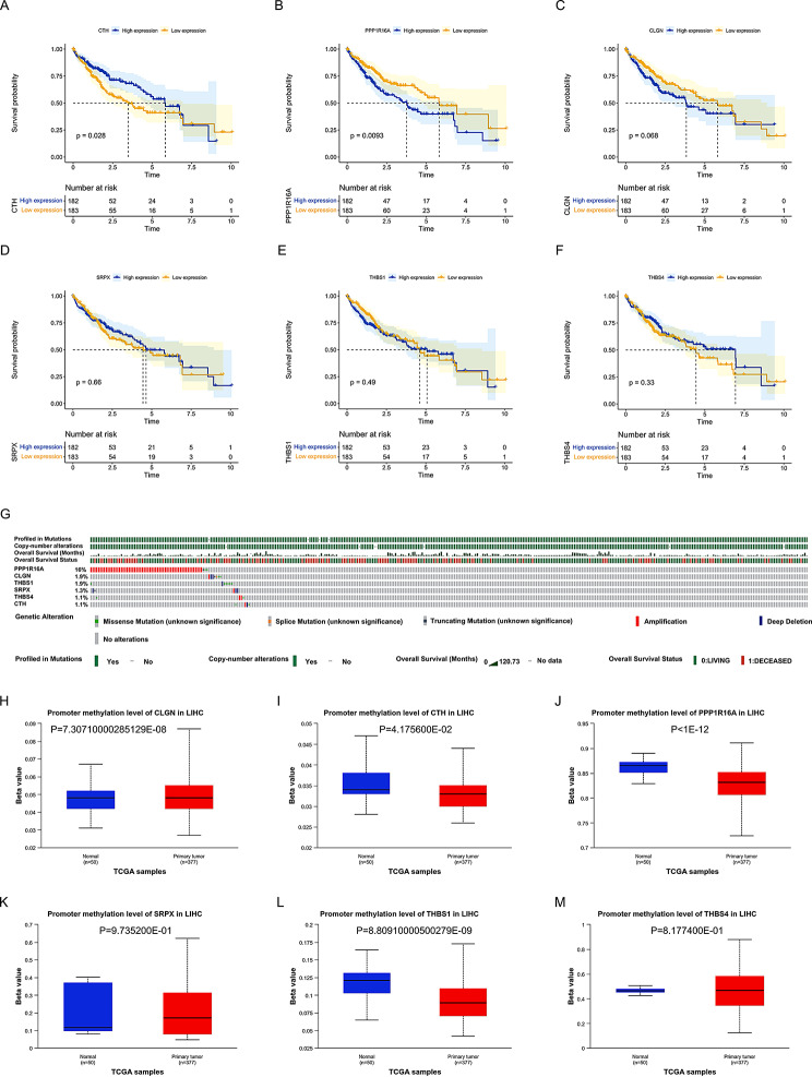
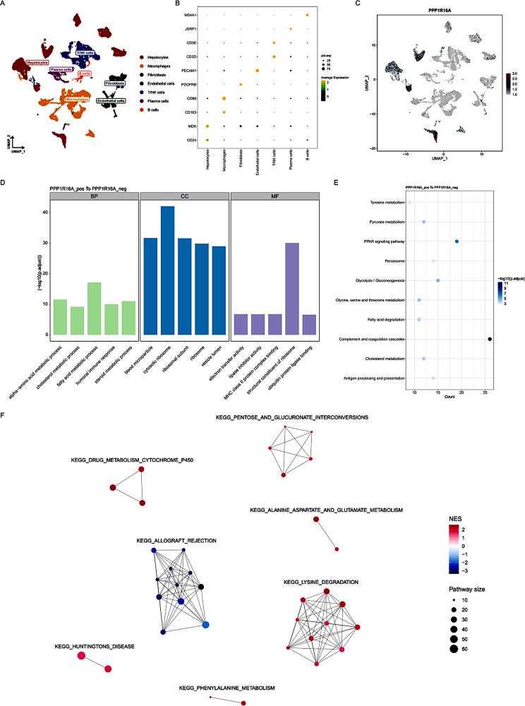
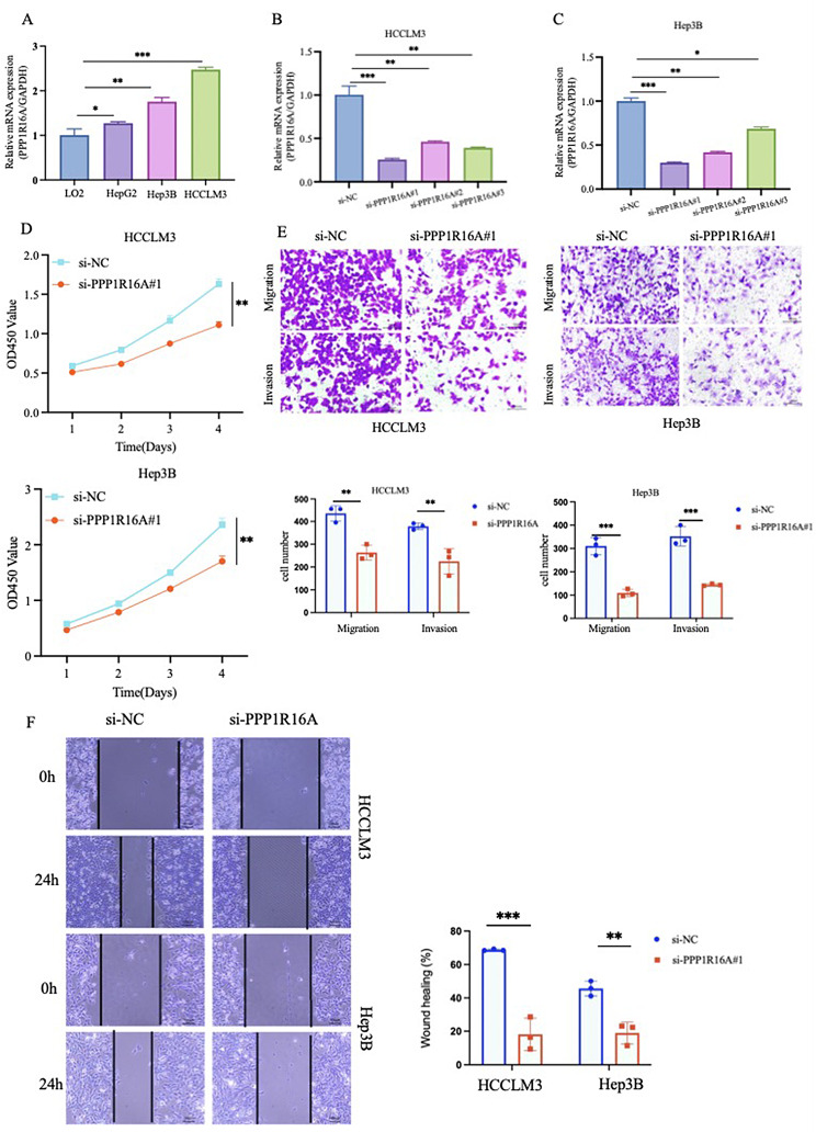
Results: An ANN related to the ERS model was constructed based on SRPX, THBS4, CTH, PPP1R16A, CLGN, and THBS1. The area under the curve (AUC) of the model in the training set was 0.979, and the AUC values in three validation sets were 0.958, 0.936, and 0.970, respectively, indicating high reliability and effectiveness. Spearman correlation analysis suggests that the expression levels of ERSRGs are significantly correlated with immune cell infiltration and immune-related pathways, indicating their potential as important targets for immunotherapy. Mometasone was predicted to be the most promising treatment drug based on its highest binding score. Among the six ERSRGs, PPP1R16A had the highest mutation rate, predominantly copy number mutations, which may be the core gene of the ERSRGs model. Single-cell analysis and cell communication indicated that PPP1R16A is predominantly distributed in liver malignant parenchymal cells and may reshape the tumor microenvironment by enhancing macrophage migration inhibitory factor (MIF)/CD74 + CXCR4 signaling pathways. Functional experiments revealed that after siRNA knockdown, the expression of PPP1R16A was downregulated, which inhibited the proliferation, migration, and invasion capabilities of HCCLM3 and Hep3B cells in vitro.
Conclusion: The consensus of various machine learning algorithms and artificial intelligence neural networks has established a novel predictive model for the diagnosis of liver cancer associated with ERS. This study offers a new direction for the diagnosis and treatment of HCC.
Keywords: Artificial neural network; Endoplasmic reticulum stress; Hepatocellular carcinoma; Immune infiltration; Prognosis.
© 2024. The Author(s).
Conflict of interest statement
The authors declare that there are no competing interests.
Figures


References
-
- Cronin KA, Scott S, Firth AU, Sung H, Henley SJ, Sherman RL, Siegel RL, Anderson RN, Kohler BA, Benard VB, Negoita S, Wiggins C, Cance WG, Jemal A. Annual report to the nation on the status of cancer, part 1: National cancer statistics. Cancer. 2022;128:4251–84. doi: 10.1002/cncr.34479. - DOI - PMC - PubMed
MeSH terms
Grants and funding
- Innovative Research Group Project of the National Natural Science Foundation of China/Innovative Research Group Project of the National Natural Science Foundation of China
- This work was supported by grants provided by the National Natural Science Foundation of China (No. 81070125, 81270213, 81670306); the Science and Technology Foundation in Guangdong Province (No. 2010B031600032, 2014A020211002); the National Natural Scienc/This work was supported by grants provided by the National Natural Science Foundation of China (No. 81070125, 81270213, 81670306); the Science and Technology Foundation in Guangdong Province (No. 2010B031600032, 2014A020211002); the National Natural Scienc
LinkOut - more resources
Full Text Sources
Medical
Miscellaneous
