Review
. 2024 Jul 1:15:1418516.
doi: 10.3389/fphar.2024.1418516. eCollection 2024.

Recent advances in the study of zika virus structure, drug targets, and inhibitors

Affiliations
Review

Recent advances in the study of zika virus structure, drug targets, and inhibitors

Yingqi Feng. Front Pharmacol. .

Abstract

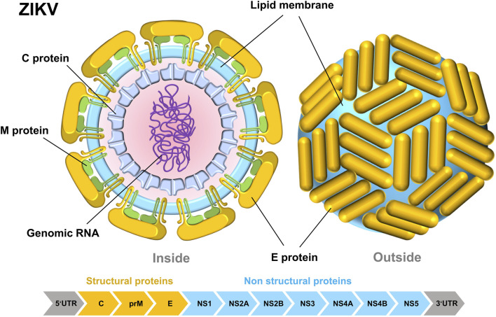
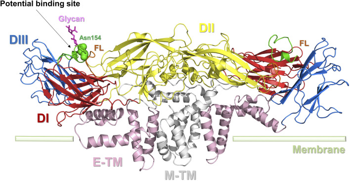
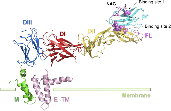
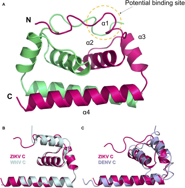
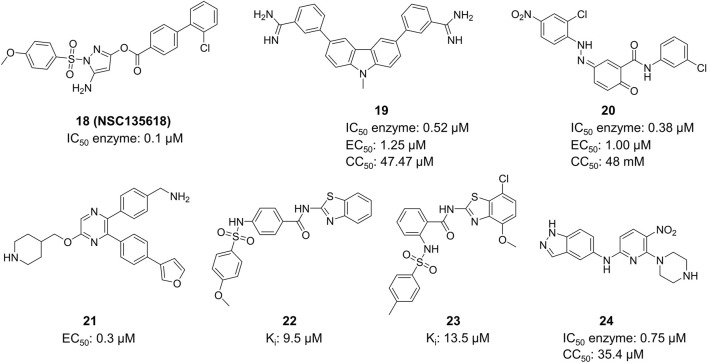
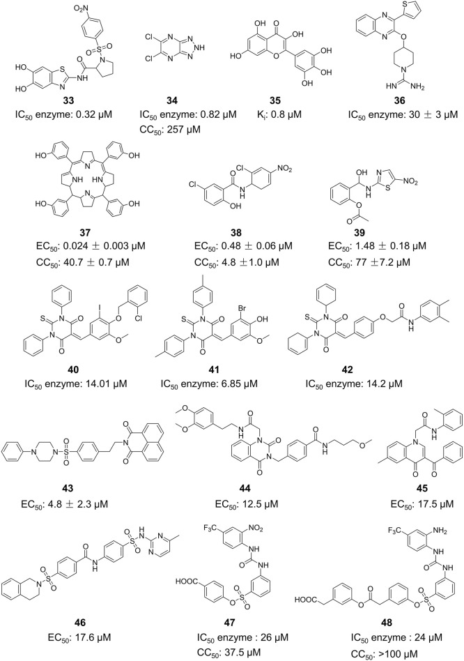
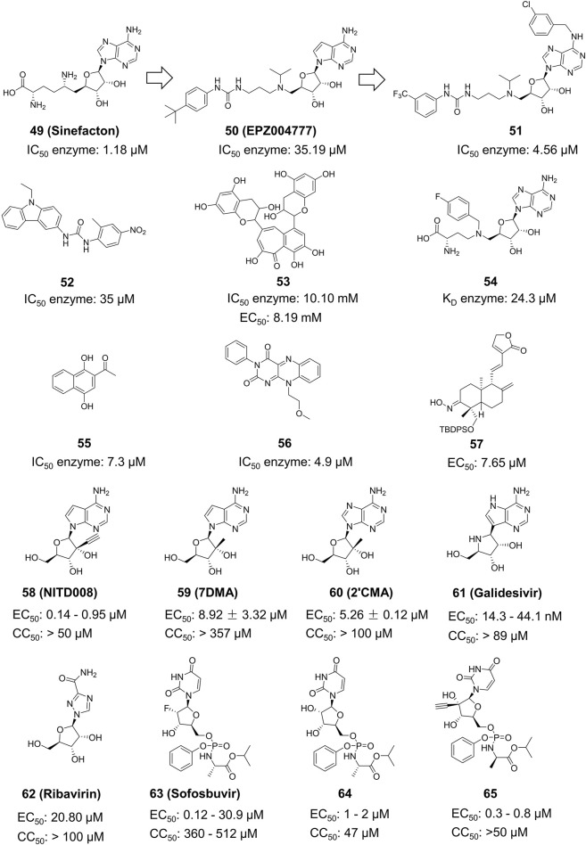
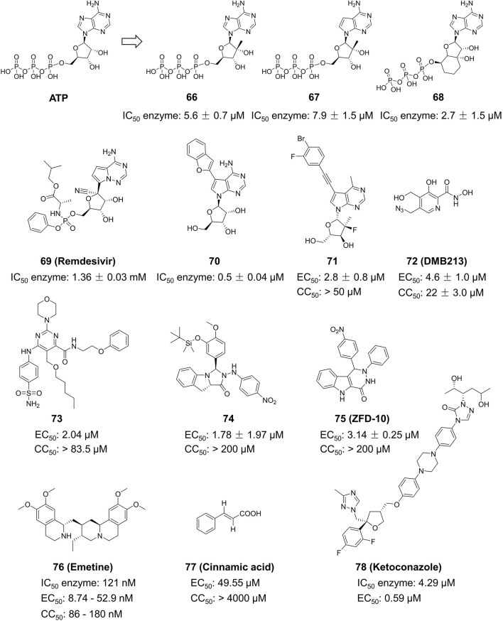
Zika Virus (ZIKV) is a positive-strand RNA virus that can lead to Guillain-Barré syndrome or encephalitis in some individuals and hence presents a serious public health risk. Since the first outbreak of ZIKV in Brazil in 2015, no effective clinical inhibitors have been developed, making the development of effective ZIKV drugs an urgent issue that needs to be addressed. ZIKV belongs to the Flaviviridae family, and its structure includes three structural proteins, namely, capsular (C), premembrane (prM), and envelope (E) proteins, as well as seven nonstructural proteins, namely, NS1, NS2A, NS2B, NS3, NS4A, NS4B, and NS5. To provide a reference for the development of future ZIKV drugs, this paper reviews the structure of the ZIKV based on recent literature reports, analyzes the potential therapeutic targets of various proteins, and proposes feasible drug design strategies. Additionally, this paper reviews and classifies the latest research progress on several protease inhibitors, such as E protein inhibitors, NS2B-NS3 inhibitors, and NS5 inhibitors, so that researchers can quickly understand the current status of development and the interconnections among these inhibitors.

Keywords: MTase; RdRp; ZIKV; drug target; inhibitor.


Conflict of interest statement

The author declares that the research was conducted in the absence of any commercial or financial relationships that could be construed as a potential conflict of interest.

Figures

FIGURE 1
FIGURE 1
Internal structural diagram of ZIKV (left) and surface structure diagram (right). The schematic pieces were provided by Smart Medical Art and adapted from (http://www.servier.com). Servier Medical Art by Servier is licensed under a Creative Commons Attribution 3.0 Unported License.
FIGURE 2
FIGURE 2
Life cycle of ZIKV. The schematic pieces were provided by Smart Medical Art and adapted from (http://www.servier.com). Servier Medical Art by Servier is licensed under a Creative Commons Attribution 3.0 Unported License.
FIGURE 3
FIGURE 3
Dimer structure of the E protein of ZIKV. E protein domain I (DI): red; domain II (DII): yellow; domain III (DIII): blue; the transmembrane portion of the E protein (E-TM): pink; the transmembrane portion of the M protein (M-TM): gray; FL: brown. The key amino acid Asn154 is represented by a green sphere. Potential binding sites for inhibitors are indicated.
FIGURE 4
FIGURE 4
Structure of the ZIKV M protein. E protein domain I (DI): red; domain II (DII): yellow; domain III (DIII): blue; E-TM: pink. The pr protein (light blue) and M protein (green) are located at the two ends of the E protein, respectively. Inhibitor binding sites 1 and 2 are represented by spheres.
FIGURE 5
FIGURE 5
(A) Dimer structure of the C protein of ZIKV (5YGH). The two monomers are colored magenta and green. (B) Structural alignment of ZIKV C protein (magenta) and WNV C protein (light blue). (C) Structural alignment of ZIKV C protein (magenta) and DENV C protein (purple). Potential binding sites for inhibitors are indicated.
FIGURE 6
FIGURE 6
Ribbon representation of the ZIKV NS1 dimer (5GS6). The β-hairpin domains (residues 1–29) are shown in red and blue, respectively, for distinction; the Wing domain (residues 30–180) is shown in green; the β-ladder domain (residues 181–352) is shown in purple. Potential binding sites 1 and 2 of inhibitors are represented by spheres.
FIGURE 7
FIGURE 7
The X-ray crystal structure of the ZIKV virus protease NS2B-NS3 in its catalytically active conformation (PDB ID: 5LC0, ribbon diagram). NS2B is colored blue, and NS3 is pink. The S1 site (catalytic triad) is highlighted in green, and the S2 site (allosteric pocket) is highlighted in blue.
FIGURE 8
FIGURE 8
(A) The structure and active sites of the ZIKV Mtase domain. The central active site, GTP-binding pocket, and SAM-binding pocket are respectively represented in pink, yellow, and green. GTP and SAH molecules are bound to the GTP-binding pocket and SAM-binding pocket, respectively. (B) Structure and active sites of the ZIKV RdRp domain. The palm, fingers, and thumb regions of the RdRp domain are denoted in pink, green, and dark blue, respectively. The Central channel, Template channel, and NTP channel are as marked in the figure.
FIGURE 9
FIGURE 9
Chemical structures and bioactivities of Compound 110.
FIGURE 10
FIGURE 10
Chemical structures and bioactivities of Compound 1824.
FIGURE 11
FIGURE 11
Chemical structures and bioactivities of Compound 3348.
FIGURE 12
FIGURE 12
Chemical structures and bioactivities of Compound 4965.
FIGURE 13
FIGURE 13
Chemical structures and bioactivities of Compound 6678.

Similar articles

Cited by

References

    1. Agrelli A., de Moura R. R., Crovella S., Brandão L. A. C. (2019). ZIKA virus entry mechanisms in human cells. Infect. Genet. Evol. 69, 22–29. 10.1016/j.meegid.2019.01.018 - DOI - PubMed
    1. Alcon S., Talarmin A., Debruyne M., Falconar A., Deubel V., Flamand M. (2002). Enzyme-linked immunosorbent assay specific to Dengue virus type 1 nonstructural protein NS1 reveals circulation of the antigen in the blood during the acute phase of disease in patients experiencing primary or secondary infections. J. Clin. Microbiol. 40 (2), 376–381. 10.1128/JCM.40.02.376-381.2002 - DOI - PMC - PubMed
    1. Bernatchez J. A., Coste M., Beck S., Wells G. A., Luna L. A., Clark A. E., et al. (2019). Activity of selected nucleoside analogue ProTides against zika virus in human neural stem cells. Viruses 11 (4), 365. 10.3390/v11040365 - DOI - PMC - PubMed
    1. Bernatchez J. A., Tran L. T., Li J., Luan Y., Siqueira-Neto J. L., Li R. (2020). Drugs for the treatment of zika virus infection. J. Med. Chem. 63 (2), 470–489. 10.1021/acs.jmedchem.9b00775 - DOI - PubMed
    1. Boonyasuppayakorn S., Padmanabhan R. (2014). Construction of plasmid, bacterial expression, purification, and assay of dengue virus type 2 NS5 methyltransferase. Methods Mol. Biol. 1138, 361–373. 10.1007/978-1-4939-0348-1_22 - DOI - PubMed

LinkOut - more resources