. 2024 Jun 25;16(3):1626-1638.
doi: 10.14336/AD.2024.0405.

SARS-CoV-2 S Protein Reduces Cytoprotective Defenses and Promotes Human Endothelial Cell Senescence

Affiliations

SARS-CoV-2 S Protein Reduces Cytoprotective Defenses and Promotes Human Endothelial Cell Senescence

Alicia Villacampa et al. Aging Dis. .

Abstract

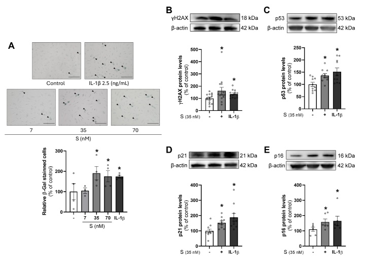
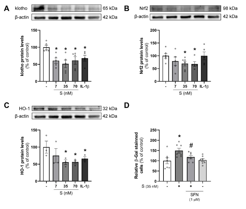
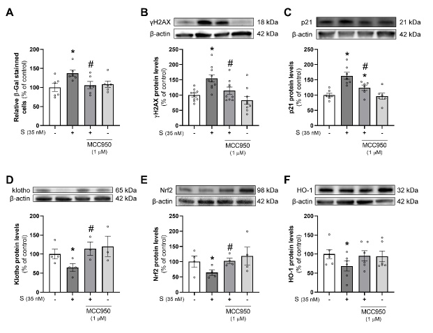
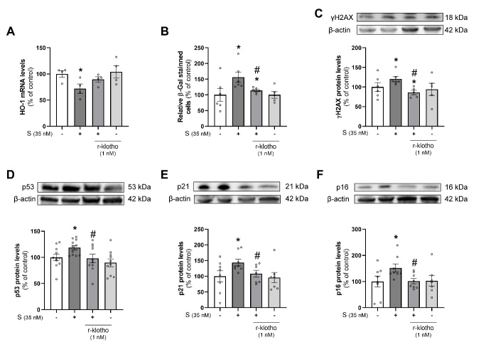
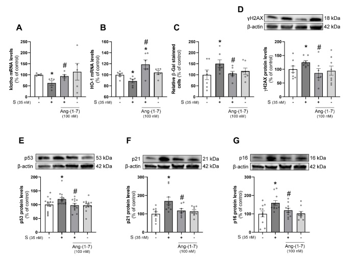
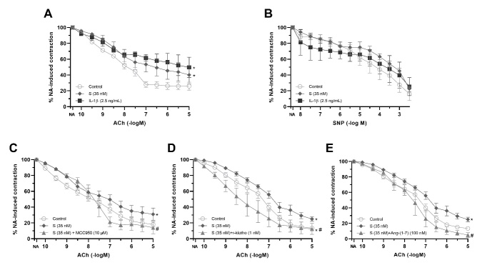
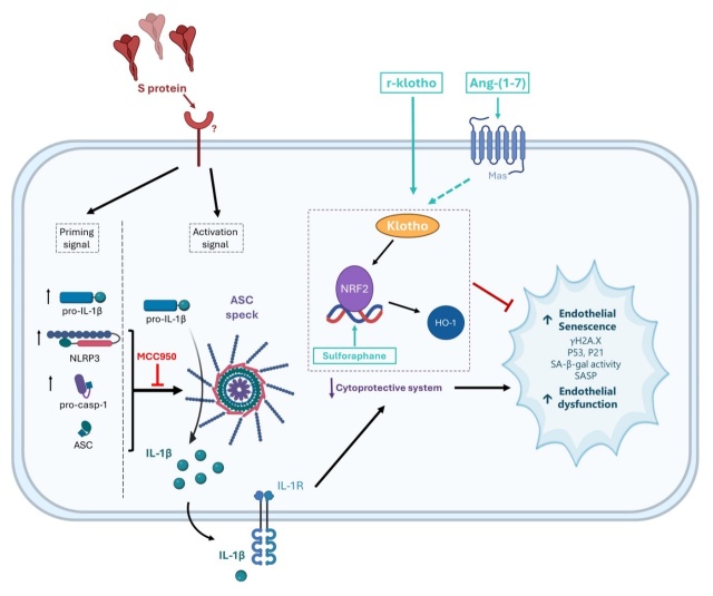
Premature vascular aging and endothelial cell senescence are major risk factors for cardiovascular diseases and atherothrombotic disturbances, which are main complications of both acute and long COVID-19. The S protein of SARS-CoV2, which acts as the receptor binding protein for the viral infection, is able to induce endothelial cells inflammation and it has been found as an isolated element in the circulation and in human tissues reservoirs months after infection. Here, we investigated whether the S protein is able to directly induce endothelial cell senescence and deciphered some of the mechanisms involved. In primary cultures of human umbilical vein endothelial cells (HUVEC), SARS-CoV-2 S protein enhanced in a concentration-dependent manner the cellular content of senescence and DNA damage response markers (senescence-associated-β galactosidase, γH2AX), as well as growth-arrest effectors (p53, p21, p16). In parallel, the S protein reduced the availability of cytoprotective proteins, such as the anti-aging protein klotho, Nrf2 or heme oxygenase-1, and caused functional harm by impairing ex vivo endothelial-dependent vasorelaxation in murine microvessels. These effects were prevented by the pharmacological inhibition of the NLRP3 inflammasome with MCC950. Furthermore, the supplementation with either recombinant klotho or angiotensin-(1-7), equally protected against the pro-senescence, pro-inflammatory and pro-oxidant action of the S protein. Globally, this study proposes novel mechanisms of disease in the context of COVID-19 and its vascular sequelae and provides pharmacological clues in order to prevent such complications.


Figures

Figure 1.
Figure 1.
SARS-CoV-2 S protein induces cellular senescence in endothelial cells. Human umbilical vein endothelial cells (HUVEC) were treated for 24 h with S protein (S; 7, 35, or 70 nM), or IL-1β (2.5 ng/mL) after which (A) SA-β-Gal staining was performed and quantified (n=4, except n=3 for S 7 nM). Black arrows point to positive SA-β-gal-stained cells in a representative experiment. Scale bar represents 150 µm. Similarly, after exposure to S protein (35 nM) or IL-1β (2.5 ng/mL) for 18 h, the protein levels of senescence markers such as (B) γH2AX histone (n=11), (C) p53 (n=10), (D) p21 (n=10) and (E) p16 (n=7, except n=6 for S 35 nM) were determined by Western blot. Representative gels are shown on top of the corresponding graphs, with β-actin used as a loading control. All bar graphs represent the mean ± SEM. Statistical differences were tested with t-test (A, C, D) or Mann-Whitney (B, E). * p < 0.05 versus control.
Figure 2.
Figure 2.
SARS-CoV-2 S protein reduces cellular antioxidant and anti-senescence defenses. Human umbilical vein endothelial cells (HUVEC) were treated for 24 h with S protein (S; 7, 35, or 70 nM), or IL-1β (2.5 ng/mL) for 18 h after which the protein levels of (A) klotho (n=5), (B) Nrf2 (n=5, except n=6 for untreated control and S 35 nM) and (C) heme oxygenase-1 (HO-1) (n=3) were determined by Western blot. (D) In another set of experiments SA-β-gal staining (n=7) was performed in HUVEC stimulated for 24 h S (35 nM) with or without the Nrf2 activator sulforaphane (SFN; 1 µM) for 24 h. Representative gels are shown on top of the corresponding graphs, with β-actin used as a loading control. All bar graphs represent the mean ± SEM. Statistical differences were tested with t-test (B, C, D) or Mann-Whitney (A). * p < 0.05 versus control. # p< 0.05 versus S protein.
Figure 3.
Figure 3.
NLRP3 inflammasome inhibition prevents the decrease in cytoprotective proteins and cellular senescence induced by SARS-CoV-2 S protein. Human umbilical vein endothelial cells (HUVEC) were treated for 18-24 h with S protein (S, 35 nM), MCC950 (1 µM) or both. Thereafter, (A) SA-β-gal staining (n=6) was performed and the protein levels of (B) γH2AX histone (n=10, except n=9 for S + MCC950 and MCC950 alone), (C) p21 (n=7, except n=6 for S + MCC950 and MCC950 alone), (D) klotho (n=4, except n=3 for S + MCC950 and MCC950 alone), (E) Nrf2 (n=4) and (F) heme oxygenase-1 (HO-1) (n=6) were determined by Western blot (WB). Representative gels are shown on top of the corresponding graphs for WB with β-actin used as a loading control. All bar graphs represent the mean ± SEM. Statistical differences were tested with t-test (A, B, C, D, E) or Mann-Whitney (F). * p < 0.05 versus control. # p < 0.05 versus to S protein.
Figure 4.
Figure 4.
r-klotho prevents deficient cytoprotection and endothelial cell senescence induced by SARS-CoV-2 S protein. Human umbilical vein endothelial cells (HUVEC) were treated for 18-24 h with S protein (S, 35 nM), r-klotho (1 nM) or both. Afterwards, (A) heme oxygenase-1 (HO-1) expression (n=4) was determined by RT-qPCR using 18S housekeeping gene for normalization, (B) SA-β-gal staining (n=6, except n=5 for r-klotho alone) was performed and protein levels of (C) γH2AX histone (n=6, except n=5 for S + r-klotho and r-klotho alone), (D) p53 (n=11, except n=10 for S + r-klotho and r-klotho alone), (E) p21 (n=8, except n=7 for S + r-klotho and r-klotho alone) and (F) p16 (n=8, except n=7 for r-klotho alone) were determined by Western blot (WB). Representative gels are shown on top of the corresponding graphs for WB with β-actin used as a loading control. All bar graphs represent the mean ± SEM. Statistical differences were tested with t-test (B, C, D, F) or Mann-Whitney (A, E). * p < 0.05 versus control. # p < 0.05 versus to S protein.
Figure 5.
Figure 5.
Ang-(1-7) prevents deficient cytoprotection and endothelial cell senescence induced by SARS-CoV-2 S protein. Human umbilical vein endothelial cells (HUVEC) were treated for 18-24 h with S protein (S; 35 nM), Ang-(1-7) (100 nM) or both. Afterwards, (A) klotho (n= 5) and (B) heme oxygenase-1 (HO-1) expression (n=6) were determined by RT-qPCR using 18S housekeeping gene for normalization, (C) SA-β-gal staining (n=7) was performed and protein levels of (D) γH2AX histone (n=8, except n=7 for S + Ang-(1-7)), (E) p53 (n=14, except n=12 for S + Ang-(1-7) and n= 13 for Ang-(1-7) alone), (F) p21 (n=9, except n=8 for S + Ang-(1-7)) and (G) p16 (n=11, except n=9 for Ang-(1-7) alone and n=10 for S + Ang-(1-7)) were determined by Western blot (WB). Representative gels are shown on top of the corresponding graphs for WB with β-actin used as a loading control. All bar graphs represent the mean ± SEM. Statistical differences were tested with t-test (A, B, F, G) or Mann-Whitney (C, D, E). * p < 0.05 versus control. # p < 0.05 versus to S protein.
Figure 6.
Figure 6.
The endothelium-dependent relaxation impaired by SARS-CoV-2 S protein is prevented by NLRP3 inflammasome blockade or the supplementation with r-klotho or Ang-(1-7). Isolated mesenteric microvessels from C57BL/6J mice were preincubated for 1 h with IL-1β (2.5 ng/mL) or S protein (35 nM) after which vascular relaxations in response to increasing concentrations of (A) acetylcholine (ACh; 0.1 nM to 10 μM), n=5-7 segments from 5 mice; or (B) sodium nitroprusside (SNP; 10 nM to 3 mM), n=3-5 segments from 5 mice; were assessed. In selected experiments, (C) the inflammasome inhibitor MCC 950 (10 µM), n=4-19 segments from 4-8 mice; (C) r-klotho (1 nM), n=4-12 segments from 4 mice; or (D) Ang-(1-7) (100 nM), n=3-12 segments from 3-4 mice; were added during the preincubation period. Results are presented as the percentage of contraction to noradrenaline (NA; 2 µM). More details on the exact number of mice and segments per treatment can be found in Supplementary Tables 2-5. Data are represented as mean ± SEM. Statistical differences were tested with two-way ANOVA. * p<0.05 versus control; # p<0.05 versus S protein.
Figure 7.
Figure 7.
Graphical abstract summarizing the direct actions of SARS-CoV-2 S protein reported in the present study. SARS-CoV-2 S protein promotes by itself human endothelial cell senescence and DNA damage response, determined by the induction of markers such as senescence-associated β-galactosidase (SA-β-gal) or histone γH2AX and growth-arrest effectors like p53, p21 and p16. This effect is paralleled by ex vivo endothelial dysfunction in terms of impaired endothelial-dependent relaxation of murine microvessels. Such deleterious effects of the isolated viral protein are associated to a reduction of anti-aging and cytoprotective proteins, such as klotho, Nrf2 and heme oxygenase-1 (HO-1). In fact, activating Nrf2 with the drug sulforaphane or supplementing with recombinant klotho (r-klotho) or the klotho inducer angiotensin (Ang)-(1-7) prevents the pro-senescence action and the defective vasorelaxation induced by SARS-CoV-2 S protein. Moreover, blocking the activation of the NLRP3 inflammasome by means of MCC950 protects against the restricted cytoprotective protein availability, cellular senescence and microvascular dysfunction directly caused by SARS-CoV-2 S protein.

References

    1. Gu SX, Tyagi T, Jain K, Gu VW, Lee SH, Hwa JM, et al.. (2021). Thrombocytopathy and endotheliopathy: crucial contributors to COVID-19 thromboinflammation. Nat Rev Cardiol, 18:194-209. - PMC - PubMed
    1. Varga Z, Flammer AJ, Steiger P, Haberecker M, Andermatt R, Zinkernagel AS, et al.. (2020). Endothelial cell infection and endotheliitis in COVID-19. The Lancet, 395:1417-1418. - PMC - PubMed
    1. Martínez-Salazar B, Holwerda M, Stüdle C, Piragyte I, Mercader N, Engelhardt B, et al.. (2022). COVID-19 and the Vasculature: Current Aspects and Long-Term Consequences. Front Cell Dev Biol, 10:1-23. - PMC - PubMed
    1. Zanini G, Selleri V, Roncati L, Coppi F, Nasi M, Farinetti A, et al.. (2023). Vascular “Long COVID”: A New Vessel Disease? Angiology, 75:1-7. - PMC - PubMed
    1. Saz-Lara A, Cavero-Redondo I, Pascual-Morena C, Martínez-García I, Rodríguez-Gutiérrez E, Lucerón-Lucas-Torres M, et al.. (2023). Early vascular aging as an index of cardiovascular risk in healthy adults: confirmatory factor analysis from the EVasCu study. Cardiovasc Diabetol, 22:1-8. - PMC - PubMed

Substances

LinkOut - more resources