. 2024 Jul 16;134(17):e174199.
doi: 10.1172/JCI174199.

DRD2 activation inhibits choroidal neovascularization in patients with Parkinson's disease and age-related macular degeneration

Affiliations

DRD2 activation inhibits choroidal neovascularization in patients with Parkinson's disease and age-related macular degeneration

Thibaud Mathis et al. J Clin Invest. .

Abstract

Neovascular age-related macular degeneration (nAMD) remains a major cause of visual impairment and puts considerable burden on patients and health care systems. l-DOPA-treated Parkinson's disease (PD) patients have been shown to be partially protected from nAMD, but the mechanism remains unknown. Using murine models that combine 1-methyl-4-phenyl-1,2,3,6-tetrahydropyridine-induced (MPTP-induced) PD and laser-induced nAMD with standard PD treatment of l-DOPA/DOPA-decarboxylase inhibitor or specific dopamine receptor inhibitors, we here demonstrate that l-DOPA treatment-induced increase of dopamine-mediated dopamine receptor D2 (DRD2) signaling inhibits choroidal neovascularization independently of MPTP-associated nigrostriatal pathway lesion. Analyzing a retrospective cohort of more than 200,000 patients with nAMD receiving anti-VEGF treatment from the French nationwide insurance database, we show that DRD2 agonist-treated PD patients have a significantly delayed age of onset of nAMD and reduced need for anti-VEGF therapies, similar to the effects of the l-DOPA treatment. While providing a mechanistic explanation for an intriguing epidemiological observation, our findings suggest that systemic DRD2 agonists might constitute an adjuvant therapy to delay and reduce the need for anti-VEGF therapy in patients with nAMD.

Keywords: Neurodegeneration; Neuroscience; Ophthalmology; Parkinson disease; Retinopathy.


Figures
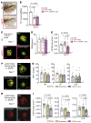
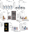
Figure 1
Figure 1. l-DOPA/DDI treatment prevents choroidal neovascularization independently of subretinal inflammation and loss of central dopaminergic neurons in mice.
(A and B) TH-stained SNpc (A) and quantification of dopaminergic TH+ neurons in SNpc (B) of 9-week-old C57BL/6J mice that received PBS or MPTP intoxication at 6 weeks of age and laser injury at 8 weeks and were treated with intraperitoneal PBS or l-DOPA (30 mg/kg/d) plus benserazide (bens) (12 mg/kg/d) from 1 week before injury until 7 days after injury. One-way ANOVA/Bonferroni’s test. (CE) IBA-1 (green) and CD102 (red) stained choroidal flat mounts (C), quantification of number of subretinal IBA-1+ mononuclear phagocytes (MPs) counted on the RPE at a distance of 0–500 μm to CD102+ CNV (D), and quantification of CD102+ CNV area (E), 7 days after laser injury of mice that received MPTP intoxication and treatment regimens as for A. One-way ANOVA/Bonferroni’s test; control-PBS (Ctl-PBS) vs. MPTP-PBS P = nonsignificant. (F) IBA-1 (green) and CD102 (red) stained choroidal flat mounts 7 days after laser injury of 8-week-old C57BL/6J mice treated with PBS, benserazide (12 mg/kg/d), or l-DOPA (30 mg/kg/d) plus benserazide (12 mg/kg/d) from 1 week before injury until sacrifice. (G) Quantification of subretinal IBA-1+ MPs counted on the RPE at a distance of 0–500 μm to CD102+ CNV, 4, 7, and 10 days after laser injury of 8-week-old C57BL/6J mice treated with PBS, benserazide (12 mg/kg/d), or l-DOPA (30 mg/kg/d) plus benserazide (12 mg/kg/d) from 1 week before laser injury until sacrifice. One-way ANOVA/Bonferroni’s test; P = nonsignificant for each group. (H) CD102 (red) stained choroidal flat mounts 7 days after laser injury of 8-week-old C57BL/6J mice treated with PBS, benserazide (12 mg/kg/d), or l-DOPA (30 mg/kg/d) plus benserazide (12 mg/kg/d) from 1 week before injury until sacrifice. (I) CD102+ CNV area 4, 7, and 10 days after laser injury of 8-week-old C57BL/6J mice treated with PBS, benserazide (12 mg/kg/d), or l-DOPA (30 mg/kg/d) plus benserazide (12 mg/kg/d) from 1 week before injury until sacrifice. One-way ANOVA/Bonferroni’s test. n is indicated in each column for each group. Scale bars: 200 μm.
Figure 2
Figure 2. The decarboxylation of l-DOPA to dopamine is necessary for its anti-angiogenic effect on CNV ex vivo and in vitro.
(A) Quantitative RT-PCR of mouse DOPA-decarboxylase (Ddc) mRNAs normalized with β-actin of fresh neuroretina, RPE/choroid, and choroid from 8-week-old C57BL/6J mice. (B) Quantitative RT-PCR of human Ddc mRNAs normalized with β-actin of fresh human RPE from donor eye and HCECs. (C) Representative microphotographs of choroidal explants from 2-week-old C57BL/6J pups at day 6 after 3-day treatment with PBS, l-DOPA (1 μM), or dopamine (1 μM). Scale bars: 1 mm. (D) Quantification of vascular sprouting area between days 3 and 6 from choroidal explants prepared from 2-week-old C57BL/6J pups and treated with dopamine (1 μM), benserazide (10 μM), or l-DOPA (1 μM) or preincubated 1 hour with benserazide (10 μM) before addition of l-DOPA (1 μM). One-way ANOVA/Bonferroni’s test. (E) Quantification of vascular sprouting area between days 3 and 6 from choroidal explants prepared from 2-week-old C57BL/6J pups and treated with increasing concentrations of dopamine. (F) Representative microphotographs of DAPI+ HCECs after a 24-hour treatment with PBS, l-DOPA (1 μM), or dopamine (1 μM) with or without VEGF exposure (10 ng/mL). Scale bars: 200 μm. (G) Quantification of DAPI+ HCECs after a 24-hour treatment with PBS, l-DOPA (1 μM), or dopamine (1 μM) with VEGF exposure (10 ng/mL). One-way ANOVA/Bonferroni’s test, PBS vs. l-DOPA P = nonsignificant. (H) Volcano plot of differentially expressed genes and scatterplot of DUSP4 read counts of FACS-sorted CD31+CD11b+ endothelial cells from day 4 choroidal explants that had or had not been incubated with 1 μM dopamine for 24 hours. The volcano plot shows the log2(fold change) (x axis) versus the significance [–log10(P value); y axis] of the 2,276 genes with a log2(fold change) greater than ±1.0. Vertical lines indicate the cutoff of fold change = ±1.0. Adjusted P value, indicated for the difference in DUSP4 transcription, was calculated by Benjamini and Hochberg false discovery rate correction method. (I) Representative microphotographs and quantification of vascular sprouting area between days 3 and 6 of choroidal explants from 2-week-old C57BL/6J Dusp4+/+ and Dusp4–/– pups. Mann-Whitney U test. Scale bars: 1 mm. hRPE, fresh human retinal pigment epithelium; ND, not detected. n is indicated in each column for each group.
Figure 3
Figure 3. DRD2 activation mediates the CNV inhibition observed under l-DOPA/DDI treatment in vivo.
Quantitative RT-PCR of dopamine receptor (DR1–DR5) mRNAs normalized with β-actin of 8-week-old C57BL/6J mouse retina (A) or choroid (B) 7 days after laser injury compared with control mice. Mann-Whitney U test DRD2 in CNV group vs. DRD2 in Ctl group; P value indicated in the figure when statistically significant. (C) Quantitative RT-PCR of DRD2 mRNAs normalized with β-actin of fresh RPE from donor eye and HCECs. (D) Representative microphotographs of choroidal explants of 2-week-old C57BL/6J pups at day 6 after 3-day treatment with PBS or dopamine (1 μM) or pretreated 1 hour with eticlopride (10 μM) before addition of dopamine (1 μM). Scale bars: 1 mm. (E) Quantification of choroidal vascular sprouting from 2-week-old C57BL/6J pups at day 6 after 3 days of treatment with PBS or dopamine (1 μM) or pretreated for 1 hour with eticlopride (10 μM) before addition of dopamine (1 μM), quinpirole (10 μM), SKF38393 (10 μM), or PD168077 (10 μM). One-way ANOVA/Bonferroni’s test; P values indicated in the figure, PBS vs. SKF38393 and PD168077 P = nonsignificant. (F) IBA-1 (green) and CD102 (red) stained choroidal flat mounts 7 days after laser injury of 8-week-old C57BL/6J mice treated with PBS or quinpirole (5 mg/kg) from 1 week before injury until sacrifice. Scale bars: 200 μm. (G) Quantification of subretinal IBA-1+ MPs counted on the RPE at a distance of 0–500 μm to CD102+ CNV 7 days after laser injury of 8-week-old C57BL/6J mice and treated with PBS, l-DOPA (30 mg/kg) plus benserazide (12 mg/kg), l-DOPA (30 mg/kg) plus benserazide (12 mg/kg) plus eticlopride (1 mg/kg), or quinpirole (5 mg/kg) from 1 week before injury until sacrifice. One-way ANOVA/Bonferroni’s test; P = nonsignificant for each group. (H) CD102+ CNV area 7 days after laser injury of 8-week-old C57BL/6J mice and treated with PBS, l-DOPA (30 mg/kg) plus benserazide (12 mg/kg), l-DOPA (30 mg/kg) plus benserazide (12 mg/kg) plus eticlopride (1 mg/kg), or quinpirole (5 mg/kg) from 1 week before injury until sacrifice. One-way ANOVA/Bonferroni’s test; P values indicated in the figure. hRPE, fresh human retinal pigment epithelium. n is indicated in each column for each group.
Figure 4
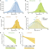
Figure 4. Incidence of nAMD and frequency of anti-VEGF injections are reduced in PD patients treated with l-DOPA/DDI and DRD2 agonists.
(AC) Number of patients (y axis) receiving the first IVT as a function of age (x axis) of the 193,512 nAMD control patients (A), of the 5,650 parkinsonian nAMD patients receiving DRD2 agonists (B), and of the 1,570 parkinsonian nAMD patients receiving l-DOPA/DDI therapy (C). The mean age for each group is indicated by the dotted line. (D) Distribution curves normalized for each group (percentage of cases of the group) of patients receiving the first IVT as a function of age (ANOVA test; control patients with nAMD vs. DRD2 agonist or l-DOPA/DDI nAMD patients P < 0.0001). (E and F) Correlation of DRD2 agonist (E) or l-DOPA/DDI (F) treatment group with the number of IVTs during the second year of nAMD treatment (anti-VEGF–naive patients with nAMD in France are generally initiated with a fixed proactive regimen during the first year and the treatment regimen is only then adapted to pathology activity) (univariate linear generalized model; control patients with nAMD vs. DRD2 agonist nAMD patients only, P < 0.0001 [E], or vs. l-DOPA/DDI nAMD patients only, P = 0.02 [F]).

References

    1. de Lau LML, Breteler MMB. Epidemiology of Parkinson’s disease. Lancet Neurol. 2006;5(6):525–535. doi: 10.1016/S1474-4422(06)70471-9. - DOI - PubMed
    1. Colijn JM, et al. Prevalence of age-related macular degeneration in Europe: the past and the future. Ophthalmology. 2017;124(12):1753–1763. doi: 10.1016/j.ophtha.2017.05.035. - DOI - PMC - PubMed
    1. Wong WL, et al. Global prevalence of age-related macular degeneration and disease burden projection for 2020 and 2040: a systematic review and meta-analysis. Lancet Glob Health. 2014;2(2):e106–e116. doi: 10.1016/S2214-109X(13)70145-1. - DOI - PubMed
    1. Klein R, et al. The epidemiology of age-related macular degeneration. Am J Ophthalmol. 2004;137(3):486–495. doi: 10.1016/j.ajo.2003.11.069. - DOI - PubMed
    1. Sarks SH. Ageing and degeneration in the macular region: a clinico-pathological study. Br J Ophthalmol. 1976;60(5):324–341. doi: 10.1136/bjo.60.5.324. - DOI - PMC - PubMed