. 2024 Jul 23;121(30):e2303642121.
doi: 10.1073/pnas.2303642121. Epub 2024 Jul 16.

AKT-dependent nuclear localization of EPRS1 activates PARP1 in breast cancer cells

Affiliations

AKT-dependent nuclear localization of EPRS1 activates PARP1 in breast cancer cells

Isaac Zin et al. Proc Natl Acad Sci U S A. .

Abstract

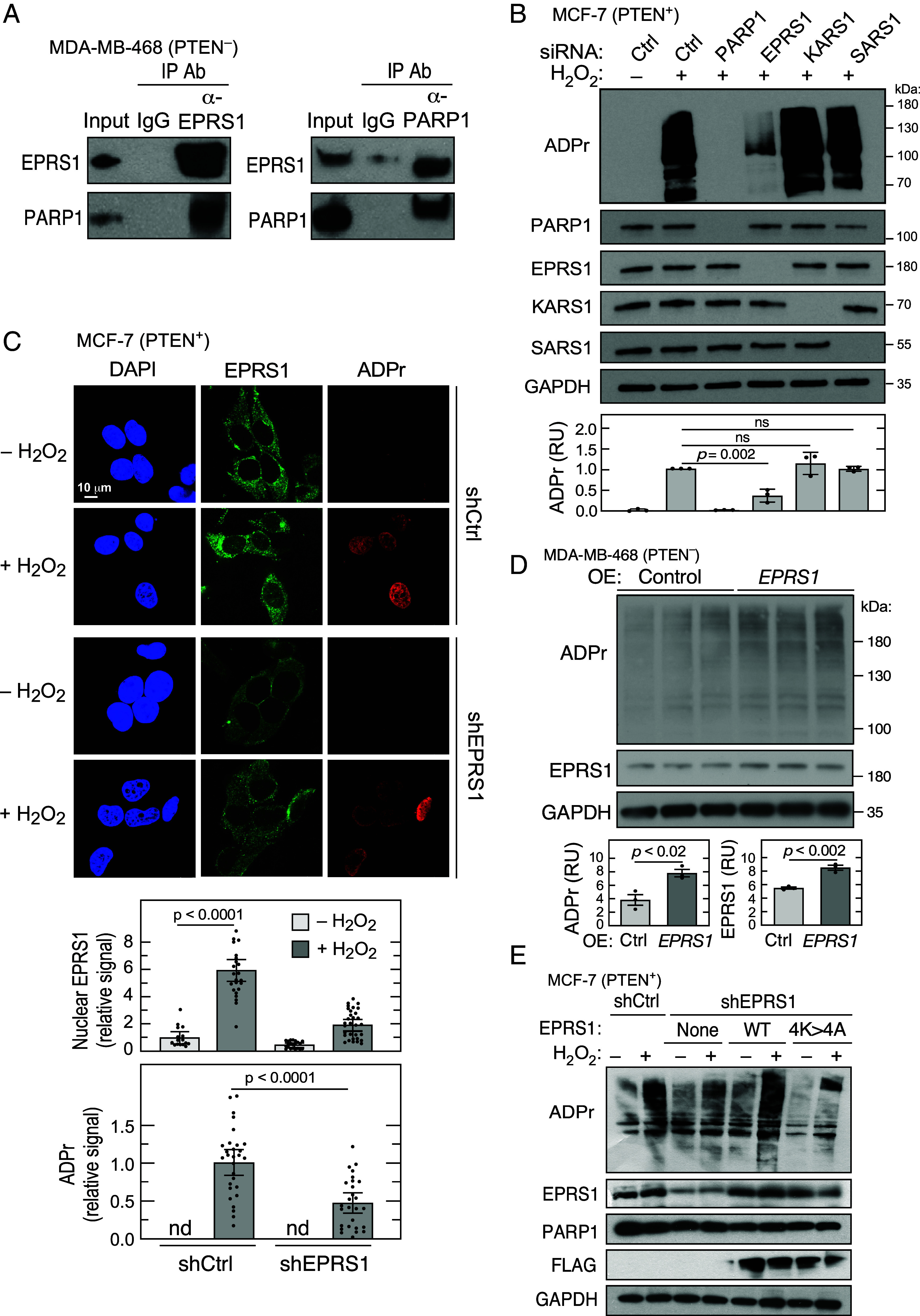
Glutamyl-prolyl-tRNA synthetase (EPRS1) is a bifunctional aminoacyl-tRNA-synthetase (aaRS) essential for decoding the genetic code. EPRS1 resides, with seven other aaRSs and three noncatalytic proteins, in the cytoplasmic multi-tRNA synthetase complex (MSC). Multiple MSC-resident aaRSs, including EPRS1, exhibit stimulus-dependent release from the MSC to perform noncanonical activities distinct from their primary function in protein synthesis. Here, we show EPRS1 is present in both cytoplasm and nucleus of breast cancer cells with constitutively low phosphatase and tensin homolog (PTEN) expression. EPRS1 is primarily cytosolic in PTEN-expressing cells, but chemical or genetic inhibition of PTEN, or chemical or stress-mediated activation of its target, AKT, induces EPRS1 nuclear localization. Likewise, preferential nuclear localization of EPRS1 was observed in invasive ductal carcinoma that were also P-Ser473-AKT+. EPRS1 nuclear transport requires a nuclear localization signal (NLS) within the linker region that joins the catalytic glutamyl-tRNA synthetase and prolyl-tRNA synthetase domains. Nuclear EPRS1 interacts with poly(ADP-ribose) polymerase 1 (PARP1), a DNA-damage sensor that directs poly(ADP-ribosyl)ation (PARylation) of proteins. EPRS1 is a critical regulator of PARP1 activity as shown by markedly reduced ADP-ribosylation in EPRS1 knockdown cells. Moreover, EPRS1 and PARP1 knockdown comparably alter the expression of multiple tumor-related genes, inhibit DNA-damage repair, reduce tumor cell survival, and diminish tumor sphere formation by breast cancer cells. EPRS1-mediated regulation of PARP1 activity provides a mechanistic link between PTEN loss in breast cancer cells, PARP1 activation, and cell survival and tumor growth. Targeting the noncanonical activity of EPRS1, without inhibiting canonical tRNA ligase activity, provides a therapeutic approach potentially supplementing existing PARP1 inhibitors.

Keywords: ADP-ribosylation; AKT; EPRS1; PARP1; aminoacyl-tRNA synthetase.


Conflict of interest statement

Competing interests statement:The authors declare no competing interest.

Figures

Fig. 1.
Fig. 1.
Nuclear localization of EPRS1 in breast cancer cells. (A) Isolation of cytosolic and nuclear fractions of PTEN+ and PTEN breast cancer cell lines and EPRS1 immunoblot. Nuclear fractions were detected with anti-p84 antibody and cytoplasmic fractions with anti-α-tubulin antibody. (B) Immunoblot determination of EPRS1 in subcellular fractions of MDA-MB-468 cells. Cell fractions were detected as in A; anti-histone H3 and anti-Na+/K+-ATPase detected nuclear chromatin and membranes, respectively. (C) Size-exclusion chromatography of cytosolic and nuclear lysates of MDA-MB-468 cells. (D) Schematic of major mammalian EPRS1 domains (Top). Intracellular localization of EPRS1 domains expressed in HEK293T cells (Bottom) was detected with nuclear (anti-HDAC) and cytosolic (anti-α-tubulin) marker. (E) Sequence alignment of potential EPRS1 NLS in multiple species (Top). Nuclear localization of FLAG-EPRS1 bearing NLS mutation 4K>4A in HEK293T cells (Bottom).
Fig. 2.
Fig. 2.
AKT-dependent nuclear localization of EPRS1. (A) siRNA-mediated knockdown of PTEN in MCF-7 cells induced AKT Ser473 phosphorylation (Right) and EPRS1 nuclear localization (Left). (B) A PTEN inhibitor, bpV (HOpic) (20 mM) induced EPRS1 nuclear localization in MCF-7 cells. (C) Treatment of MCF-7 cells with 400 µM H2O2 for 10 min induced EPRS1 nuclear localization. (D) Heat shock treatment of MCF-7 cells at 43 °C for 1 h induced nuclear localization of EPRS1, but not other MSC (Left) and non-MSC (Right) aaRSs. (E) Nuclear localization was determined in MEFs from WT (Ptenf1/f1, Top) or Pten−/− knockout (Bottom) mice after 43 °C heat shock for 1 h. (F) Heat-shocked MCF-7 cells were treated for 24 h with the pan-PI3K inhibitor BKM120 (100 nM, Left) or the pan-AKT inhibitor MK2206 (100 nM, Right) (or with DMSO solvent control), and EPRS1 nuclear localization was determined. (G) The effect of AKT activation on EPRS1 nuclear localization was determined following treatment with insulin and SC79, a small-molecule activator of AKT. (H) Cellular localization of EPRS1 and P-Ser473-AKT was determined by immunofluorescence in breast cancer and tumor-adjacent tissue (Left). Nuclear fluorescence intensity of EPRS1 was quantitated in seven tumor samples and six tumor-adjacent samples (mean ± SEM, P < 0.01, Right). Nuclear fractions were detected with anti-p84 and cytoplasmic fractions with anti-α-tubulin antibodies.
Fig. 3.
Fig. 3.
EPRS1 contributes to PARP1-mediated ADP-ribosylation and gene expression. (A) Reciprocal coimmunoprecipitation experiments show interaction of EPRS1 and PARP1 in nuclear fractions. (B) Determination of ADP-ribosylation (ADPr) in total cell extracts of MCF-7 cells subjected to siRNA-mediated knockdown of PARP1, EPRS1, KARS1, and SARS1. The cells were incubated with H2O2 for 10 min and subjected to immunoblot analysis. (C) Immunofluorescence detection of ADPr and EPRS1 localization in shControl and shEPRS1 MCF-7 cells treated with H2O2. Image Pro Plus software was used to analyze EPRS1 and ADPr fluorescence signal (n = 16 to 30 cells, mean ± SEM). (D) Effect of EPRS1 over-expression on ADPr in MDA-MB-468 cells. (E) Role of EPRS1 NLS in activation of PARP1 ADP-ribosylation activity in shEPRS1 knockdown cells overexpressing FLAG-tagged WT and 4K>4A EPRS1 cells.
Fig. 4.
Fig. 4.
Mechanism of EPRS1-mediated activation of PARP1. (A) Following siRNA-mediated knockdown of EPRS1, MCF-7 cells were incubated with H2O2, and NAD+ (Left) and NADH (2nd panel) were determined by colorimetric assay; total cellular dinucleotide (3rd panel) and their ratio (4th panel) were calculated (mean ± SEM, n = 4). (B) Effect of siEPRS1 knockdown on PARP1 binding to chromatin in the nuclear-insoluble fraction in H2O2-treated MCF-7 cells. Whole-cell lysate and cytoplasm samples were 1% of total; nuclear fractions were 10% of the total. (C) In vitro PARP1-mediated ADP-ribosylation of immobilized histone in the presence of recombinant EPRS1 was determined by modification by biotinylated PAR; (n = 3, mean ± SEM). (D) PARP1 auto-PARylation was determined in MDA-MB-468 cells (Left) and H2O2-treated MCF-7 cells (Right) by IP with anti-PARP1 antibody followed by immunoblot with anti-ADPr antibody. PARP1 Ser499 MAR was detected by immunoblot. (E) Effect of EPRS1 knockdown on H3S10-ADPr in H2O2-treated MCF-7 cells.
Fig. 5.
Fig. 5.
Role of AKT in PARP1-mediated ADP-ribosylation. (A) Role of AKT in PARP1-mediated ADP-ribosylation was determined using the AKT inhibitor, MK2206, in H2O2-treated MCF-7 cells. (B) Sufficiency of AKT activation for PARP1-mediated ADP-ribosylation was shown using the AKT activator, SC79, and ADPr immunoblot was quantitated by densitometry.
Fig. 6.
Fig. 6.
EPRS1 promotes DNA DSB repair and cell survival. (A) Effect of EPRS1 knockdown on mRNA expression of selected PARP1 target genes in shEPRS1 MCF-7 cells treated with H2O2 (n = 3; ns = not significant, t = trend). (B) Alkaline COMET assay assessing DNA DSB formation in shControl, shPARP1, shEPRS1, and shLARS1 MCF-7 cells treated with H2O2 (Left). Quantitative evaluation of tail DNA percentage and tail moment (tail% × tail length) of MCF-7 knockdown cells (Right; mean ± SEM; n = 4). (C) Clonogenic survival assay of shControl, shPARP1, shEPRS1, and shLARS1 in MCF-7 cells treated with MMS for 1 h. (D) Formation of tumor spheres by shControl, shEPRS1, and shPARP1 MCF-7 cells treated with 400 µM H2O2 and then incubated for 14 d (n = 3, mean ± SEM, P < 0.05). (E) MDA-MB-468 cells were subjected to siEPRS1-mediated knockdown and incubated with talazoparib for 72 h. ADPr formation was determined by immunoblot (Left) and quantitated by densitometry (Right); mean ± SEM, n = 3. Talazoparib IC50 was calculated by nonlinear regression of inhibitor concentration versus response (three-parameter fit) using GraphPad. For siCtrl, IC50 = 1.23, R2 = 0.96; for siEPRS1, IC50 = 0.41, R2 = 0.98. IC50’s are illustrated by dashed lines. (F) Effect of EPRS1 knockdown on cell sensitivity to talazoparib was determined by MTT cell survival assay. Rescue of survival by transfection with WT or NLS mutant EPRS1 was determined. For panels C, E, and F: mean ± SEM, n = 3, ns = not significant; **P < 0.01; ***P < 0.001, ****P < 0.0001).
Fig. 7.
Fig. 7.
Schematic of enhanced PARP1-mediated DNA repair by EPRS1.

References

    1. Eswarappa S. M., Potdar A. A., Sahoo S., Sankar S., Fox P. L., Metabolic origin of the fused aminoacyl-tRNA synthetase, glutamyl-prolyl-tRNA synthetase. J. Biol. Chem. 293, 19148–19156 (2018). - PMC - PubMed
    1. Sampath P., et al. , Noncanonical function of glutamyl-prolyl-tRNA synthetase: Gene-specific silencing of translation. Cell 119, 195–208 (2004). - PubMed
    1. Khan K., Baleanu-Gogonea C., Willard B., Gogonea V., Fox P. L., 3-Dimensional architecture of the human multi-tRNA synthetase complex. Nucleic Acids Res. 48, 8740–8754 (2020). - PMC - PubMed
    1. Ray P. S., et al. , A stress-responsive RNA switch regulates VEGFA expression. Nature 457, 915–919 (2009). - PMC - PubMed
    1. Arif A., et al. , EPRS is a critical mTORC1-S6K1 effector that influences adiposity in mice. Nature 542, 357–361 (2017). - PMC - PubMed

MeSH terms