. 2025 Jan 7;148(1):119-132.
doi: 10.1093/brain/awae234.

Brain inflammation co-localizes highly with tau in mild cognitive impairment due to early-onset Alzheimer's disease

Affiliations

Brain inflammation co-localizes highly with tau in mild cognitive impairment due to early-onset Alzheimer's disease

Johanna Appleton et al. Brain. .

Abstract

Brain inflammation, with an increased density of microglia and macrophages, is an important component of Alzheimer's disease and a potential therapeutic target. However, it is incompletely characterized, particularly in patients whose disease begins before the age of 65 years and, thus, have few co-pathologies. Inflammation has been usefully imaged with translocator protein (TSPO) PET, but most inflammation PET tracers cannot image subjects with a low-binder TSPO rs6971 genotype. In an important development, participants with any TSPO genotype can be imaged with a novel tracer, 11C-ER176, that has a high binding potential and a more favourable metabolite profile than other TSPO tracers currently available. We applied 11C-ER176 to detect brain inflammation in mild cognitive impairment (MCI) caused by early-onset Alzheimer's disease. Furthermore, we sought to correlate the brain localization of inflammation, volume loss, elevated amyloid-β (Aβ)and tau. We studied brain inflammation in 25 patients with early-onset amnestic MCI (average age 59 ± 4.5 years, 10 female) and 23 healthy controls (average age 65 ± 6.0 years, 12 female), both groups with a similar proportion of all three TSPO-binding affinities. 11C-ER176 total distribution volume (VT), obtained with an arterial input function, was compared across patients and controls using voxel-wise and region-wise analyses. In addition to inflammation PET, most MCI patients had Aβ (n = 23) and tau PET (n = 21). For Aβ and tau tracers, standard uptake value ratios were calculated using cerebellar grey matter as region of reference. Regional correlations among the three tracers were determined. Data were corrected for partial volume effect. Cognitive performance was studied with standard neuropsychological tools. In MCI caused by early-onset Alzheimer's disease, there was inflammation in the default network, reaching statistical significance in precuneus and lateral temporal and parietal association cortex bilaterally, and in the right amygdala. Topographically, inflammation co-localized most strongly with tau (r = 0.63 ± 0.24). This correlation was higher than the co-localization of Aβ with tau (r = 0.55 ± 0.25) and of inflammation with Aβ (0.43 ± 0.22). Inflammation co-localized least with atrophy (-0.29 ± 0.26). These regional correlations could be detected in participants with any of the three rs6971 TSPO polymorphisms. Inflammation in Alzheimer's disease-related regions correlated with impaired cognitive scores. Our data highlight the importance of inflammation, a potential therapeutic target, in the Alzheimer's disease process. Furthermore, they support the notion that, as shown in experimental tissue and animal models, the propagation of tau in humans is associated with brain inflammation.

Keywords: 11C-ER176 PET; TSPO; early-onset Alzheimer’s disease; inflammation; mild cognitive impairment.


Conflict of interest statement

J.C.M. received research funding from Eli Lilly, parent company of Avid Radiopharmaceuticals, manufacturer of flortaucipir. The rest of the authors declare no competing interests.

Figures

Figure 1
Figure 1
11C-ER176 PET in all three genotypes. Examples of 11C-ER176 signals in high- (HAB), mixed- (MAB) and low-affinity (LAB) binders from our mild cognitive impairment group. Bars on the right provide the thresholds at which total distribution volume (VT) values for each image are shown. The low-affinity binder had posterior cortical atrophy, with greatest involvement of posterior temporal cortex. The uptake in thalamus and brainstem is also present in normal controls. TSPO = translocator protein; y/o = years old.
Figure 2
Figure 2
Inflammation, cortical thickness, amyloid-β and tau in mild cognitive impairment early-onset Alzheimer’s disease (MCI-EOAD). (A) Surface-based ANCOVA comparing 11C-ER176 uptake in 25 MCI-EOAD patients and 23 cognitively unimpaired (CU) controls. Coloured vertices on the cortical surface map indicate areas where MCI-EOAD patients had greater inflammation than controls. (B) Cortical thickness maps comparing 25 MCI-EOAD subjects and 23 CU controls. Coloured vertices indicate areas where patients showed greater cortical atrophy than controls. (C) Surface-based ANOVA comparing 18F-florbetaben uptake in 21 MCI-EOAD patients and 19 CU controls. Coloured vertices on the cortical surface map indicate areas where MCI-EOAD patients had greater amyloid-β (Aβ) than controls. (D) Surface-based ANOVA comparing 18F-flortaucipir uptake in 21 MCI-EOAD patients and 23 CU controls. Coloured vertices on the cortical surface map indicate areas where MCI-EOAD patients had greater tau than controls. FDR = false discovery rate.
Figure 3
Figure 3
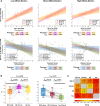
Inflammation and tau in the Tau-PET Braak-stage regions. In tau-PET Braak regions, modified from Pascoal et al., box and whisker plots show values of 11C-ER176 (top) and 18F-flortaucipir (bottom) uptake in cognitively unimpaired (CU) controls and mild cognitive impairment early-onset Alzheimer’s disease (MCI-EOAD). P-values for group comparisons are adjusted using family-wise error (FWE) correction. SUVR = standardized uptake value ratio; VT = total distribution volume.
Figure 4
Figure 4
Correlations across regional inflammation, cortical thickness, amyloid-β and tau. (A) Scatter plots showing the pairwise relationships between inflammation, amyloid-β (Aβ) and tau PETs (top), as well as each PET tracer’s relationships with volume (bottom) across all three TSPO affinity groups. Data are standardized so that regression slopes equal correlation coefficients. Inflammation and tau exhibit the strongest of all pairwise correlations within each affinity. (B) Box plots display Pearson correlation values for each pairwise PET correlation as well as each PET tracer’s correlation with volume on an individual subject basis. P-values for comparisons are family-wise error (FWE)-corrected. (C) The matrix indicates the average, denoted as ravg, of all single Pearson intra-individual correlation values in Mild Cognitive Impairment caused by early-onset Alzheimer’s disease (MCI-EOAD) subjects. Among individual FWE-corrected correlations, inflammation and tau were significant in 16/21 subjects, Aβ and tau in 15/21, inflammation and Aβ in 11/22, tau and volume in 13/21, inflammation and volume in 7/25 and Aβ and volume in 2/22. ER176 = 11C-ER176 inflammation PET; SUVR = standardized uptake value ratio.
Figure 5
Figure 5
Correlation between the topography of 11C-ER176 PET inflammation uptake and neuropsychological scores. (A) Maps show the Pearson correlation coefficients between inflammation in Hammers’ atlas regions and cognitive scores from the DemTect and Mini-Mental Status Examination (MMSE) in the mild cognitive impairment caused by early-onset Alzheimer’s disease (MCI-EOAD) sample, not including cognitively unimpaired (CU) controls. No correlations were statistically significant. (B) Maps show uncorrected P-values for Hammer’s atlas regions with statistically significant Pearson correlations (P < 0.001) between inflammation and the neuropsychological scores on DemTect, MMSE and immediate and delayed recall sub-tests of the DemTect in the entire sample, including patients and controls. FWE = family-wise error.

References

    1. Bettcher BM, Tansey MG, Dorothée G, Heneka MT. Peripheral and central immune system crosstalk in Alzheimer disease—A research prospectus. Nat Rev Neurol. 2021;17:689–701. - PMC - PubMed
    1. Chen X, Firulyova M, Manis M, et al. . Microglia-mediated T cell infiltration drives neurodegeneration in tauopathy. Nature. 2023;615:668–677. - PMC - PubMed
    1. Lee CY, Landreth GE. The role of microglia in amyloid clearance from the AD brain. J Neural Transm (Vienna). 2010;117:949–960. - PMC - PubMed
    1. Odfalk KF, Bieniek KF, Hopp SC. Microglia: Friend and foe in tauopathy. Prog Neurobiol. 2022;216:102306. - PMC - PubMed
    1. McAlpine CS, Park J, Griciuc A, et al. . Astrocytic interleukin-3 programs microglia and limits Alzheimer's disease. Nature. 2021;595:701–706. - PMC - PubMed

MeSH terms