. 2024 Sep;11(35):e2400024.
doi: 10.1002/advs.202400024. Epub 2024 Jul 16.

mRNA-Engineered CD5-CAR-γδTCD5- Cells for the Immunotherapy of T-Cell Acute Lymphoblastic Leukemia

Affiliations

mRNA-Engineered CD5-CAR-γδTCD5- Cells for the Immunotherapy of T-Cell Acute Lymphoblastic Leukemia

Zhixiong Zhu et al. Adv Sci (Weinh). 2024 Sep.

Abstract

Clinical trials of Chimeric Antigen Receptor T-cell (CAR-T) therapy have demonstrated remarkable success in treating both solid tumors and hematological malignancies. Nanobodies (Nbs) have emerged as promising antigen-targeting domains for CARs, owing to their high specificity, robust stability, and strong affinity, leading to significant advancements in the field of Nb-CAR-T. In the realm of T-cell acute lymphoblastic leukemia (T-ALL) targets, CD5 stands out as a potentially excellent candidate for T-cell-based CAR therapy, due to its distinct expression on the surface of malignant T-ALL cells. To mitigate graft-versus-host disease associated with allogeneic CAR-T, γδT cells are selected and stimulated from peripheral blood mononuclear cells, and γδT cells are engineered via CRISPR/Cas9 to eliminate fratricide, enabling the creation of fratricide-resistant CAR-γδTCD5- cells. In vitro transcribed (IVT) mRNA is used to construct CAR-T, presenting a safer, faster, and cost-effective method compared to traditional viral vector approaches. In this study, a CD5-VHH library is constructed, and specific CD5-nanobodies are screened for subsequent use in CD5-CAR-γδTCD5- therapy. IVT-mRNA-CD5-CAR-γδTCD5- cells exhibited favorable functional characteristics and demonstrated antitumor efficacy against malignant T cell lines, underlining the potential for advancing mRNA-CD5-CAR-γδTCD5- therapy.

Keywords: CAR‐T; IVT‐mRNA; T‐ALL; γδT cells.


Conflict of interest statement

The authors declare no conflict of interest.

Figures

Figure 1
Figure 1
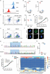
CD5‐VHH library construction, specific nanobodies screening, and characterization of the CD5‐Nbs. a) Schematic presentation of nanobody screening. b) Immunological valence analysis. c) Enrichment analysis of phage particles against CD5 protein‐specific nanobodies using polyclonal indirect ELISA. d) Identification of recombinant VHH‐gpIII proteins from the 96 clones specifically binding with CD5 protein. e) Analysis of the specific binding ability of recombinant monovalent nanobodies using indirect ELISA. f, g) Measurement of the binding ability of recombinant monovalent nanobodies using indirect ELISA. h) Real‐time binding profile of recombinant monovalent nanobodies with CD5 by SPR. i) Immunofluorescence for verifying the binding ability of CD5‐27‐Nb with CD5‐HeLa by CLSM. The scale bar represents 10 µm. j) Analysis of the binding ability of CD5‐27‐Nb with CCRF‐CEM and Molt‐4 by flow cytometry. The image of the schematic presentation of screening nanobodies is created with BioRender.com. ELISA, enzyme‐linked immunosorbent assay.
Figure 2
Figure 2
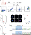
Effective disruption of CD5 gene in T cells with CRISPR/Cas9 system prevents fratricide without compromising T cell function. a, b) Flow cytometric analysis of surface expression of CD3 and CD5 in bulk peripheral blood mononuclear cells from healthy donors: CD3+CD5+ (mean, 90.8% ± 6.0%) versus CD3+CD5(mean, 9.5% ± 5.7%) (n = 5, < 0.0001, T‐test) and T‐ALL patients: CD3+CD5+ (mean, 87.95% ± 7.85%) and CD3+CD5 (mean, 9.75% ± 5.55%) (n = 5, p < 0001, T‐test). c) Flow cytometric analysis of surface expression of CD3 and CD5 in CD5‐CAR‐T cells after fratricide: CD3+CD5+ (mean, 40.75% ± 7.5%) and CD3+CD5 (mean, 53.5% ± 13.5%) (n = 5, p = ns, T‐test). d) Expansion kinetics and viability of CD5‐CAR‐T cells. CD5‐CAR‐T cells had decreased viability compared with CD19‐CAR‐T and NT‐T cells (n = 3, p < 0.01, 2‐way ANOVA). e) Flow cytometric analysis of T cells before and after CD5 depletion. f) Immunofluorescence for verifying depletion of CD5 by CLSM. The scale bar represents 5 µm. g) Sequencing analysis of the CD5 gene loss. h) Flow cytometric analysis of CD5‐KO efficacy of T cells: CD3+CD5+ (mean, 7.6% ± 4.4%) versus CD3+CD5 (mean, 92.4% ± 4.4%) (n = 5, p < 0.0001, T‐test). i) Expansion kinetics and viability of CD5‐CAR‐TCD5− cells. CD5‐CAR‐TCD5− cells had similar expansion to NT‐T cells (n = 3, p = ns, 2‐way ANOVA). j) RNA‐seq analysis of functional‐related genes in T cells. T‐test was used to compare the statistical difference between 2 groups, and one‐way or two‐way analysis of variance with Sidak's or Tukey's multiple comparison tests was used to compare 3 or more groups (* p < 0.05, ** p < 0.01, *** p < 0.005, **** p < 0.0001). ns, not significant.
Figure 3
Figure 3
Generation of fratricide‐resistant γδTCD5− cells. a) Schematic presentation of γδT cell enrichment. b) Gate strategy of γδT cells. c) Flow cytometric analysis of γδ−1 and γδ−2 expression in γδT cells stimulated from PBMCs: γδ−1 (mean, 10.05% ± 4.15%) versus γδ−2 (mean, 89.1% ± 4.5%) (n = 5, < 0001, T‐test). d) Immunofluorescence for verifying γδ−2 expression in γδT cells by CLSM. Scale bar represents 5 µm. e) Flow cytometric analysis of surface expression of CD3 and CD5 in γδT cells. CD3+CD5+ (mean, 90.9% ± 4.6%) versus CD3+CD5 (mean, 4.5% ± 4.6%) (n = 5, p < 0.0001, T‐test). f) Flow cytometric analysis CD5 expression of γδT cells before and after CD5 depletion. CD3+CD5+ (mean, 9.1% ± 4.6%) versus CD3+CD5 (mean, 91.1% ± 4.8%) (n = 5, p < 0.0001, T‐test). g) Sequencing analysis of the CD5 gene loss in γδT cells. T‐test was used to compare the statistical difference between 2 groups (* p < 0.05, ** p < 0.01, *** p < 0.005, **** p < 0.0001).
Figure 4
Figure 4
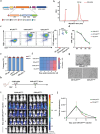
Fratricide‐resistant CD5‐CAR γδTCD5− cells constructed by anti‐CD5‐CAR‐mRNA. a) Schematic representation of in vitro transcribed mRNA encoding anti‐CD5‐VHH CAR. b) Assessment of the integrity and length of IVT CAR‐mRNA. c) Representative flow cytometry analysis showing CD5‐CAR expression after transduction of anti‐CD5‐VHH CAR mRNA. (mean transduction efficiency 88.25% ± 5.95%, n = 5, p < 0.0001, T‐test). d) Evaluation of CAR expression of CD5‐CAR‐γδTCD5− by flow cytometry analysis from 6 h to 6 days. e) Immunophenotype of CD5‐CAR‐γδTCD5− cells post transduction. f) Expansion kinetics and viability of CD5‐CAR‐γδTCD5− cells. g) Absence of fratricide observed in CD5‐CAR‐γδTCD5− under the light microscope. h) Schematic of CD5‐CAR‐γδTCD5− in vivo persistence experiment: NSG mice were injected with 1 × 105 CCRF‐CEM cells 5 days prior to a single dose of 5 × 10⁶ CD5‐CAR‐γδTCD5−.ffLuc cells. i) Corresponding bioluminescence imaging. j) Quantitative bioluminescence data. T‐test was used to compare the statistical difference between 2 groups (* p < 0.05, ** p < 0.01, *** p < 0.005, **** p < 0.0001).
Figure 5
Figure 5
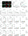
In vitro antitumor efficacy of CD5‐CAR γδTCD5− cells. a) Immunofluorescence for verifying the specific binding ability of CD5‐CAR γδTCD5− cells (green) with CCRF‐CEM‐mCherry (red) by CLSM. Scale bar represents 5 µm. b) Detection of the ability of TCD5− cells, γδT cells, and γδTCD5− cells to lyse target cells (CD5‐HeLa) using RTCA. c) Detection of the ability of CD5‐CAR‐γδTCD5− cells, CD19‐CAR‐γδTCD5− cells, NT‐γδTCD5− cells to lyse target cells (CD5‐HeLa) using RTCA. d) CD5‐CAR‐γδTCD5− specifically killed CCRF‐CEM and MOLT‐4 cells under different E:T ratios. (e‐g) CD5‐CAR‐γδTCD5− cells, CD19‐CAR‐γδTCD5− cells, NT‐γδTCD5− cells, and NT‐γδT cells were co‐cultured with media, Molm13, K562, CCRF, or MOLT‐4 at a 2:1 E:T ratio. Supernatants were harvested after 24 h and analyzed for IFN‐γ, IL‐2, and TNF‐α by ELISA (n = 3, 1‐way ANOVA, *** p < 0.005). ns, not significant.
Figure 6
Figure 6
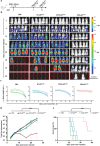
Antitumor activity of CD5‐CAR‐γδTCD5− cells in CCRF‐CEM xenograft model. a) Schematic representation of CD5‐CAR‐γδTCD5− in vivo treatment for CCRF‐CEM.ffLuc xenograft model: NSG mice were intravenously injected with 3 × 105 CCRF‐CEM.ffLuc cells on day 0 and double doses of 5 × 106 CD5‐CAR‐γδTCD5− cells on day 7 and 11, respectively. b) Corresponding bioluminescence imaging (n = 5). c) Body weight curve. d) Quantitative bioluminescence data (n = 5, 2‐way ANOVA). Mice treated with CD5‐CAR‐γδTCD5− cells exhibited significantly decreased bioluminescence (**** p < 0.0001) compared to the CD5‐CAR‐γδTCD5−, NT‐γδTCD5− and PBS‐treated groups. e) Survival curve (**** p < 0.0001, Mantel‐Cox log‐rank test). Mice treated with CD5‐CAR‐γδTCD5− cells showed significantly increased survival (< 0.0001) compared to CD19‐CAR‐γδTCD5−, NT‐γδTCD5− and PBS‐treated groups.
Figure 7
Figure 7
Antitumor activity of CD5‐CAR‐γδTCD5− cells in MOLT‐4 xenograft model. a) Schematic of CD5‐CAR‐γδTCD5− in vivo treatment of MOLT‐4 xenograft model: NSG mice were intravenously injected with 3 × 105 MOLT‐4.ffLuc cells on day 0 and double doses of 5 × 106 CD5‐CAR‐γδTCD5− cells on day 7 and 11, respectively. b) Corresponding bioluminescence imaging (n = 5). c) Body weight curve. d) Quantitative bioluminescence data (n = 5, 2‐way ANOVA). Mice treated with CD5‐CAR‐γδTCD5− cells exhibited significantly decreased bioluminescence (**** p < 0.0001) compared to CD5‐CAR‐γδTCD5−, NT‐γδTCD5− and PBS‐treated groups. e) Survival curve (**** p < 0.0001, Mantel‐Cox log‐rank test). Mice treated with CD5‐CAR‐γδTCD5− cells showed significantly increased survival (p < 0.0001) compared to CD5‐CAR‐γδTCD5−, NT‐γδTCD5− and PBS‐treated groups.

References

    1. Zhang C., Liu J., Zhong J. F., Zhang X., Biomark Res 2017, 5, 22. - PMC - PubMed
    1. June C. H., O'Connor R. S., Kawalekar O. U., Ghassemi S., Milone M. C., Science 2018, 359, 1361. - PubMed
    1. Hamers‐Casterman C., Atarhouch T., Muyldermans S., Robinson G., Hamers C., Songa E. B., Bendahman N., Hamers R., Nature 1993, 363, 446. - PubMed
    1. Harmsen M. M., Ruuls R. C., Nijman I. J., Niewold T. A., Frenken L. G., de Geus B., Mol. Immunol. 2000, 37, 579. - PubMed
    1. Xin Yu J., Hubbard‐Lucey V. M., Tang J., Nat. Rev. Drug Discov. 2019, 18, 821. - PubMed

MeSH terms

LinkOut - more resources