Meta-Analysis
. 2024 Jul;14(7):230419.
doi: 10.1098/rsob.230419. Epub 2024 Jul 17.

Genetic evidence for serum amyloid P component as a drug target in neurodegenerative disorders

Affiliations
Meta-Analysis

Genetic evidence for serum amyloid P component as a drug target in neurodegenerative disorders

A Floriaan Schmidt et al. Open Biol. 2024 Jul.

Abstract

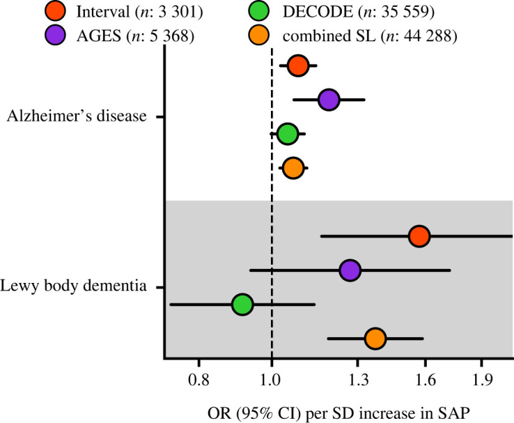
The mechanisms responsible for neuronal death causing cognitive loss in Alzheimer's disease (AD) and many other dementias are not known. Serum amyloid P component (SAP) is a constitutive plasma protein, which is cytotoxic for cerebral neurones and also promotes formation and persistence of cerebral Aβ amyloid and neurofibrillary tangles. Circulating SAP, which is produced exclusively by the liver, is normally almost completely excluded from the brain. Conditions increasing brain exposure to SAP increase dementia risk, consistent with a causative role in neurodegeneration. Furthermore, neocortex content of SAP is strongly and independently associated with dementia at death. Here, seeking genomic evidence for a causal link of SAP with neurodegeneration, we meta-analysed three genome-wide association studies of 44 288 participants, then conducted cis-Mendelian randomization assessment of associations with neurodegenerative diseases. Higher genetically instrumented plasma SAP concentrations were associated with AD (odds ratio 1.07, 95% confidence interval (CI) 1.02; 1.11, p = 1.8 × 10⁻³), Lewy body dementia (odds ratio 1.37, 95%CI 1.19; 1.59, p = 1.5 × 10⁻⁵) and plasma tau concentration (0.06 log2(ng l-1) 95%CI 0.03; 0.08, p = 4.55 × 10⁻⁶). These genetic findings are consistent with neuropathogenicity of SAP. Depletion of SAP from the blood and the brain, by the safe, well tolerated, experimental drug miridesap may thus be neuroprotective.

Keywords: Alzheimer's disease; C-reactive protein‌; Lewy body dementia; genome-wide association study; miridesap; serum amyloid P component.


Conflict of interest statement

A.F.S. and C.F. have received funding from New Amsterdam Pharma for unrelated work. M.B.P. is the inventor on expired patents on SAP depletion by miridesap (CPHPC). GlaxoSmithKline's abandoned patents on an experimental miridesap prodrug were assigned to UCL spinout company, Pentraxin Therapeutics Ltd, founded and directed by M.B.P; but are no longer maintained. The other authors have no competing interests.

Figures

Figure 1.
Figure 1.
Manhattan plot of SAP genome-wide association study. The −log10(p-value) of genetic variants is shown on the y-axis and GRCh37 base pair position within chromosomes on the x-axis. The horizontal dashed line is at p-value 5.8 × 10⁻⁸. The lead variants are labelled with the putative causal genes assigned by V2G.
Figure 2.
Figure 2.
Stacked locus-view comparing the overlap between genetic variants for plasma SAP values and plasma CRP concentration. The −log10(p-value) of the genetic association with SAP and CRP plotted (y-axis) against the genomic location (x-axis). The lead variant for each trait is indicated by a purple diamond. Linkage disequilibrium with the lead variant is indicated by coloured dots, with the R2 estimated from the UKB and 1000 genomes EUR reference. Gene locations were queried from Ensembl v109 (GRCh37).
Figure 3.
Figure 3.
Mapping of the trans signals for variants associated with plasma SAP value to putative causal genes. First column, the overall V2G score; second column, distance in base pairs from the lead variant to the canonical gene transcription start site (TSS). The other columns show the presence or absence of x-axis criteria, specifically, whether there were quantitative trait loci (QTL) linking the gene to proteomics, transcriptomics, or splice site QTL, and PCHi-C (promoter capture Hi-C) chromatin interaction experiments linking the genetic variant to the indicated gene, from Jung et al. [52] and Javierre et al. [53]. Electronic supplementary material, tables S3–S8, show the whole V2G output.
Figure 4.
Figure 4.
Estimates of the cis-Mendelian randomization effect of plasma SAP values on Alzheimer's disease and Lewy body dementia. The univariable MR effects estimated from the GWAS meta-analysis of SomaLogic (SL) plasma SAP values from the Interval, AGES and DECODE studies are shown individually, together with the combined result of all three studies. We used the Alzheimer's disease GWAS from Kunkle et al. [38] which consisted of 35 274 cases and 59 163 controls, the Lewy body dementia GWAS from Chia et al. [39] consisted of 2981 cases and 2173 controls. OR, odds ratio; 95%CI, 95%CI. The figure illustrates the data in electronic supplementary material, table S10.
Figure 5.
Figure 5.
Comparison of the cis-Mendelian randomization effect estimates of plasma SAP values and CRP concentration. Effect direction, magenta for positive and green for negative, is shown by the −log10(p-value). Open diamonds, p < 0.05; closed stars, p < 2.78 × 10⁻³. Left block, SAP; right column, CRP. ALT, alanine transaminase; AST, aspartate transaminase; CHD, coronary heart disease; DBP, diastolic blood pressure; GGT, gamma-glutamyl transferase; IPF, idiopathic pulmonary fibrosis; SBP, systolic blood pressure; SLE, systemic lupus erythematosus; T2DM, type 2 diabetes; WMH, white matter hyperintensities. The figure illustrates the data in electronic supplementary material, tables S11–S12.

Update of

References

    1. Nelson SR, Tennent GA, Sethi D, Gower PE, Ballardie FW, Amatayakul-Chantler S, Pepys MB. 1991. Serum amyloid P component in chronic renal failure and dialysis. Clin. Chim. Acta 200, 191-199. ( 10.1016/0009-8981(91)90090-y) - DOI - PubMed
    1. Hawkins PN, Rossor MN, Gallimore JR, Miller B, Moore EG, Pepys MB. 1994. Concentration of serum amyloid P component in the CSF as a possible marker of cerebral amyloid deposits in Alzheimer's disease. Biochem. Biophys. Res. Commun. 201, 722-726. ( 10.1006/bbrc.1994.1760) - DOI - PubMed
    1. Kolstoe SE, et al. 2009. Molecular dissection of Alzheimer's disease neuropathology by depletion of serum amyloid P component. Proc. Natl Acad. Sci. USA 106, 7619-7623. ( 10.1073/pnas.0902640106) - DOI - PMC - PubMed
    1. Veszelka S, et al. 2013. Efflux transport of serum amyloid P component at the blood-brain barrier. Eur. J. Microbiol. Immunol. 3, 281-289. ( 10.1556/EuJMI.3.2013.4.8) - DOI - PMC - PubMed
    1. Pepys MB, Booth DR, Hutchinson WL, Gallimore JR, Collins IM, Hohenester E. 1997. Amyloid P component. A critical review. Amyloid 4, 274-295. ( 10.3109/13506129709003838) - DOI

Publication types

MeSH terms