. 2024 Sep 10;57(9):2061-2076.e11.
doi: 10.1016/j.immuni.2024.06.013. Epub 2024 Jul 15.

Deep mutational scanning reveals functional constraints and antibody-escape potential of Lassa virus glycoprotein complex

Affiliations

Deep mutational scanning reveals functional constraints and antibody-escape potential of Lassa virus glycoprotein complex

Caleb R Carr et al. Immunity. .

Abstract

Lassa virus is estimated to cause thousands of human deaths per year, primarily due to spillovers from its natural host, Mastomys rodents. Efforts to create vaccines and antibody therapeutics must account for the evolutionary variability of the Lassa virus's glycoprotein complex (GPC), which mediates viral entry into cells and is the target of neutralizing antibodies. To map the evolutionary space accessible to GPC, we used pseudovirus deep mutational scanning to measure how nearly all GPC amino-acid mutations affected cell entry and antibody neutralization. Our experiments defined functional constraints throughout GPC. We quantified how GPC mutations affected neutralization with a panel of monoclonal antibodies. All antibodies tested were escaped by mutations that existed among natural Lassa virus lineages. Overall, our work describes a biosafety-level-2 method to elucidate the mutational space accessible to GPC and shows how prospective characterization of antigenic variation could aid the design of therapeutics and vaccines.

Keywords: Arevirumab; Arevirumab-3; GPC; Lassa virus; antibody escape; antigenic variation; arena virus; deep mutational scanning; global epistasis.


Conflict of interest statement

Declaration of interests J.D.B. is on the scientific advisory boards of Apriori Bio, Invivyd, Aerium Therapeutics, and the Vaccine Company. K.G.A. is a consultant to and on the scientific advisory board of Invivyd. J.D.B. and K.H.D.C. receive royalty payments as inventors on Fred Hutch licensed patents related to viral deep mutational scanning. N.P.K. is a co-founder, shareholder, paid consultant, and chair of the scientific advisory board of Icosavax, Inc. The King lab has received unrelated sponsored research agreements from Pfizer and GSK. M.M. is currently an employee of Seagen, though his contributions to this manuscript were performed when he was an employee of the Institute for Protein Design.

Figures

Figure 1.
Figure 1.. Pseudovirus deep mutational scanning of Lassa GPC
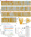
A Lentivirus backbone used for GPC deep mutational scanning. The backbone contains full-length 5′ and 3′ long terminal repeat (LTR) sequences. GPC is under control of a doxycycline-inducible TRE3GS promoter and linked to a random 16-nucleotide barcode (BC) downstream of the stop codon. A CMV promoter drives the expression of zsGreen. Other backbone components include the lentiviral Psi/Rev response element (RRE), woodchuck hepatitis virus post-transcriptional regulatory element (WPRE), and a repaired 3′-LTR to allow re-activation of integrated genomes., B Approach for producing genotype-phenotype linked GPC-pseudotyped lentivirus libraries. GPC-encoding lentivirus backbone, vesicular stomatitis virus G protein (VSV-G) expression plasmid, and lentivirus helper plasmids are first transfected into 293T cells to produce a VSV-G-pseudotyped variant library with no genotype-phenotype link. To create a genotype-phenotype link, the VSV-G-pseudotyped variant library is used to infect reverse tetracycline-controlled transactivator (rtTA) expressing 293T cells at low multiplicity of infection (MOI) so infected cells typically are transduced with just one lentivirus genome. Transduced cells are selected based on expression of zsGreen. Finally, GPC mutant viruses with a genotype-phenotype link are generated from the transduced cells by inducing GPC expression with doxycydine and transfecting the lentivirus helper plasmids. The library composition can be assessed by separately transfecting the transduced cells with VSV-G alongside the helper plasmids, which creates VSV-G-pseudotyped viruses that infect cells regardless of the functionality of the GPC variant encoded in the lentivirus genome. C Phylogenetic tree of representative Lassa GPC sequences. Tree tips are colored by the country from which the virus was collected. The major Lassa lineages I, II, III, IV, V, VI, and VII are labeled. The GPC from the Josiah strain used for deep mutational scanning is labeled in large bold font. Other GPC sequences used later in this paper are labeled in smaller font. Percent amino-acid identity with respect to the Josiah strain GPC is shown for all sequences above the tree.
Figure 2.
Figure 2.. Effects of GPC mutations on cell entry
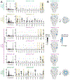
A Effects of mutations on entry into 293T cells as measured by deep mutational scanning. Each square in the heatmap represents a different mutation, with mutations that impair cell entry colored orange and those that improve cell entry colored blue. The wildtype amino acid in the parental Josiah strain at each site is indicated with a x. The overlay bar denotes the stable signal peptide (SSP), glycoprotein 1 (GP1) domain, glycoprotein 2 (GP2) domain, transmembrane domain (TM), and cytoplasmic tail (CT). See the interactive version of the heatmap at https://dms-vep.org/LASV_Josiah_GP_DMS/cell_entry.html for more effective visualization of the data. B Surface representation of GPC colored by the average effect of all amino-acid mutations at each site on cell entry (PDB: 7PUY). C Effects of mutations on cell entry for different GPC regions. Each point represents a different mutation, and medians are shown for each region as vertical lines. D Correlation of effects on cell entry measured by deep mutational scanning and the fold-change in titer of individual GPC pseudovirus mutants relative to unmutated Josiah GPC. Each point represents a biological replicate. The number of biological replicates is indicated in the legend for each mutant. The Pearson correlation (r) is indicated.
Figure 3.
Figure 3.. Mapping the effects of mutations on antibody escape
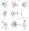
A Workflow for antibody-escape mapping. The GPC pseudovirus library is mixed with a VSV-G-pseudotyped “standard” that is not neutralized by anti-Lassa antibodies. The pseudovirus pool is incubated with different antibody concentrations, then used to infect cells. Viral genomes are recovered from infected cells ~12 hours post infection, and barcodes are sequenced. Sequencing counts are normalized to the VSV-G standard to compute neutralization. B Escape from the antibody 8.9F visualized as a line plot showing summed effects of all escape mutations at each site, or a logo plot where the height of each letter indicates the escape caused by that mutation. Letters are colored by effects of mutations on cell entry in the absence of antibody, with mutations that impair entry in lighter gray. The sites of mutations chosen for validation as described in C and D are shown in the logo plot and highlighted pink below the line plot. See https://dms-vep.org/LASV_Josiah_GP_DMS/htmls/89F_mut_effect.html for a zoomable interactive map of escape mutations across the entirety of GPC. C Validation pseudovirus neutralization assays for the indicated GPC mutants against antibody 8.9F. Error bars indicate the standard error for two technical replicates. D Correlation of escape scores measured by deep mutational scanning and the IC50 measured by pseudovirus neutralization assays. The dashed line represents the highest antibody concentration tested, and so IC50s for points on the dashed line are lower bounds. Points are colored as in C. The Pearson correlation (r) is indicated.
Figure 4.
Figure 4.. Complete escape maps for six human monoclonal antibodies
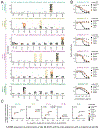
A Escape maps for each antibody. Line plots show site summed effects of all escape mutations at a site. The top 15 escape sites for each antibody are highlighted pink below the line plot and shown in logo plots where the height of each letter indicates escape caused by that mutation. Letters are colored by mutational effects on cell entry in the absence of antibody, with mutations that impair entry shown in lighter gray. Sites that contact the antibody (within 4 Å) are highlighted in yellow. The antibody escape maps are grouped by the epitope classification in Robinson et al. See https://github.com/dms-vep/LASV_Josiah_GP_DMS/tree/main/results/simplified_filtered_antibody_escape_CSVs for CSV files with all escape mutations for each antibody. B Surface representation of Lassa GPC (PDB: 7PUY) colored by summed site escape for each antibody. Blue indicates the site with the most escape from that antibody, and white indicates sites with no escape. See beginning of “Methods” for links to more detailed interactive versions of each escape map.
Figure 5.
Figure 5.. Escape mutations are usually in or near the antibody structural footprint
Surface representation of Fab-bound GPC colored by site escape as measured in deep mutational scanning, with the Fab shown in a colored cartoon representation. Because GPC is a homo-trimer, escape is colored only on sites in the monomer that is closest to the antibody shown for the structure. Blue indicates the GPC site with the most escape from that antibody, and white indicates sites with no escape. The Fab bound antibody structures shown here come from prior crystal and cryo-EM structures (25.10C PDB: 7TYV, 12.1F PDB: 7UOV, 37.7H PDB: 5VK2, 25.6A PDB: 6P95, 37.2D PDB: 7UOT, and 8.9F PDB: 7UOT). The antibodies are grouped by epitope as described in Robinson et al.
Figure 6.
Figure 6.. Some antibody escape mutations mapped by deep mutational scanning are found in natural Lassa virus sequences
A Frequency of mutations that escape antibody neutralization in natural Lassa GPC sequences. The escape for each mutation as measured in deep mutational scanning is plotted versus the mutation’s frequency in all 572 high-quality Lassa GPC sequences. Mutations chosen for validation with pseudovirus neutralization assays are labeled. B Number of natural GPC sequences with escape mutations stratified by host from which the sequences were collected. Mutations were classified as being escape mutations if they were in the top 2% of escape mutations for an antibody. Percentage of sequences with escape mutations relative to total number of sequences from a given host is indicated. C Summary of escape mutations and representative sequence containing each mutation that were chosen for validation experiments in Figures 7 and S7. For each mutation, the following are indicated: number of sequences containing that mutation, number of sequences containing any non-Josiah amino-acid identity at that site, and the natural sequence whose GPC contains the mutation that was chosen for testing. N89D is marked with an asterisk because the LM395 is the only sequence with a mutation at that site, and the deep sequencing data show site 89 is polymorphic between D and N (Table S3). All mutations shown in this figure are defined relative to the parental amino acid at that site in the Josiah strain GPC.
Figure 7.
Figure 7.. Validation that antibody escape mutations are found in natural GPC variants
A Deep mutational scanning antibody escape maps for top 10 escape sites that differ between the Josiah strain GPC and the indicated sequence’s GPC for each antibody. The height of the letter corresponds to the strength of escape, and amino acids representing mutations that are found in the indicated sequence’s GPC are colored orange (e.g., the third plot from top indicates H398K is found in the GA391 sequence). Sites that contact the antibody in structures of the GPC-Fab complex (within 4 Å) are highlighted yellow. The antibody escape maps are grouped by the antibody epitope classification of Robinson et al. B Validation pseudovirus neutralization assays for the indicated Josiah GPC mutants and natural sequence GPCs. Unmutated Josiah GPC is colored black, single mutant Josiah GPC is colored orange, and natural GPC sequence is colored gray. Error bars indicate the standard error for two technical replicates. C Correlation of escape predicted for natural GPC sequences by summing of the effects measured in deep mutational scanning for all mutations in that GPC versus the actual IC50 measured by pseudovirus neutralization assays. Squares indicate that the antibody did not neutralize at the highest concentration tested, and so the reported IC50 is a lower bound. *N89D is marked because the LM395 sequence with the N89D mutation is polymorphic at site 89 (Table S3) and is the only sequence with a mutation at site 89.

Update of

References

    1. Frame JD, Baldwin JM, Gocke DJ, and Troup JM (1970). Lassa Fever, a New Virus Disease of Man from West Africa I. Clinical Description and Pathological Findings. Am J Trop Med Hyg 19, 670–676. 10.4269/ajtmh.1970.19.670. - DOI - PubMed
    1. Leifer E, Gocke DJ, and Bourne H (1970). Lassa Fever, a New Virus Disease of Man from West Africa II. Report of a Laboratory-Acquired Infection Treated with Plasma from a Person Recently Recovered from the Disease. Am J Trop Med Hyg 19, 677–679. 10.4269/ajtmh.1970.19.677. - DOI - PubMed
    1. Buckley SM, and Casals J (1970). Lassa Fever, a New Virus Disease of Man from West Africa III. Isolation and Characterization of the Virus. Am J Trop Med Hyg 19, 680–691. 10.4269/ajtmh.1970.19.680. - DOI - PubMed
    1. Speir RW, Wood O, Liebhaber H, and Buckley SM (1970). Lassa Fever, a New Virus Disease of Man from West Africa IV. Electron Microscopy of Vero Cell Cultures Infected with Lassa Virus. Am J Trop Med Hyg 19, 692–694. 10.4269/ajtmh.1970.19.692. - DOI - PubMed
    1. McCormick JB, Webb PA, Krebs JW, Johnson KM, and Smith ES (1987). A Prospective Study of the Epidemiology and Ecology of Lassa Fever. Journal of Infectious Diseases 155, 437–444. 10.1093/infdis/155.3.437. - DOI - PubMed

Publication types

LinkOut - more resources