. 2025 Jan 16;96(2):140-149.
doi: 10.1136/jnnp-2024-333403.

Linking LRP12 CGG repeat expansion to inherited peripheral neuropathy

Affiliations

Linking LRP12 CGG repeat expansion to inherited peripheral neuropathy

Takahiro Hobara et al. J Neurol Neurosurg Psychiatry. .

Abstract

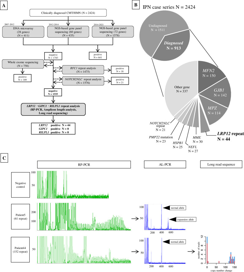
Background: The causative genes for over 60% of inherited peripheral neuropathy (IPN) remain unidentified. This study endeavours to enhance the genetic diagnostic rate in IPN cases by conducting screenings focused on non-coding repeat expansions.

Methods: We gathered data from 2424 unrelated Japanese patients diagnosed with IPN, among whom 1555 cases with unidentified genetic causes, as determined through comprehensive prescreening analyses, were selected for the study. Screening for CGG non-coding repeat expansions in LRP12, GIPC1 and RILPL1 genes was conducted using PCR and long-read sequencing technologies.

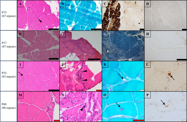
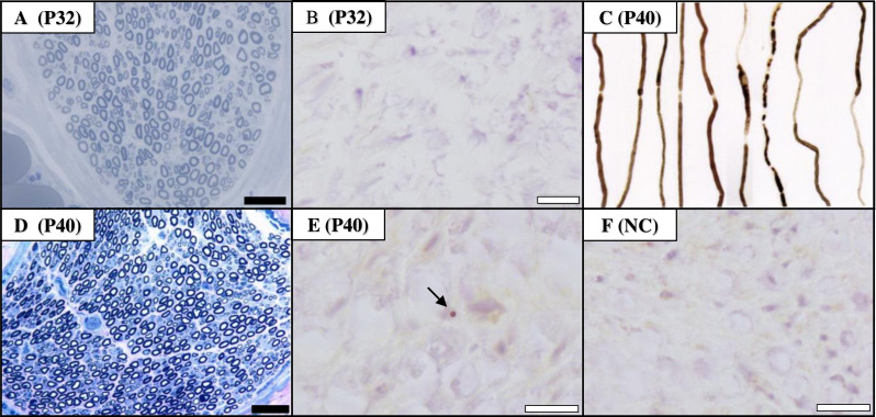
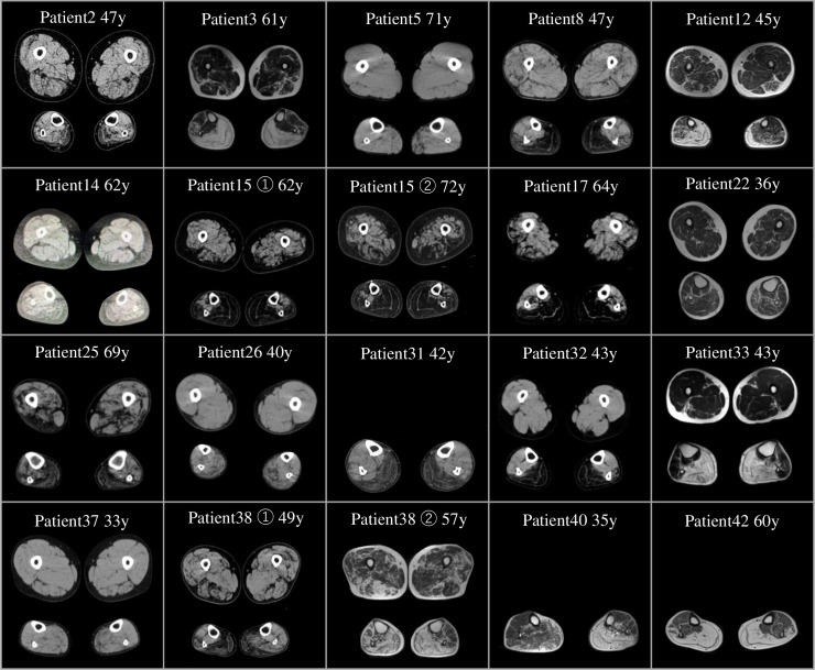
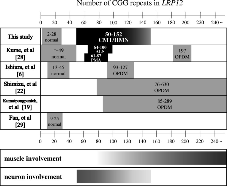
Results: We identified CGG repeat expansions in LRP12 from 44 cases, establishing it as the fourth most common aetiology in Japanese IPN. Most cases (29/37) exhibited distal limb weakness, without ptosis, ophthalmoplegia, facial muscle weakness or bulbar palsy. Neurogenic changes were frequently observed in both needle electromyography (97%) and skeletal muscle tissue (100%). In nerve conduction studies, 28 cases primarily showed impairment in motor nerves without concurrent involvement of sensory nerves, consistent with the phenotype of hereditary motor neuropathy. In seven cases, both motor and sensory nerves were affected, resembling the Charcot-Marie-Tooth (CMT) phenotype. Importantly, the mean CGG repeat number detected in the present patients was significantly shorter than that of patients with LRP12-oculopharyngodistal myopathy (p<0.0001). Additionally, GIPC1 and RILPL1 repeat expansions were absent in our IPN cases.

Conclusion: We initially elucidate LRP12 repeat expansions as a prevalent cause of CMT, highlighting the necessity for an adapted screening strategy in clinical practice, particularly when addressing patients with IPN.

Keywords: HMSN (CHARCOT-MARIE-TOOTH); NEUROGENETICS; NEUROMUSCULAR; NEUROPATHY.


Conflict of interest statement

Competing interests: None declared.

Figures

Figure 1
Figure 1. Flow chart and genetic findings in our Japanese case series of IPN. (A) Genetic analysis workflow conducted in 2424 patients with CMT/HMN (IPN case series). Following a series of prescreening studies, 1555 genetically unidentified cases were processed to CGG repeat screening in the genes of LRP12, GIPC1 and RILPL1 and repeat expansions were only detected in LRP12 from 44 cases. (B) LRP12 repeat expansion is the fourth most common causative reason in the present IPN case series in Japan, following MFN2, GJB1 and MPZ. (C) Representative results of RP-PCR, fluorescence AL-PCR and long read sequencing by GridION. AL-PCR, amplicon-length PCR; CMT, Charcot-Marie-Tooth disease; HMN, hereditary motor neuropathy; IPN, inherited peripheral neuropathy; NGS, next-generation sequencing; RP-PCR, repeat-primed PCR.
Figure 2
Figure 2. Representative pathological findings in skeletal muscle. H&E staining (A, E, F, I, J, M, N), ATP-ase (pH=10.5) (C), Gomori-Trichrome staining (B, K, O), NADH-TR staining (G), anti-p62 staining (D, H, L, P). (A–H) (Patients 15 and 17): fibre type grouping, small group atrophy and pyknotic nuclear clumps are observed, indicating neurogenic changes; rimmed vacuoles or p62-positive inclusions are not found. (I–P) (Patients 32 and 40): rimmed vacuoles, grouping atrophy, small angular fibres, pyknotic nuclear clumps and p62-positive deposition are observed. Black arrows indicate rimmed vacuole or p62-positive inclusions. Black triangles indicate pyknotic nuclear clumps. Black bar, 200 µm; red bar, 50 µm; white bar 20 µm.
Figure 3
Figure 3. Pathological findings of peripheral nerves from Patients 32 and 40. (A, D): Toluidine blue staining indicates that both large and small myelinated fibres are preserved. (C): Teased fibre analysis shows segmental and paranodal demyelination, as well as mild axonal impairment. (B, E): Anti-p62 staining shows deposition of p62 in Patient 40, but not in Patient 32. (F): Anti-p62 staining in sural nerve of a control sample. Black arrows indicate p62-positive inclusions. Black bar, 50 µm; white bar, 20 µm. NC, normal control.
Figure 4
Figure 4. CT or MRI images show lower limb muscle atrophy from 18 patients. Images of thighs (upper panel) and legs (lower panel). Muscle atrophy can be more prominently observed in the legs than in the thighs. In both regions, atrophy and fatty replacement are notably more pronounced on the posterior than anterior lower limbs. There is a tendency for more pronounced atrophy on the lateral sides compared with the medial sides in both regions. Atrophy of the posterior leg muscles, including the gastrocnemius and soleus muscles, is consistently noted across all cases. Follow-up imaging of patients 15 and 38 shows progressive atrophy over time. Patients 31, 40 and 42 did not undergo examination of their upper legs.
Figure 5
Figure 5. Diagram showing correlation between CGG repeat numbers in LRP12 and phenotypes in current and previous studies. Each square displays the disease phenotype along with the number of CGG repeats in LRP12. CGG repeat numbers have been found shorter in our patients (50–152), patients with LRP12-ALS (64–100) and LRP12-PMA (61–87), in contrast to the longer repeat range observed in patients with LRP12-OPDM (76–630). All reports consistently show that the count of CGG repeats within the LRP12 gene among control individuals does not exceed 50. CMT, Charcot-Marie-Tooth disease; HMN, hereditary motor neuropathy; OPDM, oculopharyngodistal myopathy; PMA, progressive muscular atrophy.

Similar articles

Cited by

References

    1. Higuchi Y, Takashima H. Clinical Genetics of Charcot–Marie–Tooth disease. J Hum Genet. 2023;68:199–214. doi: 10.1038/s10038-022-01031-2. - DOI - PubMed
    1. Pipis M, Rossor AM, Laura M, et al. Next-generation sequencing in Charcot-Marie-Tooth disease: opportunities and challenges. Nat Rev Neurol. 2019;15:644–56. doi: 10.1038/s41582-019-0254-5. - DOI - PubMed
    1. Murphy SM, Laura M, Fawcett K, et al. Charcot-Marie-Tooth disease: frequency of genetic subtypes and guidelines for genetic testing. J Neurol Neurosurg Psychiatry. 2012;83:706–10. doi: 10.1136/jnnp-2012-302451. - DOI - PMC - PubMed
    1. Renton AE, Majounie E, Waite A, et al. A Hexanucleotide repeat expansion in C9ORF72 is the cause of chromosome 9p21-linked ALS-FTD. Neuron. 2011;72:257–68. doi: 10.1016/j.neuron.2011.09.010. - DOI - PMC - PubMed
    1. Elden AC, Kim H-J, Hart MP, et al. Ataxin-2 intermediate-length polyglutamine expansions are associated with increased risk for ALS. Nature. 2010;466:1069–75. doi: 10.1038/nature09320. - DOI - PMC - PubMed

Substances

Supplementary concepts