. 2024 Jul 16;15(1):5984.
doi: 10.1038/s41467-024-50390-1.

Divergent evolution of male-determining loci on proto-Y chromosomes of the housefly

Affiliations

Divergent evolution of male-determining loci on proto-Y chromosomes of the housefly

Xuan Li et al. Nat Commun. .

Abstract

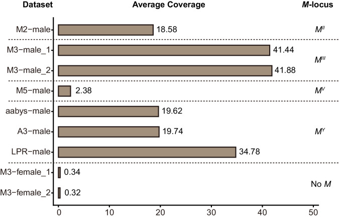
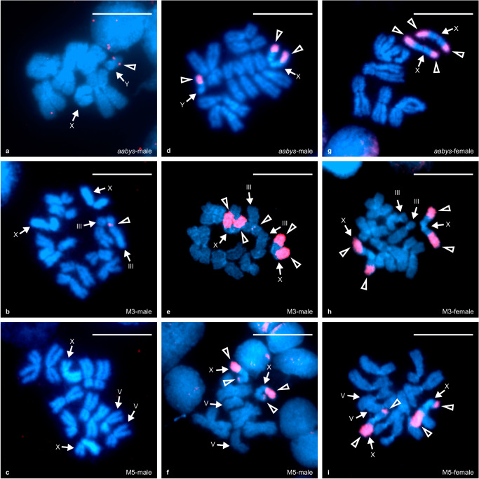
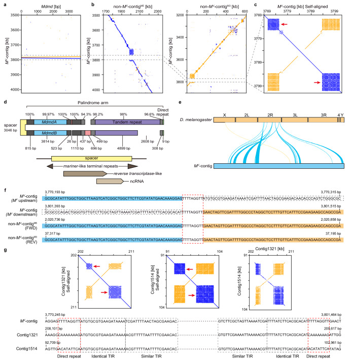
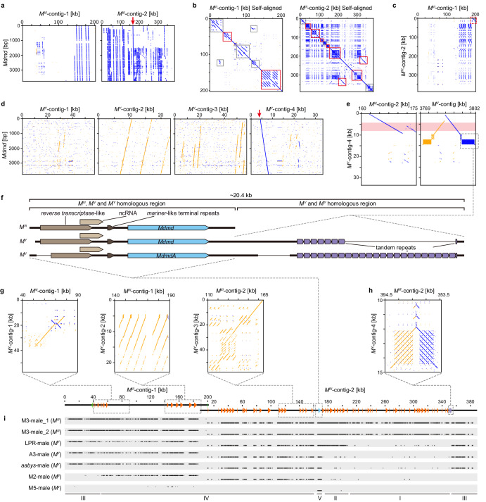
Houseflies provide a good experimental model to study the initial evolutionary stages of a primary sex-determining locus because they possess different recently evolved proto-Y chromosomes that contain male-determining loci (M) with the same male-determining gene, Mdmd. We investigate M-loci genomically and cytogenetically revealing distinct molecular architectures among M-loci. M on chromosome V (MV) has two intact Mdmd copies in a palindrome. M on chromosome III (MIII) has tandem duplications containing 88 Mdmd copies (only one intact) and various repeats, including repeats that are XY-prevalent. M on chromosome II (MII) and the Y (MY) share MIII-like architecture, but with fewer repeats. MY additionally shares MV-specific sequence arrangements. Based on these data and karyograms using two probes, one derives from MIII and one Mdmd-specific, we infer evolutionary histories of polymorphic M-loci, which have arisen from unique translocations of Mdmd, embedded in larger DNA fragments, and diverged independently into regions of varying complexity.


Conflict of interest statement

The authors declare no competing interests.

Figures

Fig. 1
Fig. 1. The average coverages of Mdmd gene in different datasets.
Coverage rates in female genomes are included to account for off-target mapping to the paralogous gene Mdncm and the calculated average coverages in two M3 female Illumina datasets turned out to be negligible. Average coverages demonstrate that the number of Mdmd sequences are highest in MIII, intermediate in MY and MII, and lowest in MV. Source data are provided as a Source Data file.
Fig. 2
Fig. 2. FISH localization of M-loci and sex chromosome-associated repeat regions.
a, b Using an Mdmd-specific probe, MY and MIII were localized to pericentromeric regions of the Y chromosome and chromosome III respectively. c MV was not detected by the Mdmd probe due to insufficient gene copy numbers. di Using a probe containing a mixture of amplified MIII sequences including Mdmd and non-Mdmd intervening sequences, the M-locus and the M and sex chromosome-located (MAS) regions of the XY chromosome pair were localized. The signals of the mixed probe on the Y chromosome cover most parts of the short arm and merge with the MY signal. The signal of the mixed probe on the X chromosome mark at the ends of both arms. Positive signals are shown in red and indicated by open triangles, chromosomes are indicated by arrows. Metaphase chromosomes are shown in blue. Signals were only considered as a successful hybridization if they were observed with consistent chromosomal locations on at least 20 metaphase nuclei on each slide. For each strain, 2–3 individuals were tested to ensure reproducibility. Scale bar: 10 μm.
Fig. 3
Fig. 3. The genomic structure and transposon-like signatures of MV.
a Dotplot visualizations of Mdmd presence in inverse orientation on MV-contig indicated by blue (forward) and orange (reverse) lines that represent longer stretches of sequence similarity. b Alignments between MV-contig and non-MV-contigs show MV was inserted in a tandem repetitive region, indicated by the gap. c Self-alignment of MV demonstrates that the main part of MV is a palindrome with a non-palindromic spacer sequence of 3046 bp in the middle. Two blocks of tandem repeats exist on each end of MV (indicated by red arrows). d Schematic drawing indicates the sequence contents of MV and percentage identities between palindromic arms. The spacer sequence shows homology to a reverse transcriptase sequence and an ncRNA. The red blocks and the missing parts represent insertions/deletions. The small green blocks indicate the existence of 9 bp direct repeats at the MV borders. e Single-copy BUSCOs in the MV-contig mainly correspond to those on chromosome 2 R (Muller element C, chromosome V in the housefly) in Drosophila melanogaster. f The 9-bp direct repeats (TTTTAGGTT) are found with one copy in non-MV-contigs at insertion sites, indicated by the red dashed-line box. In non-MV-contigs, the upstream and downstream sequences of TTTTAGGTT match with the upstream and downstream sequence of MV (indicated by the blue and orange shading). g Two examples of other genomic regions that contain the same tandem repeat blocks as MV. The self-alignment figures demonstrate MV-like palindromic structures in Contig1514 and Contig1321. Alignment between Contig1514 and Contig1321 shows sequence similarity only for the palindromic region but not for the palindrome flanking sequences. The TIRs in MV are also found in Contig1514 and Contig1321 palindromes, and direct repeats with the same 9-bp length are flanking them.
Fig. 4
Fig. 4. Genomic structure of MIII, MY and in comparison to MII, MV loci.
a Visualization of Mdmd sequences distribution in MIII-contigs. Only one complete Mdmd copy is found (blue line indicated by the red arrow). b Self-alignments of MIII-contigs show many tandem duplications clustering together (short blue lines, indicated by solid-lined boxes). Most of the duplications are Mdmd copies and their flanking sequences (red solid-lined boxes). Duplications of non-Mdmd sequences also exist (indicated by gray solid-lined boxes). c The end part of MIII-contig-1 shares homology to the beginning part of MIII-contig-2 (indicated by the red dashed-lined box). d Visualization of Mdmd sequences distribution in MY-contigs. Only one complete Mdmd copy is found in MY-contig-4 (blue line indicated by the red arrow). e MY-contig-4 show homology to regions of MIII- and MV-contigs that contain intact Mdmd gene which is indicated by red shading. f Schematic drawing of homologous regions that contain intact Mdmd in MIII, MV and MY. MV and MY both have a block of tandem repeats is located downstream (~4 kb apart) of the intact Mdmd. g MY-contigs show homology to various parts of MIII-contigs. h Tandem repeats that are adjacent to intact Mdmd in MV and MY are also found in MIII, but exist near one end of MIII-contig-2. i The coverage for MIII-specific regions in various male genomic datasets that contain sequences of MII, MIII, MV, and MY loci respectively. Schematic drawing shows Mdmd distribution (complete copy blue, truncated copies orange) in MIII-contigs. Other M-loci show various similarities to the MIII. I: Specific to MIII; II: Shared between MIII and LPR MY; III: Shared among MIII and all three MY; IV: Shared among MII, MIII and all MY; V: Shared among all tested M-loci.

References

    1. Wilkins AS. Moving up the hierarchy: a hypothesis on the evolution of a genetic sex determination pathway. Bioessays. 1995;17:71–77. doi: 10.1002/bies.950170113. - DOI - PubMed
    1. Saccone G. A history of the genetic and molecular identification of genes and their functions controlling insect sex determination. Insect Biochem. Mol. Biol. 2022;151:103873. doi: 10.1016/j.ibmb.2022.103873. - DOI - PubMed
    1. Steinemann M, Steinemann S. Enigma of Y chromosome degeneration: neo-Y and neo-X chromosomes of Drosophila miranda a model for sex chromosome evolution. Genetica. 1998;102:409. doi: 10.1023/A:1017058119760. - DOI - PubMed
    1. Charlesworth B, Charlesworth D. The degeneration of Y chromosomes. Philos. Trans. R. Soc. Lond. B Biol. Sci. 2000;355:1563–1572. doi: 10.1098/rstb.2000.0717. - DOI - PMC - PubMed
    1. Carvalho AB, Koerich LB, Clark AG. Origin and evolution of Y chromosomes: drosophila tales. Trends Genet. 2009;25:270–277. doi: 10.1016/j.tig.2009.04.002. - DOI - PMC - PubMed

LinkOut - more resources