. 2024 Jul 16;25(1):282.
doi: 10.1186/s12931-024-02907-x.

Altered hypoxia-induced cellular responses and inflammatory profile in lung fibroblasts from COPD patients compared to control subjects

Affiliations

Altered hypoxia-induced cellular responses and inflammatory profile in lung fibroblasts from COPD patients compared to control subjects

Ryde Martin et al. Respir Res. .

Abstract

Background: Chronic obstructive pulmonary disease (COPD) is a heterogeneous disease characterized by chronic bronchitis, emphysema and vascular remodelling. The disease is associated with hypoxia, inflammation and oxidative stress. Lung fibroblasts are important cells in remodelling processes in COPD, as main producers of extracellular matrix proteins but also in synthesis of growth factors and inflammatory mediators.

Methods: In this study we aimed to investigate if there are differences in how primary distal lung fibroblasts obtained from COPD patients and healthy subjects respond to hypoxia (1% O2) and pro-fibrotic stimuli with TGF-β1 (10 ng/mL). Genes and proteins associated with oxidative stress, endoplasmic reticulum stress, remodelling and inflammation were analysed with RT-qPCR and ELISA.

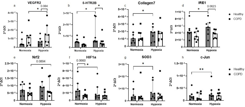
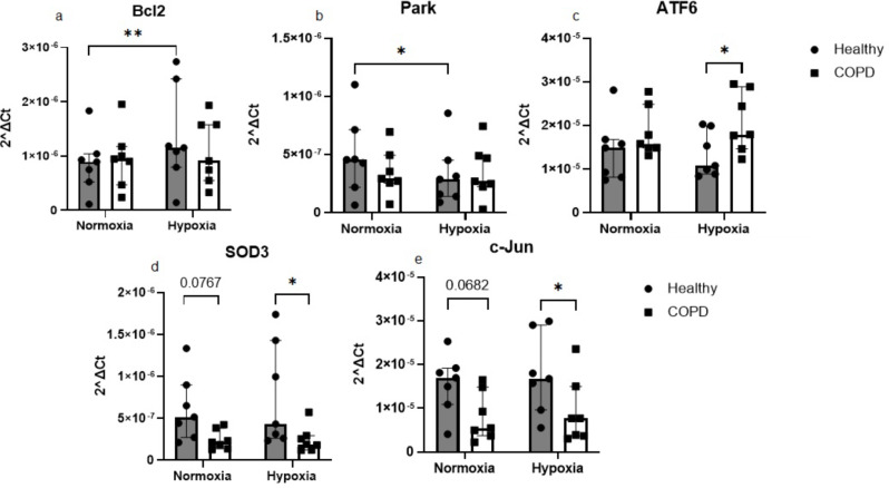
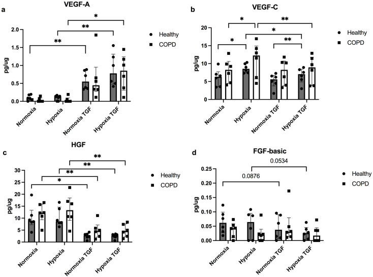
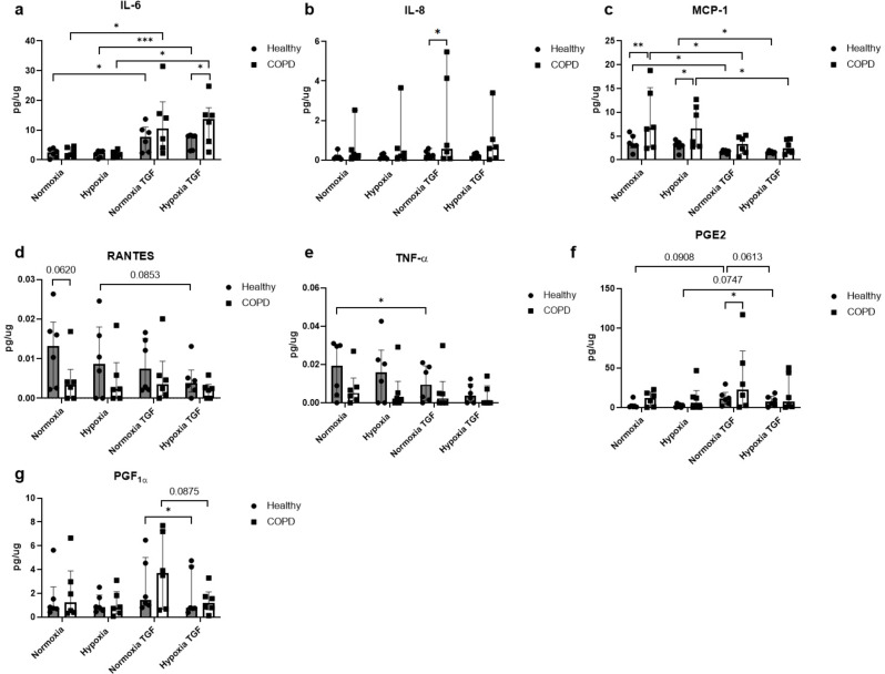
Results: Hypoxia induced differences in expression of genes involved in oxidative stress (SOD3 and HIF-1α), ER stress (IRE1, PARK and ATF6), apoptosis (c-Jun and Bcl2) and remodelling (5HTR2B, Collagen7 and VEGFR2) in lung fibroblasts from COPD subjects compared to control subjects, where COPD fibroblasts were in general less responsive. The release of VEGF-C was increased after hypoxia, whereas TGF-β significantly reduced the VEGF response to hypoxia and the release of HGF. COPD fibroblasts had a higher release of IL-6, IL-8, MCP-1 and PGE2 compared to lung fibroblasts from control subjects. The release of inflammatory mediators was less affected by hypoxia, whereas TGFβ1 induced differences in inflammatory profile between fibroblasts from COPD and control subjects.

Conclusion: These results suggest that there is an alteration of gene regulation of various stress responses and remodelling associated mediator release that is related to COPD and hypoxia, where fibroblasts from COPD patients have a deficient response.

Keywords: COPD; ER stress; Gene expression; Hypoxia; Inflammation; Lung fibroblasts; Oxidative stress; Remodelling.


Conflict of interest statement

The authors declare no competing interests.

Figures

Fig. 1
Fig. 1
Effect of 4 h of hypoxia exposure on gene expression levels. Genes related to remodelling: VEGFR2 (a), 5-HTR2B (b) and Collagen7 (c); cellular stress: IRE1 (d) and c-Jun (h), oxidative stress: Nrf2 (e), HIF-1α (f) and SOD3 (g) were measured by RT-qPCR in primary distal lung fibroblasts obtained from healthy subjects (n = 7) and COPD patients (n = 7) after 4 h of exposure to normoxic (21% O2) or hypoxic (1% O2) conditions. Beta-actin and 18 S were used as housekeeping genes. The data is presented as median with interquartile range. Ordinary two-way ANOVA or RM two-way ANOVA were used for unpaired and paired comparisons and the post-hoc test Fisher’s LSD was used for statistical analysis *p < 0.05, **p < 0.01
Fig. 2
Fig. 2
Effects of 24 h of hypoxia on gene expression levels. Genes related to cell death: Bcl2 (a), metabolic or mitochondrial stress responses: Park (b), cellular stress: IRE1 (c) and c-Jun (e) and oxidative stress: SOD3 (d) were measured by RT-qPCR in primary distal lung fibroblasts obtained from healthy subjects (n = 7) and COPD patients (n = 7) after 24 h of exposure to normoxic (21% O2) or hypoxic (1% O2) conditions. Beta-actin and 18 S were used as housekeeping genes. The data is presented as median with interquartile range. Ordinary two-way ANOVA or RM two-way ANOVA were used for unpaired and paired comparisons and the post-hoc test Fisher’s LSD was used for statistical analysis. *p < 0.05, **p < 0.01
Fig. 3
Fig. 3
Effects of hypoxia and profibrotic stimuli on mediators linked to remodelling. Release of the following growth factors vascular endothelial growth factors (VEGF)-A (a), VEGF-C (b), hepatocyte growth factor (HGF), (c) and fibroblast growth factor (FGF)-basic (d) from lung fibroblasts from healthy subjects (n = 6) and COPD patients (n = 6) at normoxic (21% O2) and hypoxic (1% O2) conditions with and without stimuli with transforming growth factor TGF-β1 (10 ng/mL). Total protein levels were used to normalize each sample. The data is presented as median with interquartile range. Ordinary two-way ANOVA or RM two-way ANOVA were used for unpaired and paired comparisons and the post-hoc test Fisher’s LSD was used for statistical analysis. *p < 0.05, **p < 0.01
Fig. 4
Fig. 4
Effects of hypoxia and profibrotic stimuli on inflammatory mediators. Release of inflammatory mediator interleukin (IL)-6 (a), IL-8 (b), monocyte chemoattractant protein (MCP)-1 (c), Regulated upon Activation, Normal T Cell Expressed and Secreted (RANTES) (d), Tumor necrosis factor (TNF)-α (e), prostaglandin (PG) E2 (f) and PGF1α (g) from lung fibroblasts from healthy subjects (n = 6) and COPD patients (n = 6) at normoxic (21% O2) and hypoxic (1% O2) conditions with and without stimuli with transforming growth factor TGF-β1 (10 ng/mL). Total protein levels were used to normalize each sample. The data is presented as mean with min to max. Ordinary two-way ANOVA or RM two-way ANOVA were used for unpaired and paired comparisons and the post-hoc test Fisher’s LSD was used for statistical analysis. *p < 0.05, **:p < 0.01, ***:p < 0.001
Fig. 5
Fig. 5
Immunocytochemistry staining and quantification of HIF-2α. Immunofluorescent staining of HIF-2α in distal lung fibroblasts from healthy (a and b) and COPD (c and d) subjects at normoxic (a and c) and hypoxic (b and d) conditions. The blue stain indicates DAPI and the red stain indicates HIF-2α. Scale bar indicates 200 μm. Quantification of the % positive cells (e). Repeated measures ANOVA was used for paired samples and 2-way ANOVA was used for unpaired samples. **p < 0.01, ***p < 0.001

References

    1. Yang IA, Jenkins CR, Salvi SS. Chronic obstructive pulmonary disease in never-smokers: risk factors, pathogenesis, and implications for prevention and treatment. Lancet Respir Med. 2022;10(5):497–511. doi: 10.1016/S2213-2600(21)00506-3. - DOI - PubMed
    1. Barnes PJ. Oxidative stress-based therapeutics in COPD. Redox Biol. 2020;33:101544. doi: 10.1016/j.redox.2020.101544. - DOI - PMC - PubMed
    1. Fu X, Zhang F. Role of the HIF-1 signaling pathway in chronic obstructive pulmonary disease. Exp Ther Med. 2018;16(6):4553–61. - PMC - PubMed
    1. Harkness LM, Kanabar V, Sharma HS, Westergren-Thorsson G, Larsson-Callerfelt AK. Pulmonary vascular changes in asthma and COPD. Pulm Pharmacol Ther. 2014;29(2):144–55. doi: 10.1016/j.pupt.2014.09.003. - DOI - PubMed
    1. Eapen MS, Hansbro PM, Larsson-Callerfelt AK, Jolly MK, Myers S, Sharma P, et al. Chronic obstructive Pulmonary Disease and Lung Cancer: underlying pathophysiology and New Therapeutic modalities. Drugs. 2018;78(16):1717–40. doi: 10.1007/s40265-018-1001-8. - DOI - PubMed

MeSH terms

Substances

LinkOut - more resources