. 2024 Jul 16;16(1):91.
doi: 10.1186/s13148-024-01709-8.

H3K27ac acts as a molecular switch for doxorubicin-induced activation of cardiotoxic genes

Affiliations

H3K27ac acts as a molecular switch for doxorubicin-induced activation of cardiotoxic genes

Yu Hong et al. Clin Epigenetics. .

Abstract

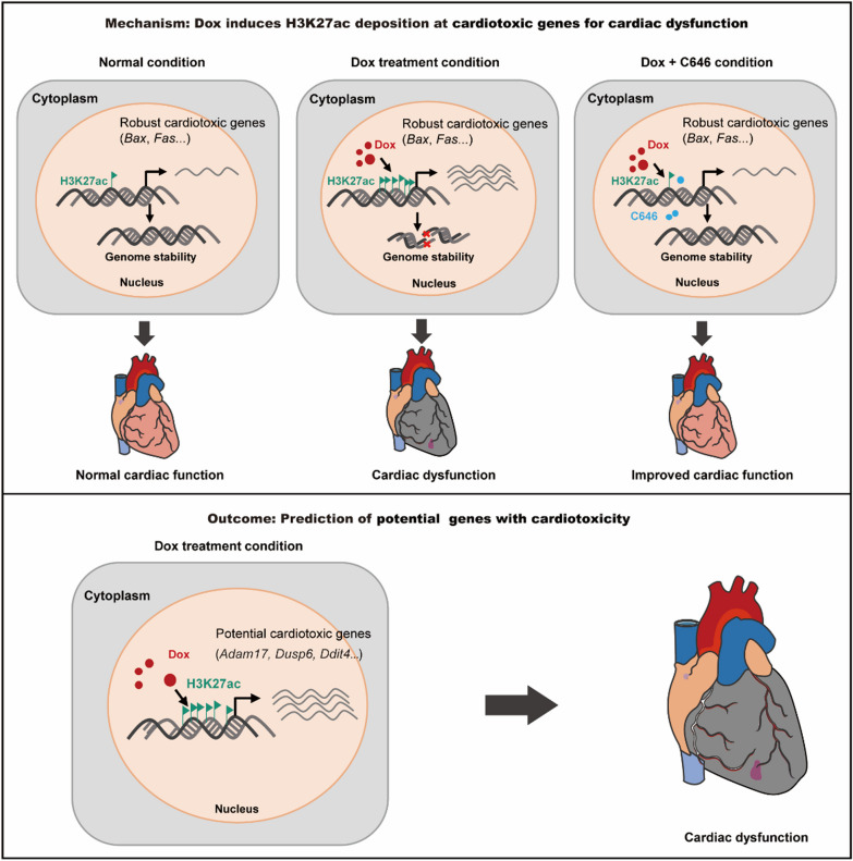
Background: Doxorubicin (Dox) is an effective chemotherapeutic drug for various cancers, but its clinical application is limited by severe cardiotoxicity. Dox treatment can transcriptionally activate multiple cardiotoxicity-associated genes in cardiomyocytes, the mechanisms underlying this global gene activation remain poorly understood.

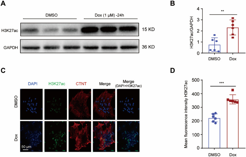
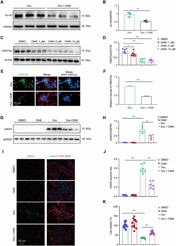
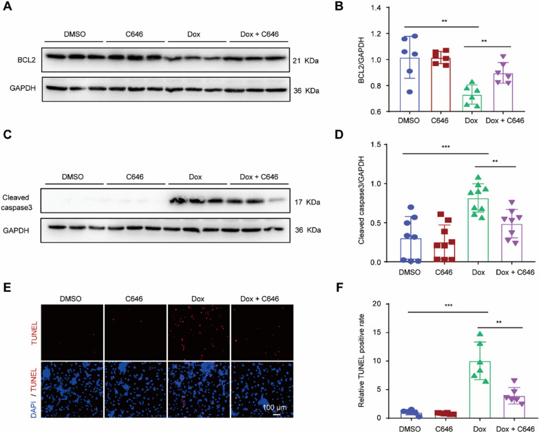
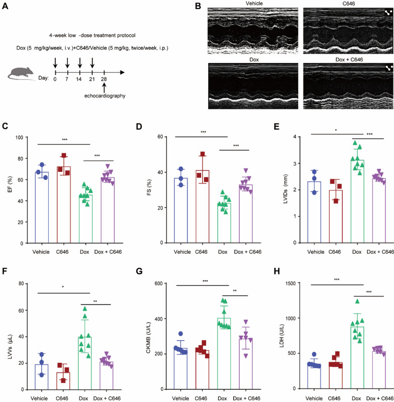
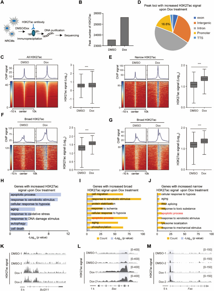
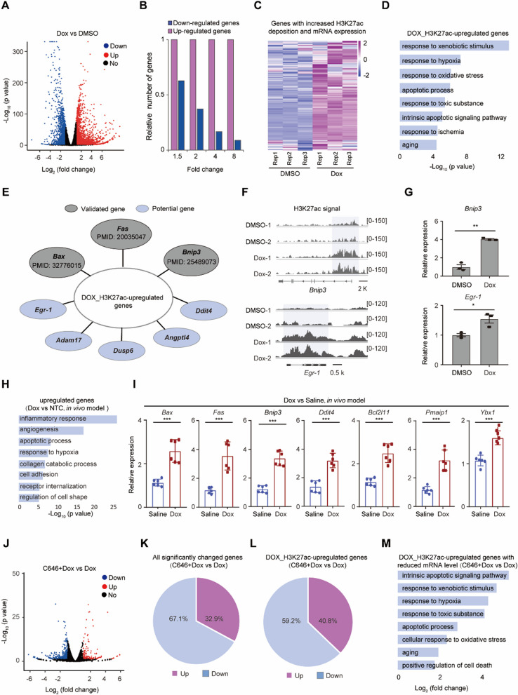
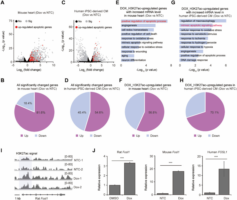
Methods and results: Herein, we integrated data from animal models, CUT&Tag and RNA-seq after Dox treatment, and discovered that the level of H3K27ac (a histone modification associated with gene activation) significantly increased in cardiomyocytes following Dox treatment. C646, an inhibitor of histone acetyltransferase, reversed Dox-induced H3K27ac accumulation in cardiomyocytes, which subsequently prevented the increase of Dox-induced DNA damage and apoptosis. Furthermore, C646 alleviated cardiac dysfunction in Dox-treated mice by restoring ejection fraction and reversing fractional shortening percentages. Additionally, Dox treatment increased H3K27ac deposition at the promoters of multiple cardiotoxic genes including Bax, Fas and Bnip3, resulting in their up-regulation. Moreover, the deposition of H3K27ac at cardiotoxicity-related genes exhibited a broad feature across the genome. Based on the deposition of H3K27ac and mRNA expression levels, several potential genes that might contribute to Dox-induced cardiotoxicity were predicted. Finally, the up-regulation of H3K27ac-regulated cardiotoxic genes upon Dox treatment is conservative across species.

Conclusions: Taken together, Dox-induced epigenetic modification, specifically H3K27ac, acts as a molecular switch for the activation of robust cardiotoxicity-related genes, leading to cardiomyocyte death and cardiac dysfunction. These findings provide new insights into the relationship between Dox-induced cardiotoxicity and epigenetic regulation, and identify H3K27ac as a potential target for the prevention and treatment of Dox-induced cardiotoxicity.

Keywords: Broad H3K27ac; DNA damage; Dox-induced cardiotoxicity; Histone modification.


Conflict of interest statement

The authors declare that they have no competing interests.

Figures

Fig. 1
Fig. 1
Dox treatment led to H3K27ac accumulation in cardiomyocytes. A, B Representative western blot (A) and statistical data (B) showing the levels of H3K27ac from the cardiomyocytes treated with Dox for 24 h, n = 6 per group. C, D Representative immunostaining image (C) and mean fluorescence intensity (D) for H3K27ac (green) in the cardiomyocytes treated with Dox for 24 h; CTNT denotes cardiac troponin T (red); Nuclei were stained with DAPI (blue)
Fig. 2
Fig. 2
Reducing H3K27ac accumulation via C646 ameliorated Dox-induced DNA damage in cardiomyocytes. A, B Representative western blot (A) and statistical data (B) showing the levels of Ac-H3 from the cardiomyocytes treated with Dox or Dox with C646 for 24 h. n = 3 per group. C, D Representative western blot (C) and statistical data (D) showing the levels of H3K27ac from the cardiomyocytes treated with increasing concentrations of C646 ranging from 0 to 10 μM for 24 h. n = 6 per group. E, F Representative immunostaining image (E) and relative fluorescence intensity (F) for H3K27ac (green) in the cardiomyocytes treated with Dox or Dox with C646 for 24 h; red field denotes cardiac troponin T; Nuclei were stained with DAPI (blue). G, H Representative western blot (G) and statistical data (H) showing the levels of γH2AX from Dox-treated cardiomyocytes with or without C646 for 24 h. n = 6 per group. I, J Representative immunostaining image and analysis for γH2AX (green) in Dox-treated cardiomyocytes with or without C646 for 24 h. K CCK-8 assay revealed cell viability after Dox treatment with or without C646, respectively
Fig. 3
Fig. 3
Decreased H3K27ac accumulation attenuated Dox-induced apoptosis in cardiomyocytes. A, B Representative western blot (A) and statistical data (B) showing the levels of BCL2 from Dox-treated cardiomyocytes with or without C646 for 24 h. n = 6 per group. C, D Representative western blot (C) and statistical data (D) showing the levels of cleaved caspase3 from Dox-treated cardiomyocytes with or without C646 for 24 h. n values are 8, 9, 9, 8, respectively. E, F Representative images (E) and statistical data (F) of TUNEL-positive cells (red) from Dox-treated cardiomyocytes with or without C646 for 24 h. n = 6 per group
Fig. 4
Fig. 4
H3K27ac suppression protected the hearts from the functional impairment caused by Dox in mice. A Experimental overview of present animal experiment, “i.v.” denotes Intravenous injection, “i.p.” denotes Intraperitoneal injection. B Representative echocardiographic images of Dox-treated mice heart with or without C646 for 4 weeks. C, D Ejection fraction (C) and fractional shortening (D) data of Dox-treated mice heart with or without C646 for 4 weeks. E, F Left ventricular internal diameter at end systole (E) and left ventricular volume at end systole of Dox-treated mice heart with or without C646 for 4 weeks. G, H CKMB (creatine kinase MB) (G) and LDH (lactic dehydrogenase) (H) activity from serum of mice, which were treated with Dox combined with or without C646 for 4 weeks
Fig. 5
Fig. 5
Dox increased broad H3K27ac deposition at the promoters of cardiotoxicity-related genes. A Overview of H3K27ac CUT&Tag experiment. B Peak number of H3K27ac in DMSO and Dox group. C Heatmap and quantification showing the global H3K27ac signal in DMSO and Dox group, two-tailed Wilcoxon test. D The distribution of signal-increased H3K27ac peak upon Dox treatment. E Heatmap and quantification showing the increased ChIP signal at narrow H3K27ac peak loci in DMSO and Dox group, two-tailed Wilcoxon test. F, G Heatmap and quantification showing the increased ChIP signal at broad H3K27ac peak loci in DMSO and Dox group, two-tailed Wilcoxon test. H Gene ontology analysis showing the biological processes for all the genes with increased H3K27ac signal upon Dox treatment. I, J Gene ontology analysis showing the biological processes for the genes with increased broad (I) and narrow H3K27ac (J) signal upon Dox treatment, and the apoptotic process was highlighted. KM The representative regions showing the H3K27ac signal around Bcl2l11, Bax and Fas genes between the DMSO and Dox group with two replicates
Fig. 6
Fig. 6
Dox treatment upregulated cardiotoxicity-related genes. A Volcano plot showing the expression alteration of genes in rat cardiomyocytes upon Dox treatment. B Histogram showing the number of down-regulated and up-regulated genes with different fold change in cardiomyocytes upon Dox treatment. C Heatmap showing the genes with both increased H3K27ac signal and increased mRNA expression in rat cardiomyocytes upon Dox treatment. D Gene ontology analysis showing the biological processes for the genes with both increased H3K27ac signal and increased mRNA expression in rat cardiomyocytes upon Dox treatment. E The representative genes with both increased H3K27ac signal and increased mRNA expression in cardiomyocytes upon Dox treatment, several genes are reported to be associated with Dox-induced cardiotoxicity including Fas, Bax and Bnip3, other genes are potentially associated with cardiotoxicity. F, G H3K27ac signal nearby Bnip3 and Egr-1 (F) and their mRNA expression (G) in cardiomyocytes upon Dox treatment with two replicates. H Gene ontology analysis showing the biological processes for the upregulated genes in cardiomyocytes from an in vivo model upon Dox treatment (GSE223698). I Expression of apoptotic and cell death associated genes in cells from mouse heart upon Dox treatment for 24 h. J Compared to the Dox group, volcano plot showing the expression alteration of genes in cardiomyocytes upon Dox + C646 treatment. K Compared to the Dox group, the percentages of down-regulated and up-regulated genes after Dox + C646 treatment. L Compared to the Dox group, the percentage of down-regulated and up-regulated genes among Dox_H3K27ac-upregulated genes after Dox + C646 treatment. M Gene ontology analysis showing the biological processes for the Dox_H3K27ac-upregulated genes with decreased mRNA expression in cardiomyocytes upon Dox + C646 treatment
Fig. 7
Fig. 7
H3K27ac-regulated cardiotoxic genes were conservatively up-regulated upon Dox treatment across species. A, B Volcano plot showing the expression alteration of genes (A) and their percentages (B) in mouse heart (GSE226116) upon Dox treatment; black points denote the genes without significant change (No), gray points denote the genes with significant change (Sig), red points denote the apoptotic genes with significant up-regulation. C, D Volcano plot showing the expression alteration of genes (C) and their percentages (D) in human iPSC-derived cardiomyocytes (GSE230638) upon Dox treatment. CM denotes cardiomyocytes; black points denote the genes without significant change (No), gray points denote the genes with significant change (Sig), red points denote the apoptotic genes with significant up-regulation. E Gene ontology analysis showing the biological processes for the Dox_H3K27ac-upregulated genes with increased mRNA expression in mouse heart upon Dox treatment. F The percentage of down-regulated and up-regulated genes among Dox_H3K27ac-upregulated genes in mouse heart after Dox treatment. G Gene ontology analysis showing the biological processes for the Dox_H3K27ac-upregulated genes with increased mRNA expression in human iPSC-derived cardiomyocytes. H The percentage of down-regulated and up-regulated genes among Dox_H3K27ac-upregulated genes in human iPSC-derived cardiomyocytes after Dox treatment. I H3K27ac signal nearby Fosl1 in rat cardiomyocytes upon Dox treatment with two replicates. J The expression alteration of Fosl1 in rat cardiomyocytes, mouse heart and human iPSC-derived cardiomyocytes after Dox treatment
Fig. 8
Fig. 8
Model for the activation of robust cardiotoxic genes during doxorubicin-induced cardiotoxicity. Dox treatment increases the deposition of H3K27ac at promoters of robust cardiotoxic genes, and activates their transcription in cardiomyocytes. These results further promote DNA damage in the nucleus, and excessive genome instability leads to heart failure. C646 treatment reduces the expression of doxorubicin-induced cardiotoxic genes, and improves the cardiac function

Similar articles

Cited by

References

    1. Chabner BA, Roberts TG. Chemotherapy and the war on cancer. Nat Rev Cancer. 2005;5(1):65–72. doi: 10.1038/nrc1529. - DOI - PubMed
    1. Wu WC, Pu YY, Shi JL. Nanomedicine-enabled chemotherapy-based synergetic cancer treatments. J Nanobiotechnol. 2022;20(1):4. doi: 10.1186/s12951-021-01181-z. - DOI - PMC - PubMed
    1. Yeasmin S, Azharuddin M. Effect of exercise on gait and postural control in patients with chemotherapy induced peripheral neuropathy: a systematic review. Sport Sci Health. 2024. 10.1007/s11332-024-01168-x
    1. Burstein HJ, Mangu PB, Somerfield MR, Schrag D, Samson D, Holt L, Zelman D, Ajani JA. American society of clinical oncology clinical practice guideline update on the use of chemotherapy sensitivity and resistance assays. J Clin Oncol. 2011;29(24):3328–3330. doi: 10.1200/JCO.2011.36.0354. - DOI - PubMed
    1. Rivankar S. An overview of doxorubicin formulations in cancer therapy. J Cancer Res Ther. 2014;10(4):853–858. doi: 10.4103/0973-1482.139267. - DOI - PubMed

LinkOut - more resources