

Impaired glymphatic system in genetic frontotemporal dementia: a GENFI study
- PMID: 39015769
- PMCID: PMC11249959
- DOI: 10.1093/braincomms/fcae185
Impaired glymphatic system in genetic frontotemporal dementia: a GENFI study
Abstract
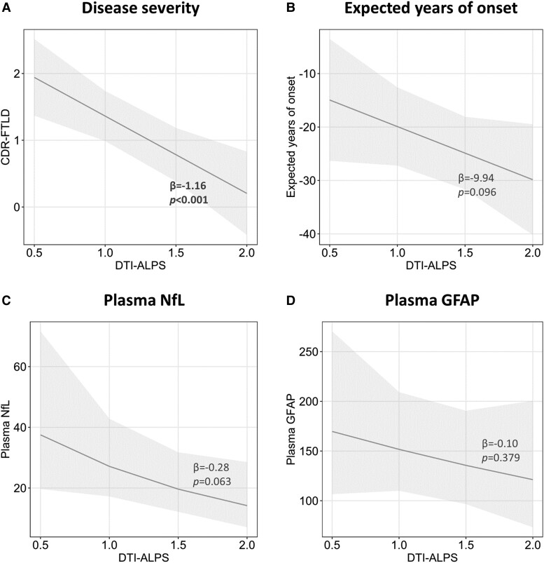
The glymphatic system is an emerging target in neurodegenerative disorders. Here, we investigated the activity of the glymphatic system in genetic frontotemporal dementia with a diffusion-based technique called diffusion tensor image analysis along the perivascular space. We investigated 291 subjects with symptomatic or presymptomatic frontotemporal dementia (112 with chromosome 9 open reading frame 72 [C9orf72] expansion, 119 with granulin [GRN] mutations and 60 with microtubule-associated protein tau [MAPT] mutations) and 83 non-carriers (including 50 young and 33 old non-carriers). We computed the diffusion tensor image analysis along the perivascular space index by calculating diffusivities in the x-, y- and z-axes of the plane of the lateral ventricle body. Clinical stage and blood-based markers were considered. A subset of 180 participants underwent cognitive follow-ups for a total of 640 evaluations. The diffusion tensor image analysis along the perivascular space index was lower in symptomatic frontotemporal dementia (estimated marginal mean ± standard error, 1.21 ± 0.02) than in old non-carriers (1.29 ± 0.03, P = 0.009) and presymptomatic mutation carriers (1.30 ± 0.01, P < 0.001). In mutation carriers, lower diffusion tensor image analysis along the perivascular space was associated with worse disease severity (β = -1.16, P < 0.001), and a trend towards a significant association between lower diffusion tensor image analysis along the perivascular space and higher plasma neurofilament light chain was reported (β = -0.28, P = 0.063). Analysis of longitudinal data demonstrated that worsening of disease severity was faster in patients with low diffusion tensor image analysis along the perivascular space at baseline than in those with average (P = 0.009) or high (P = 0.006) diffusion tensor image analysis along the perivascular space index. Using a non-invasive imaging approach as a proxy for glymphatic system function, we demonstrated glymphatic system abnormalities in the symptomatic stages of genetic frontotemporal dementia. Such measures of the glymphatic system may elucidate pathophysiological processes in human frontotemporal dementia and facilitate early phase trials of genetic frontotemporal dementia.
Keywords: DTI-ALPS; frontotemporal dementia; frontotemporal lobar degeneration; genetic; glymphatic system.
© The Author(s) 2024. Published by Oxford University Press on behalf of the Guarantors of Brain.
Conflict of interest statement
The authors have no competing interests.
Figures


References
-
- Neumann M, Mackenzie IRA. Review: Neuropathology of non-tau frontotemporal lobar degeneration. Neuropathol Appl Neurobiol. 2019;45:19–40. - PubMed
LinkOut - more resources
Full Text Sources
Miscellaneous