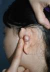
Ear keloid and epidermal cyst following auricular cartilage harvest for rhinoplasty: A case report
- PMID: 39015904
- PMCID: PMC11235563
- DOI: 10.12998/wjcc.v12.i20.4434
Ear keloid and epidermal cyst following auricular cartilage harvest for rhinoplasty: A case report
Abstract
Background: This case report highlights a rare instance of concurrent keloid and epidermal cyst development at an ear cartilage harvest site following rhinoplasty in a 25-year-old woman. Both conditions, which typically stem from skin trauma, seldom occur together, demonstrating the exceptional characteristics of this case.
Case summary: The patient underwent successful surgical removal of both the keloid and the epidermal cyst. Postoperative treatment included the use of silicone sheets, gel, and oral tranilast to reduce scarring. No recurrence was observed over a 6-mo follow-up period, indicating effective management of the condition.
Conclusion: The effective management of complex skin trauma cases underscores the need for individualized treatment strategies in plastic surgery.
Keywords: Auricular cartilage harvesting; Case report; Ear keloids; Epidermal cysts; Rhinoplasty complications.
©The Author(s) 2024. Published by Baishideng Publishing Group Inc. All rights reserved.
Conflict of interest statement
Conflict-of-interest statement: All authors declare no conflicts of interest.
Figures


References
-
- Shaheen A. Comprehensive Review of Keloid Formation. CRDOA. 2017;4:1–18.
-
- Hendricks WM. Complications of ear piercing: treatment and prevention. Cutis. 1991;48:386–394. - PubMed
-
- Handa U, Chhabra S, Mohan H. Epidermal inclusion cyst: cytomorphological features and differential diagnosis. Diagn Cytopathol. 2008;36:861–863. - PubMed
Publication types
LinkOut - more resources
Full Text Sources

