. 2024 Sep 3;30(17):3855-3867.
doi: 10.1158/1078-0432.CCR-23-3980.

Targeting AKR1B10 by Drug Repurposing with Epalrestat Overcomes Chemoresistance in Non-Small Cell Lung Cancer Patient-Derived Tumor Organoids

Affiliations

Targeting AKR1B10 by Drug Repurposing with Epalrestat Overcomes Chemoresistance in Non-Small Cell Lung Cancer Patient-Derived Tumor Organoids

Kanve N Suvilesh et al. Clin Cancer Res. .

Abstract

Purpose: Systemic treatments given to patients with non-small cell lung cancer (NSCLC) are often ineffective due to drug resistance. In the present study, we investigated patient-derived tumor organoids (PDTO) and matched tumor tissues from surgically treated patients with NSCLC to identify drug repurposing targets to overcome resistance toward standard-of-care platinum-based doublet chemotherapy.

Experimental design: PDTOs were established from 10 prospectively enrolled patients with non-metastatic NSCLC from resected tumors. PDTOs were compared with matched tumor tissues by histopathology/immunohistochemistry, whole exome sequencing, and transcriptome sequencing. PDTO growths and drug responses were determined by measuring 3D tumoroid volumes, cell viability, and proliferation/apoptosis. Differential gene expression analysis identified drug-repurposing targets. Validations were performed with internal/external data sets of patients with NSCLC. NSCLC cell lines were used for aldo-keto reductase 1B10 (AKR1B10) knockdown studies and xenograft models to determine the intratumoral bioavailability of epalrestat.

Results: PDTOs retained histomorphology and pathological biomarker expression, mutational/transcriptomic signatures, and cellular heterogeneity of the matched tumor tissues. Five (50%) PDTOs were chemoresistant toward carboplatin/paclitaxel. Chemoresistant PDTOs and matched tumor tissues demonstrated overexpression of AKR1B10. Epalrestat, an orally available AKR1B10 inhibitor in clinical use for diabetic polyneuropathy, was repurposed to overcome chemoresistance of PDTOs. In vivo efficacy of epalrestat to overcome drug resistance corresponded to intratumoral epalrestat levels.

Conclusions: PDTOs are efficient preclinical models recapitulating the tumor characteristics and are suitable for drug testing. AKR1B10 can be targeted by repurposing epalrestat to overcome chemoresistance in NSCLC. Epalrestat has the potential to advance to clinical trials in patients with drug-resistant NSCLC due to favorable toxicity, pharmacological profile, and bioavailability.


Conflict of interest statement

M.A. Ciorba reports grants from Incyte, Pfizer, and Janssen as well as personal fees from AbbVie and from Geneoscopy outside the submitted work. J.T. Kaifi reports grants from Department of Veterans Affairs during the conduct of the study as well as other support from Extract Biologics, LLC, outside the submitted work; in addition, J.T. Kaifi has a patent for US 11,890,616B2 issued and licensed to Kaifi/Kwon. No disclosures were reported by the other authors.

Figures
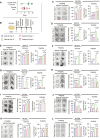
Figure 1.
Figure 1.
NSCLC PDTOs as drug testing platforms for standard-of-care chemotherapy. A, Time frame for organoids to grow to 100 μm diameter from the day of tissue resection (growth time) and doubling time between passages 1 and 2. B, Diagram showing treatment schedules. C–L, Growth comparisons of PDTOs in chemotherapy treatment (carboplatin + paclitaxel) vs. vehicle control groups, Left, Representative bright-field microscope images of PDTOs from vehicle and carboplatin + paclitaxel treatment groups in 10 patients with NSCLC tested (scale bar, 200 μm). Middle, Growth percentages of PDTOs in vehicle control and treatment groups on day 3 and day 6 in comparison to baseline on day 0. Right, ATP metabolic assay to determine the cytotoxicity of carboplatin and paclitaxel on PDTOs. Relative luminescence units (RLU) are directly proportional to the viability of PDTOs in treatment and control groups to the baseline on day 0. Data are presented as mean ± standard error of the mean (SEM) of the three biological replicates. Statistical analysis was performed using Student t test. ***, P < 0.001; **, P < 0.005; *, P < 0.05; ns, not significant.
Figure 2.
Figure 2.
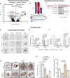
Differential gene expression analysis and drug repurposing of epalrestat to overcome chemotherapy resistance. A, Differential gene expression analysis of chemoresistant (N = 4) vs. chemosensitive (N = 5) PDTOs [arrow: aldo-keto reductase 1B10 (AKR1B10)]. (Complete list of differentially expressed genes is provided in Supplementary File S4.) B, Gene ontology analysis of upregulated genes in chemoresistant PDTOs shows enrichment of metabolite interconverting enzymes. C, Pathway enrichment analysis of upregulated genes in chemoresistant PDTOs demonstrates enrichment of biotransformation pathway critical for metabolizing chemotherapy drugs. D, Overcoming chemoresistance by repurposing AKR1B10 inhibitor drug epalrestat (Epal). Representative images of chemoresistant PDTO (MU383) treated with chemotherapy (carboplatin/paclitaxel), chemotherapy plus epalrestat (scale bar, 200 μm). E, Growth percentages of PDTOs (N = 5) in different treatment groups. F, ATP metabolic assay to determine the viability of chemoresistant PDTOs (N = 5) upon treatment with carboplatin/paclitaxel plus epalrestat (Growth measurements and viability were performed in biological triplicates and the data are represented as mean ± SEM. Statistical analysis was performed using one-way ANOVA and P value was determined by Tukey’s multiple comparisons test; *, P < 0.05; **, P < 0.01; ***, P < 0.001; ns, not significant). G, Proliferation is decreased, and apoptosis is increased in chemoresistant PDTOs treated with epalrestat. PDTOs were harvested on day 6 and analyzed by immunostaining for proliferation marker (Ki67) and apoptosis marker (Cleaved caspase 3). Representative brightfield microscope images of MU383 PDTO are presented (scale bar, 200 μm). H and I, Percentages of Ki67-positive (H) and cleaved caspase 3-positive (I) cells from different treatment groups are represented as mean ± SEM (Kruskal–Wallis multiple comparison; *, P < 0.05; **, P < 0.01).
Figure 3.
Figure 3.
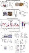
AKR1B10 expression in internal and external cohorts of tumor tissues of patients with NSCLC, NSCLC LUAC and LUSC cell lines. A, Validation of AKR1B10 expression in chemoresistant vs. chemosensitive NSCLC internal patient tumor tissues (categorized based on PDTOs chemotherapy response). Differential gene expression analysis revealed overexpression of AKR1B10 in chemoresistant patient tumor tissues in alignment with matched PDTOs (arrow: AKR1B10; a complete list of differentially expressed genes is provided in Supplementary File S4). B, qPCR measurement of mRNA expression of AKR1B10 gene in chemosensitive (n = 4) and chemoresistant (n = 4) PDTOs’ matched patient tumor tissues. C, Representative images of immunostaining of AKR1B10 in chemosensitive (n = 5) and chemoresistant (n = 5) PDTOs’ matched patient tumor tissues showing high expression in chemoresistant tumors (inset, control IgG; scale bar, 20 μm). D, Scoring of AKR1B10 staining between chemoresistant and –sensitive PDTOs’ matched patient tumor tissues performed by a pathologist blinded to the study. E, Representative images of immunostaining of AKR1B10 in additional internal validation set (n = 14) of platinum-based doublet chemotherapy resistant (n = 7) and sensitive (n = 7) patient primary tumors. F, Scoring of AKR1B10 staining between resistant and sensitive internal validation patient tumor tissues performed by a pathologist blinded to the study, scores of 0 or 1-negative, 2- moderate positive, and 3- strong positive. To qualify for 2 and 3 scores, staining of more than 10% of tumor cells had to be observed. G, Receiver operating characteristic (ROC) and area under the curve (AUC) analysis using an internal validation set of patient tumors demonstrated biomarker potential of AKR1B10 with high sensitivity (85.71%) and specificity (100%) and AUC of 0.93. H, External validation of AKR1B10 expression in larger external cohort (TCGA) of NSCLC patient’s tumor tissues. TCGA pan-cancer and NSCLC RNA expression data were analyzed to determine the expression of AKR1B10. Pan-cancer expression profile of AKR1B10 highlighting overexpression of AKR1B10 in NSCLC (both LUAC and LUSC) tumor tissues compared to normal lung tissues. I, Normalized AKR1B10 gene counts between tumor tissues of patients with NSCLC and normal lung tissues obtained from TCGA highlighting the overexpression of AKR1B10 in 58.96% of patients with NSCLC compared to normal lung tissues. J and K, Western blot (J) and densitometric analysis (K) demonstrating differential expression of AKR1B10 and NRF2 in LUAC and LUSC cell lines. L, Drug screening of LUAC and LUSC cell lines with commonly used cytotoxic agents in NSCLC demonstrates strong correlation in IC50 values of carboplatin, cisplatin, and paclitaxel with AKR1B10 expression but no correlation concerning gemcitabine and pemetrexed (IC50 values for individual cytotoxic agent against AKR1B10 high, medium and no expressing cell lines is provided in Supplementary Table S3). M and N, Western blot (M) and densitometric analysis (N) showing knockdown of AKR1B10 in A549 cell line. O, Cell viability of AKR1B10 knocked down A549 and scramble shRNA treated A549 against IC25 concentration of carboplatin, cisplatin, paclitaxel, gemcitabine, and pemetrexed showing AKR1B10 involvement in mediating resistance to platinum-based drugs. Results are presented as mean ± SD from three independent experiments. Statistical analysis was performed using Student t test. **, P < 0.005; ***, P < 0.0001.
Figure 4.
Figure 4.
Potential and bioavailability of epalrestat to overcome chemotherapy resistance in vivo. Mice (n = 4) were subcutaneously (s.c.) injected with 0.75 × 10⁶ A549 cells and treated with intraperitoneal injection of either platinum-based doublet-[carboplatin (20 mg/kg body weight) plus paclitaxel (10 mg/kg body weight)], or platinum monotherapy [cisplatin (5 mg/kg body weight)] alone, or in combination with epalrestat (10 mg/kg body weight) either through intraperitoneal (i.p.) or oral administration, and tumor growth was monitored. A, Treatment schedules. B, Representative tumor images of mice treated with carboplatin/paclitaxel doublet therapy with or without epalrestat. C, Tumor growth kinetics. D, Tumor weights at euthanasia. E, Intratumoral concentration of epalrestat quantified by mass spectrometry reveals that inhibition of tumor growth by carboplatin-paclitaxel doublet therapy is directly proportional to the intratumoral concentration of epalrestat. F, Representative tumor images of mice treated with cisplatin monotherapy with or without epalrestat. G, Tumor growth kinetics. H, Tumor weights at euthanasia. I, Intratumoral concentration of epalrestat quantified by mass spectrometry reveals that inhibition of tumor growth by cisplatin therapy is directly proportional to the intratumoral concentration of epalrestat. Statistical analysis was performed using one-way ANOVA, and P value was determined by Tukey’s multiple comparisons test; *, P < 0.05; **, P < 0.005; ***, P < 0.001; ns, not significant.

References

    1. Arriagada R, Bergman B, Dunant A, Le Chevalier T, Pignon J-P, Vansteenkiste J, et al. . Cisplatin-based adjuvant chemotherapy in patients with completely resected non-small-cell lung cancer. N Engl J Med 2004;350:351–60. - PubMed
    1. Krebs MG, Sloane R, Priest L, Lancashire L, Hou J-M, Greystoke A, et al. . Evaluation and prognostic significance of circulating tumor cells in patients with non-small-cell lung cancer. J Clin Oncol 2011;29:1556–63. - PubMed
    1. Wu Y-L, Tsuboi M, He J, John T, Grohe C, Majem M, et al. . Osimertinib in resected EGFR-mutated non-small-cell lung cancer. N Engl J Med 2020;383:1711–23. - PubMed
    1. Forde PM, Spicer J, Lu S, Provencio M, Mitsudomi T, Awad MM, et al. . Neoadjuvant nivolumab plus chemotherapy in resectable lung cancer. N Engl J Med 2022;386:1973–85. - PMC - PubMed
    1. Jin G, Wong STC. Toward better drug repositioning: prioritizing and integrating existing methods into efficient pipelines. Drug Discov Today 2014;19:637–44. - PMC - PubMed

MeSH terms