. 2024 Sep 1;9(9):826-834.
doi: 10.1001/jamacardio.2024.1874.

Lipoprotein(a) and Long-Term Plaque Progression, Low-Density Plaque, and Pericoronary Inflammation

Affiliations

Lipoprotein(a) and Long-Term Plaque Progression, Low-Density Plaque, and Pericoronary Inflammation

Nick S Nurmohamed et al. JAMA Cardiol. .

Erratum in

  • Error in Abstract.
    [No authors listed] [No authors listed] JAMA Cardiol. 2024 Sep 1;9(9):861. doi: 10.1001/jamacardio.2024.3130. JAMA Cardiol. 2024. PMID: 39259261 Free PMC article. No abstract available.

Abstract

Importance: Lipoprotein(a) (Lp[a]) is a causal risk factor for cardiovascular disease; however, long-term effects on coronary atherosclerotic plaque phenotype, high-risk plaque formation, and pericoronary adipose tissue inflammation remain unknown.

Objective: To investigate the association of Lp(a) levels with long-term coronary artery plaque progression, high-risk plaque, and pericoronary adipose tissue inflammation.

Design, setting, and participants: This single-center prospective cohort study included 299 patients with suspected coronary artery disease (CAD) who underwent per-protocol repeated coronary computed tomography angiography (CCTA) imaging with an interscan interval of 10 years. Thirty-two patients were excluded because of coronary artery bypass grafting, resulting in a study population of 267 patients. Data for this study were collected from October 2008 to October 2022 and analyzed from March 2023 to March 2024.

Exposures: The median scan interval was 10.2 years. Lp(a) was measured at follow-up using an isoform-insensitive assay. CCTA scans were analyzed with a previously validated artificial intelligence-based algorithm (atherosclerosis imaging-quantitative computed tomography).

Main outcome and measures: The association between Lp(a) and change in percent plaque volumes was investigated in linear mixed-effects models adjusted for clinical risk factors. Secondary outcomes were presence of low-density plaque and presence of increased pericoronary adipose tissue attenuation at baseline and follow-up CCTA imaging.

Results: The 267 included patients had a mean age of 57.1 (SD, 7.3) years and 153 were male (57%). Patients with Lp(a) levels of 125 nmol/L or higher had twice as high percent atheroma volume (6.9% vs 3.0%; P = .01) compared with patients with Lp(a) levels less than 125 nmol/L. Adjusted for other risk factors, every doubling of Lp(a) resulted in an additional 0.32% (95% CI, 0.04-0.60) increment in percent atheroma volume during the 10 years of follow-up. Every doubling of Lp(a) resulted in an odds ratio of 1.23 (95% CI, 1.00-1.51) and 1.21 (95% CI, 1.01-1.45) for the presence of low-density plaque at baseline and follow-up, respectively. Patients with higher Lp(a) levels had increased pericoronary adipose tissue attenuation around both the right coronary artery and left anterior descending at baseline and follow-up.

Conclusions and relevance: In this long-term prospective serial CCTA imaging study, higher Lp(a) levels were associated with increased progression of coronary plaque burden and increased presence of low-density noncalcified plaque and pericoronary adipose tissue inflammation. These data suggest an impact of elevated Lp(a) levels on coronary atherogenesis of high-risk, inflammatory, rupture-prone plaques over the long term.


Conflict of interest statement

Conflict of Interest Disclosures: Dr Nurmohamed reported grants from the Dutch Heart Foundation (Dekker 03-007-2023-0086), the European Atherosclerosis Society (2023), and Cleerly, and personal fees from Daiichi Sankyo and Novartis, outside the submitted work; and is co-founder of Lipid Tools. Dr Malkasian reported personal fees from Cleerly; in addition, Dr Malkasian patents for 18/454,462 and 18/149,006 issued. Dr Earls reported employment and equity from Cleerly during the conduct of the study. Dr Min reported employment and equity from Cleerly during the conduct of the study and outside the submitted work. Dr Danad reported membership of Cleerly’s Scientific Advisory Board during the conduct of the study. Dr van Rosendael reported membership of Cleerly’s Scientific Advisory Board during the conduct of the study. Dr Choi reported equity from Cleerly, personal fees from Siemens, and grants from the George Washington University Heart and Vascular Institute during the conduct of the study. Dr Stroes reported grants from Amgen, Novartis, and Ionis, and personal fees from Sanofi, Merck, and AstraZeneca outside the submitted work. Dr Knaapen reported grants from HeartFlow and Cleerly, outside the submitted work. No other disclosures were reported.

Figures

Figure 1.
Figure 1.. Association of Lipoprotein(a) (Lp[a]) With Plaque Burden and Coronary Computed Tomography Angiography (CCTA) Plaque Progression During Follow-Up
The whiskers show 95% CIs from the unadjusted linear mixed-effect models for the 10th (7 nmol/L), 50th (25 nmol/L), and 90th (221 nmol/L) percentiles of Lp(a) in this study for percent atheroma volume (A), percent noncalcified plaque volume (B), and percent calcified plaque volume (C) at baseline and follow-up (10.2 years).
Figure 2.
Figure 2.. Association of Lipoprotein(a) (Lp[a]) With High-Risk Plaque and Pericoronary Inflammation
Adjusted odds ratios from logistic regression models for the presence low-density plaque and presence of increased pericoronary adipose tissue attenuation (PCATa). LAD indicates left anterior descending coronary artery; OR, odds ratio; RCA, right coronary artery.
Figure 3.
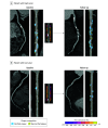
Figure 3.. Coronary Plaque Progression and Pericoronary Inflammation With High and Low Lipoprotein(a) (Lp[a])
Two case examples of a patient with high (A) and low (B) Lp(a). Shown are baseline computed tomography angiography curved reformat (left) and straightened multiplanar reformatted (right) reconstructions of the right coronary artery (RCA) and pericoronary adipose tissue attenuation (PCATa) around the RCA. At baseline, patient A (high Lp[a]) had a percent atheroma volume (PAV) of 19.4%, a percent noncalcified plaque volume of 6.1%, and a percent noncalcified plaque volume of 13.3%, while RCA PCATa was −79.1 HU, above the scanner-specific threshold, indicative of pericoronary inflammation. During the 10-year follow-up, the plaque volumes for PAV, percent noncalcified plaque volume, and percent calcified plaque volume progressed to 30.0%, 12.2%, and 15.8% at follow-up (55% PAV increase, 100% percent noncalcified plaque volume increase, and 19% percent calcified plaque volume increase), indicative of important plaque progression. Patient B (low Lp[a]) had a baseline total plaque volume of 11.6%, an noncalcified plaque volume of 5.8%, and a calcified plaque volume of 5.8%, while RCA PCATa was −94.8 HU, below the scanner-specific threshold, indicating absence of pericoronary inflammation. During the 10-year follow-up, the plaque volumes for PAV, noncalcified plaque volume, and calcified plaque volume changed to 11.2%, 4.4%, and 6.7%, respectively (3% PAV decrease, 24% noncalcified plaque volume decrease, and 19% calcified plaque volume increase), indicative of plaque stabilization.

References

    1. Kronenberg F, Mora S, Stroes ESG, et al. . Lipoprotein(a) in atherosclerotic cardiovascular disease and aortic stenosis: a European Atherosclerosis Society consensus statement. Eur Heart J. 2022;43(39):3925-3946. doi:10.1093/eurheartj/ehac361 - DOI - PMC - PubMed
    1. Mach F, Baigent C, Catapano AL, et al. ; ESC Scientific Document Group . 2019 ESC/EAS Guidelines for the management of dyslipidaemias: lipid modification to reduce cardiovascular risk. Eur Heart J. 2020;41(1):111-188. doi:10.1093/eurheartj/ehz455 - DOI - PubMed
    1. Schnitzler JG, Hoogeveen RM, Ali L, et al. . Atherogenic lipoprotein(a) increases vascular glycolysis, thereby facilitating inflammation and leukocyte extravasation. Circ Res. 2020;126(10):1346-1359. doi:10.1161/CIRCRESAHA.119.316206 - DOI - PMC - PubMed
    1. van der Valk FM, Bekkering S, Kroon J, et al. . Oxidized phospholipids on lipoprotein(a) elicit arterial wall inflammation and an inflammatory monocyte response in humans. Circulation. 2016;134(8):611-624. doi:10.1161/CIRCULATIONAHA.116.020838 - DOI - PMC - PubMed
    1. Kaiser Y, Daghem M, Tzolos E, et al. . Association of lipoprotein(a) with atherosclerotic plaque progression. J Am Coll Cardiol. 2022;79(3):223-233. doi:10.1016/j.jacc.2021.10.044 - DOI - PMC - PubMed