. 2024 Jul 23;121(30):e2407461121.
doi: 10.1073/pnas.2407461121. Epub 2024 Jul 17.

A broad survey of choanoflagellates revises the evolutionary history of the Shaker family of voltage-gated K+ channels in animals

Affiliations

A broad survey of choanoflagellates revises the evolutionary history of the Shaker family of voltage-gated K+ channels in animals

Timothy Jegla et al. Proc Natl Acad Sci U S A. .

Abstract

The Shaker family of voltage-gated K+ channels has been thought of as an animal-specific ion channel family that diversified in concert with nervous systems. It comprises four functionally independent gene subfamilies (Kv1-4) that encode diverse neuronal K+ currents. Comparison of animal genomes predicts that only the Kv1 subfamily was present in the animal common ancestor. Here, we show that some choanoflagellates, the closest protozoan sister lineage to animals, also have Shaker family K+ channels. Choanoflagellate Shaker family channels are surprisingly most closely related to the animal Kv2-4 subfamilies which were believed to have evolved only after the divergence of ctenophores and sponges from cnidarians and bilaterians. Structural modeling predicts that the choanoflagellate channels share a T1 Zn2+ binding site with Kv2-4 channels that is absent in Kv1 channels. We functionally expressed three Shakers from Salpingoeca helianthica (SheliKvT1.1-3) in Xenopus oocytes. SheliKvT1.1-3 function only in two heteromultimeric combinations (SheliKvT1.1/1.2 and SheliKvT1.1/1.3) and encode fast N-type inactivating K+ channels with distinct voltage dependence that are most similar to the widespread animal Kv1-encoded A-type Shakers. Structural modeling of the T1 assembly domain supports a preference for heteromeric assembly in a 2:2 stoichiometry. These results push the origin of the Shaker family back into a common ancestor of metazoans and choanoflagellates. They also suggest that the animal common ancestor had at least two distinct molecular lineages of Shaker channels, a Kv1 subfamily lineage predicted from comparison of animal genomes and a Kv2-4 lineage predicted from comparison of animals and choanoflagellates.

Keywords: Shaker; choanoflagellate; potassium channel; voltage-gated.


Conflict of interest statement

Competing interests statement:The authors declare no competing interest.

Figures
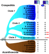
Fig. 1.
Fig. 1.
Shaker-like Kv channels in 21 choanoflagellate species. Phylogeny of 21 choanoflagellate species based on sequence comparison established by Richter et al. (38). Numbers of Shaker-like Kv channel genes without a T1 domain and true Shaker family genes with a T1 domain are shown in the 1st and 2nd columns, respectively. Only three species of Class II and Class III craspedidans have true Shaker family genes. Note that only the Class III species S. dolichothecata has both the Shaker-like Kvs and a true Shaker family gene. Sequences of the choanoflagellate channel proteins are included in SI Appendix, File S1.
Fig. 2.
Fig. 2.
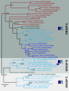
Bayesian inference phylogeny of choanoflagellate Shaker-related channel proteins. Choanoflagellate channels related to the animal Shaker family fall into three highly supported groups (A–C) in phylogenetic analysis based on an alignment of the S1 to S6 channel core. Basic subunit structure cartoons are given for each group; The true Shaker family genes with a T1 domain comprise group B. Channels are named with a five-letter Genus/Species prefix and the GenBank accession number. The sequence alignment used to generate the phylogeny can be found in SI Appendix, File S2, and the tree file is provided as SI Appendix, File S3. S. helianthica channels functionally expressed in this study were given the names SheliKvT1.1-1.3. Branches are color-coded by species clade: blue, Craspedida Class I; light blue, Craspedida Class II; Teal, Craspedida Class III; Red, Acanthoecida. All three channel groups are represented in Class II/III craspedidans, though no Class II species has all three channel types. Branch lengths show sequence divergence with a scale bar in substitutions/site. Node values indicate posterior probabilities.
Fig. 3.
Fig. 3.
Amino acid sequence alignment of the T1 domain between animal Kv1-4 channels from D. melanogaster (Bilateria, Dmela) and N. vectensis (Cnidaria, Nvect) and eight choanoflagellate Shaker family channels. Structural elements identified in Bixby et al. (35) are indicated with bars at the top of the alignment. Residues conserved (identical or chemically conservative substitution) in a majority of all channels are shaded red, while those selectively conserved in a majority of animal or choanoflagellate channels are shaded gray or black, respectively. Five residues identically conserved in all channels are marked with stars below the alignment. Residues comprising the Zn2+ binding site of animal Kv2-4 channels are highlighted in blue and marked with + signs below the alignment. Residue numbers are given at the right margin and the accession numbers for the animal sequences are M17211 (Dmela_Shaker), AF084525 (Dmela_Shab), AC005149 (Dmela_Shaw), M32660, (Dmela_Shal), JX846630 (Nvect_Shak1), KP219397 (Nvect_Shab), KP219392 (Nvect_Shaw1), and KP219396 (Nvect_Shal1).
Fig. 4.
Fig. 4.
Bayesian inference phylogeny of choanoflagellate and animal Shaker family channel proteins. The phylogeny includes Shaker family channels from choanoflagellates (Blue), the ctenophore M. leidyi (Mleid, Black), the cnidarian N. vectensis (Nvect, purple), and the bilaterians D. melanogaster and H. sapiens (Dmela and Hsapi, red). The sequence alignment is provided in SI Appendix, File S4. Branch lengths indicate substitutions/site (scale bar provided), and posterior probabilities are indicated for key nodes. The unrooted phylogeny is based on a T1 + S1 to S6 alignment and is shown with a midpoint root for display purposes. The full tree file including all node support values is included as SI Appendix, File S5. The animal Kv1-4 subfamilies and choanoflagellate Group B channels are indicated with labels (Right margin) and differential background shading. Cnidarians and bilaterians have all four animal subfamilies, while ctenophore channels cluster exclusively in the Kv1 subfamily. Choanoflagellate Group B channels form a highly supported clade with the animal Kv2-4 subfamilies with which they share the T1 Zn2+ binding site. Note that we do not find strong support for the order of subfamily divergence within the Kv2-4 subfamily clade.
Fig. 5.
Fig. 5.
Functional expression of choanoflagellate Shaker family channels in Xenopus oocytes. (A) Currents recorded from oocytes expressing single choanoflagellate channels (Top row) or combinations (Bottom row) in response to 400 ms voltage steps to the indicated membrane potentials in 20 mV increments. Tails were recorded at −40 mV. For SheliKvT1.1/1.2, voltage steps start at −80 mV, and tails were recorded at −60 mV. Only combinatorial expression of SheliKvT1.1/1.2 or SheliKvT1.1/1.3 showed significant outward K+ currents. Scale bars indicate current in μA and time in ms. Capacitive transient currents were clipped for display purposes. (B) Line and symbol current/voltage plots for indicated channel subunit combinations. Data points show mean ± SEM (N = 5 for homomers, 9 for SheliKvT1.1/1.2, 10 for SheliKvT1.1/1.3, and 8 for SheliKvT1.2/1.3). (C) Inactivation time course vs. voltage plots for SheliKvT1.1/1.2 (blue squares, N = 9) and SheliKvT1.1/1.3 (red circles, N = 10). Measurements indicate the time at which currents had decayed to one half of their peak value (Iinact0.5), and data points show mean ± SEM.
Fig. 6.
Fig. 6.
SheliKvT1.1 confers fast N-type inactivation. Example current traces recorded from oocytes expressing (A) SheliKvT1.1 Δ2–25/1.2 and (B) SheliKvT1.1 Δ2–25/1.3. Mixes using SheliKvT1 WT and truncated (Δ2–19) versions of (C) SheliKvT1.2 or (D) SheliKvT1.3 retain fast inactivation. Holding potential was −120 mV, and step depolarizations in 20 mV increments ranged from −100 mV to −20 mV for SheliKvT1.1 Δ2–25/1.2 and −60mV to 20 mV for SheliKvT1.1 Δ2–25/1.3; tails were recorded at −70 mV and −40 mV respectively. SSI and GV curves for (E) SheliKvT1.1/1.2 and SheliKvT1.1/1.3. Oocytes were held at the indicated voltages for 5 s prior to a test pulse to 0 mV to activate current. Data from individual oocytes were normalized prior to comparison, points show mean ± SEM and curves are Boltzmann fits (parameters given in Table 1). Note that the SSI data for SheliKvT1.1/1.2 are fit with a double Boltzmann. GV data were collected from isochronal tail currents using SheliKvT1.1 Δ2–25 to remove inactivation. (F and G) Normalized current traces recorded at 0 mV in 2 mM and 100 mM external K+ for SheliKvT1.1/1.2 and SheliKvT1.1/1.3. (H) Recovery from inactivation at −120 mV for both heteromers in 2 mM and 100 mM external K+. Oocytes received pairs 500 ms depolarizing pulses to 0 mV from a holding potential of −120 mV. Recovery times at −120 mV between the paired depolarizations are represented on the X-axis. Data points show mean ± SEM of N = 6 to 11. Curves show single exponential fits with time constants reported in Results. Recovery was not sufficient for SheliKvT1.1/1.2 in 2 mM K+ for exponential fitting.
Fig. 7.
Fig. 7.
Polar T1 interface bonds in structural models of SheliKvT1 tetramers. Protein backbones are color-coded blue (SheliKvT1.1), green (SheliKvT1.2), or tan (SheliKvT1.3), and structural elements from the T1 alignment in Fig. 3 are labeled on the righthand subunit of the Upper Right panel. Note that not all elements adopt the form predicted in Bixby et al. (35), consistent with variability seen in published structures. Only the two subunits contributing to the interface are shown for each panel. Background shading delineates three structural layers of the T1 interface. Residues involved in the coordination of Zn2+ (silver sphere) are shown with Zn2+ coordination bonds indicated with dashed magenta lines. Residues participating in interface polar bonds are also shown with salt bridges indicated with orange dashed lines and hydrogen bonds indicated with yellow dashed lines. Numbers indicate fractional bond occupancies in MD simulations. (A–C) T1 interface of for SheliKvT1.1-1.3 homotetramers. Only one interface is shown since all four interfaces of the tetramer are identical. (DI) T1 interfaces for 2:2 heterotetramers of SheliKvT1.1 + SheliKvT1.2 (Left column), SheliKvT1.1 + SheliKvT1.3 (Middle column), and SheliKvT1.2 + SheliKvT1.3 (Right column). Heterotetramers have two unique interfaces. Note only the two interfaces of SheliKvT1.1 + SheliKvT1.2 and SheliKvT1.1 + SheliKvT1.3 heterotetramers have polar side-chain bonds connecting adjacent T1s on all three structural levels.
Fig. 8.
Fig. 8.
Summary of the evolution of the Shaker family of voltage-gated K+ channels. A “ctenophore first” phylogeny of animals and choanoflagellates is shown with the types of Shaker family genes found in extant phyla indicated with color-coded channel icons. Channel icons at ancestral nodes indicate the Shaker channel types we infer to have been present in the ancestral species. The current study shows Kv2-4-like (Kv2-4L) Shaker family channels with a T1 Zn2+ binding site (green circle in icons) were already present in the common ancestor of choanoflagellates and animals. We now predict that the common ancestor of extant animals had at least two Shaker family lineages, the Kv1 subfamily that is present in all animal phyla and a Kv2-4L lineage that was lost in ctenophores and sponges. The Kv2-4L lineage diversified into the Kv2, Kv3, and Kv4 subfamilies in parahoxozoans. The Kv2-4L loss in ctenophores and sponges and the absence of the Kv4 subfamily in placozoans are supported by sequence analysis of only a few species and should be interpreted cautiously until more species are sequenced.

Similar articles

Cited by

References

    1. Papazian D. M., Schwarz T. L., Tempel B. L., Jan Y. N., Jan L. Y., Cloning of genomic and complementary DNA from Shaker, a putative potassium channel gene from Drosophila. Science 237, 749–753 (1987). - PubMed
    1. Kamb A., Iverson L. E., Tanouye M. A., Molecular characterization of Shaker, a Drosophila gene that encodes a potassium channel. Cell 50, 405–413 (1987). - PubMed
    1. Butler A., Wei A. G., Baker K., Salkoff L., A family of putative potassium channel genes in Drosophila. Science 243, 943–947 (1989). - PubMed
    1. Salkoff L., et al. , An essential “set” of K+ channels conserved in flies, mice and humans. Trends Neurosci. 15, 161–166 (1992). - PubMed
    1. Wei A., Jegla T., Salkoff L., Eight potassium channel families revealed by the C. elegans genome project. Neuropharmacology 35, 805–829 (1996). - PubMed

MeSH terms

Substances

LinkOut - more resources