. 2024 Jul 23;121(30):e2404778121.
doi: 10.1073/pnas.2404778121. Epub 2024 Jul 17.

Metformin synergizes with PD-1 blockade to promote normalization of tumor vessels via CD8T cells and IFNγ

Affiliations

Metformin synergizes with PD-1 blockade to promote normalization of tumor vessels via CD8T cells and IFNγ

Miho Tokumasu et al. Proc Natl Acad Sci U S A. .

Abstract

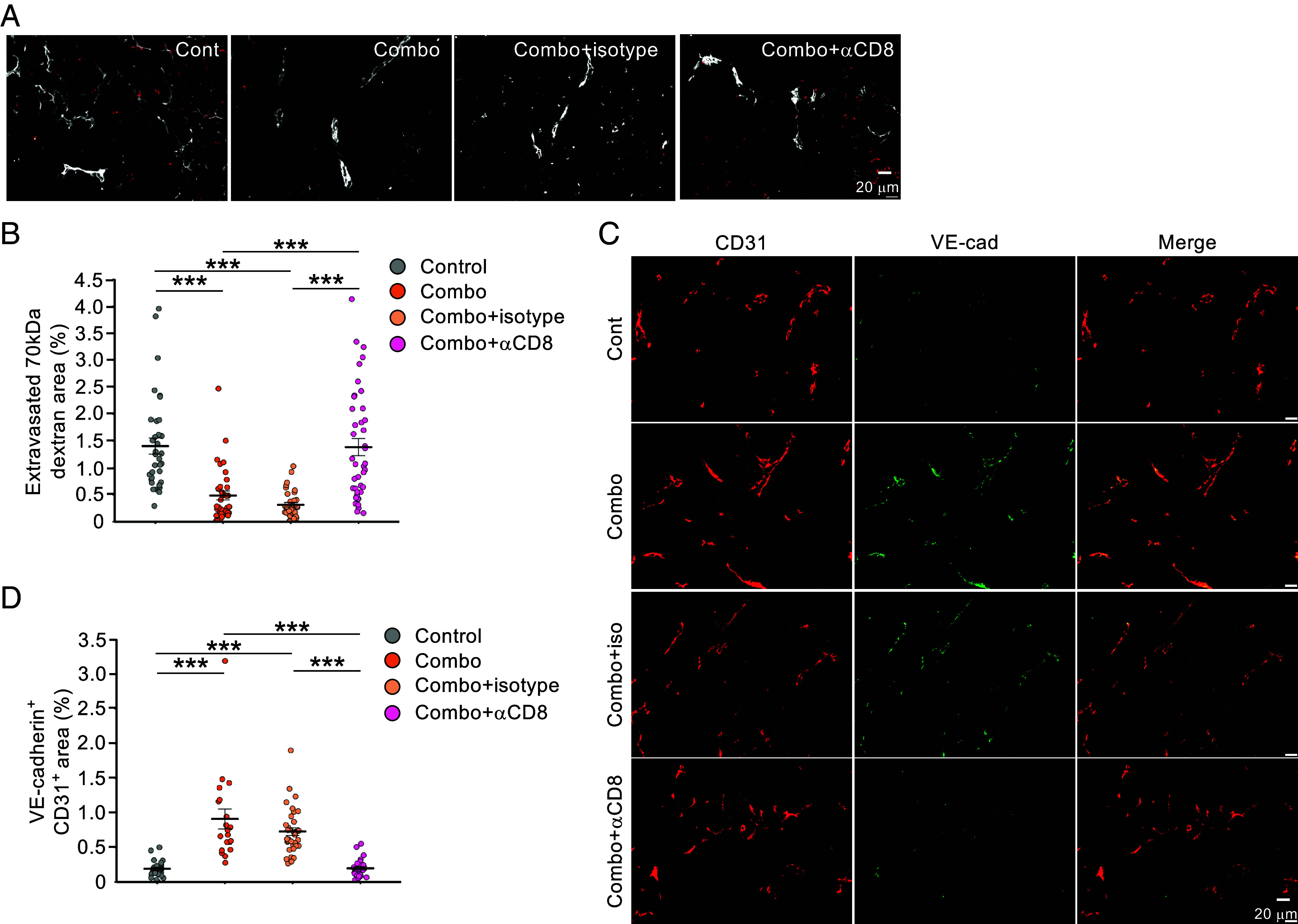
Tumor blood vessels are highly leaky in structure and have poor blood perfusion, which hampers infiltration and function of CD8T cells within tumor. Normalizing tumor vessels is thus thought to be important in promoting the flux of immune T cells and enhancing ant-tumor immunity. However, how tumor vasculature is normalized is poorly understood. Metformin (Met) combined with ant-PD-1 therapy is known to stimulate proliferation of and to produce large amounts of IFNγ from tumor-infiltrating CD8T lymphocytes (CD8TILs). We found that the combination therapy promotes the pericyte coverage of tumor vascular endothelial cells (ECs) to improve blood perfusion and that it suppresses the hyperpermeability through the increase of VE-cadherin. Peripheral node addressin(PNAd) and vascular cell adhesion molecule (VCAM)-1, both implicated to promote tumor infiltration of CD8T cells, were also increased. Importantly, tumor vessel normalization, characterized as the reduced 70-kDa dextran leakage and the enhancement of VE-cadherin and VCAM-1, were canceled by anti-CD8 Ab or anti-IFNγ Ab injection to mice. The increased CD8TILs were also abrogated by anti-IFNγ Ab injection. In vascular ECs, flow cytometry analysis revealed that pSTAT1 expression was found to be associated with VE-cadherin expression. Moreover, in vitro treatment with Met and IFNγ enhanced VE-cadherin and VCAM-1 on human umbilical vein endothelial cells (HUVECs). The Kaplan-Meier method revealed a correlation of VE-cadherin or VCAM-1 levels with overall survival in patients treated with immune checkpoint inhibitors. These data indicate that IFNγ-mediated cross talk of CD8TILs with tumor vessels is important for creating a better tumor microenvironment and maintaining sustained antitumor immunity.

Keywords: CD8T cells; IFNγ; VCAM-1; VE-cadherin; tumor vessels.


Conflict of interest statement

Competing interests statement:The authors declare no competing interest.

Figures

Fig. 1.
Fig. 1.
Met synergizes with anti-PD-1 therapy to increase the CD8TILs/Foxp3+ cell ratio in 4T1 breast tumor. (A) Growth of 4T1 tumors inoculated into BALB/c mice by intradermal injection on day 0. Met and/or anti-PD-1 antibody treatment is initiated on day 5. Tumor growth is monitored every other day. Data are presented as mean tumor volume + SEM from pooled data of two independent experiments (n = 5 × 2). (B) Representative images of immunostaining of CD8TILs (red), Foxp3+ cells (green), and DAPI (blue) on day 8 in each group. (Scale bar, 20 µm.) (CE) Quantified numbers of CD8TILs/mm2 (C), numbers of Foxp3+ cells/mm2 (D), and CD8TILs/Foxp3+ cell ratio (E) on day 8 in each group. Two to three sections per individual mouse were counted. Data are presented as mean ± SEM (n = 5). *P < 0.05, ***P < 0.001 using the Student’s t test.
Fig. 2.
Fig. 2.
Met+anti-PD-1 Ab combination therapy increases the coverage of mural cells on tumor vessels and improved blood perfusion. (A) CD31+ area is measured. Data are presented as mean ± SEM (n = 5). (B) Representative images of immunostaining for CD31 (red) and αSMA (green) on day 8 in each group. (Scale bar, 20 µm.) (C) Merged area of CD31+ and αSMA+ is measured. Data are presented as mean ± SEM (n = 5). (D) Representative images of immunostaining for CD31 (white) and fluorescence of 2MDa dextran (red) on day 8 in each group. (Scale bar, 20 µm.) (E) Quantification of 2-MDa dextran area within tumor vessels. Data are presented as mean ± SEM (n = 3, more than four areas/individual tumors were observed). *P < 0.05, **P < 0.01, ***P < 0.001 using the Student’s t test.
Fig. 3.
Fig. 3.
Met and/or anti-PD-1 Ab therapies decrease the leakage of 70kDex along with increased expression of VE-cadherin on CD31+ cells. (A) Representative images of immunostaining for CD31 (white) and fluorescence of 70kDex (red) on day 8 in each group. (Scale bar, 20 µm.) (B) Quantification of extravasated 70kDex dextran area from tumor vessels. Data are presented as mean ± SEM (n = 4, more than six areas/individual tumors were observed). (C) Representative images of immunostaining for CD31 (red) VE-cadherin (green) and merged area (yellow) on day 8 in each group. (Scale bar, 20 µm.) (D) Quantification of VE-cadherin+ CD31+ area (%). Data are presented as mean ± SEM (n = 4 to 5, more than five areas/individual tumors were observed). *P < 0.05, **P < 0.01, ***P < 0.001 using the Student’s t test.
Fig. 4.
Fig. 4.
CD8+ T cells mediate the decreased leakage of 70kDex and the increased expression of VE-cadherin on CD31+ cells in the Met+anti-PD-1 antibody combination therapy group. (A) Representative images of immunostaining for CD31 (white) and fluorescence of 70kDex (red) on day 8 in each group. (Scale bar, 20 µm.) (B) Quantification of extravasated 70kDex area from tumor vessels. Data are presented as mean ± SEM (n = 4 to 5, more than five areas/individual tumors were observed). (C) Representative images of immunostaining for CD31 (red), VE-cadherin (green), and merged area (yellow) on day 8 in each group. (Scale bar, 20 µm.) (D) Quantification of VE-cadherin+ CD31+ area (%). Data are presented as mean ± SEM (n = 4 to 5, more than five areas/individual tumors were observed). ***P < 0.001 using the Student’s t test.
Fig. 5.
Fig. 5.
IFNγ mediates the decreased leakage of 70kDex from tumor vessels in the Met+anti-PD-1 Ab combination therapy group. (A) Representative images of immunostaining for CD31 (blue) and pSTAT1 (red) on day 8 in each group. (Scale bar, 20 µm.) (BD) (B) Quantification of pSTAT1+ CD31+ area per whole tumor area (%), (C) Quantification of pSTAT1+ CD31+ area per CD31+ area (%), and (D) Quantification of pSTAT1 MFI in CD31+ cells. Data are presented as mean ± SEM (n = 5). (EG) CD31+ cells isolated from tumors are analyzed by FACS (n = 3 pooled mice in each group). (E) pSTAT1 expression level of CD31+ cells. (pSTAT1 MFI; isotype = 295, control = 373, combo + IgG = 658). (F) VE-cadherin expression level on pSTAT1-low or pSTAT1-high CD31+ cells from Met + anti-PD-1 Ab treated group (VE-cadherin MFI; pSTAT1 low = 503, pSTAT1 high = 1,996). (G) VE-cadherin expression level on CD31+ cells in each group (VE-cadherin MFI; isotype = 127, control = 591, combo + IgG = 1,174, combo + anti-IFNγ =504). (H) VE-cadherin expression on HUVECs treated with or without Met and/or recombinant IFNγ in vitro was measured by FACS. (I) Representative images of immunostaining for CD31 (white) and fluorescence of 70kDex (red) on day 8 in each group. (Scale bar, 20 µm.) (J) Quantification of the extravasated 70kDex area from tumor vessels. Data are presented as mean ± SEM (n = 4 to 5, more than five areas/individual tumors were observed). *P < 0.05, ***P < 0.001 using the Student’s t test.

References

    1. de Aguiar L. G., et al. , Metformin improves endothelial vascular reactivity in first-degree relatives of type 2 diabetic patients with metabolic syndrome and normal glucose tolerance. Diabetes Care 29, 1083–1089 (2006). - PubMed
    1. Jensterle M., et al. , Improvement of endothelial function with metformin and rosiglitazone treatment in women with polycystic ovary syndrome. Eur. J. Endocrinol. 159, 399–406 (2008). - PubMed
    1. O’Hora T. R., Markos F., Wiernsperger N. F., Noble M. I., Metformin causes nitric oxide-mediated dilatation in a shorter time than insulin in the iliac artery of the anesthetized pig. J. Cardiovasc. Pharmacol. 59, 182–187 (2012). - PubMed
    1. Jain R. K., Normalizing tumor microenvironment to treat cancer: Bench to bedside to biomarkers. J. Clin. Oncol. 31, 2205–2218 (2013). - PMC - PubMed
    1. Viallard C., Larrivee B., Tumor angiogenesis and vascular normalization: Alternative therapeutic targets. Angiogenesis 20, 409–426 (2017). - PubMed

MeSH terms

LinkOut - more resources