. 2024 Nov 1;22(11):996-1010.
doi: 10.1158/1541-7786.MCR-24-0361.

GRAIL1 Stabilizes Misfolded Mutant p53 through a Ubiquitin Ligase-Independent, Chaperone Regulatory Function

Affiliations

GRAIL1 Stabilizes Misfolded Mutant p53 through a Ubiquitin Ligase-Independent, Chaperone Regulatory Function

Paramita Ray et al. Mol Cancer Res. .

Abstract

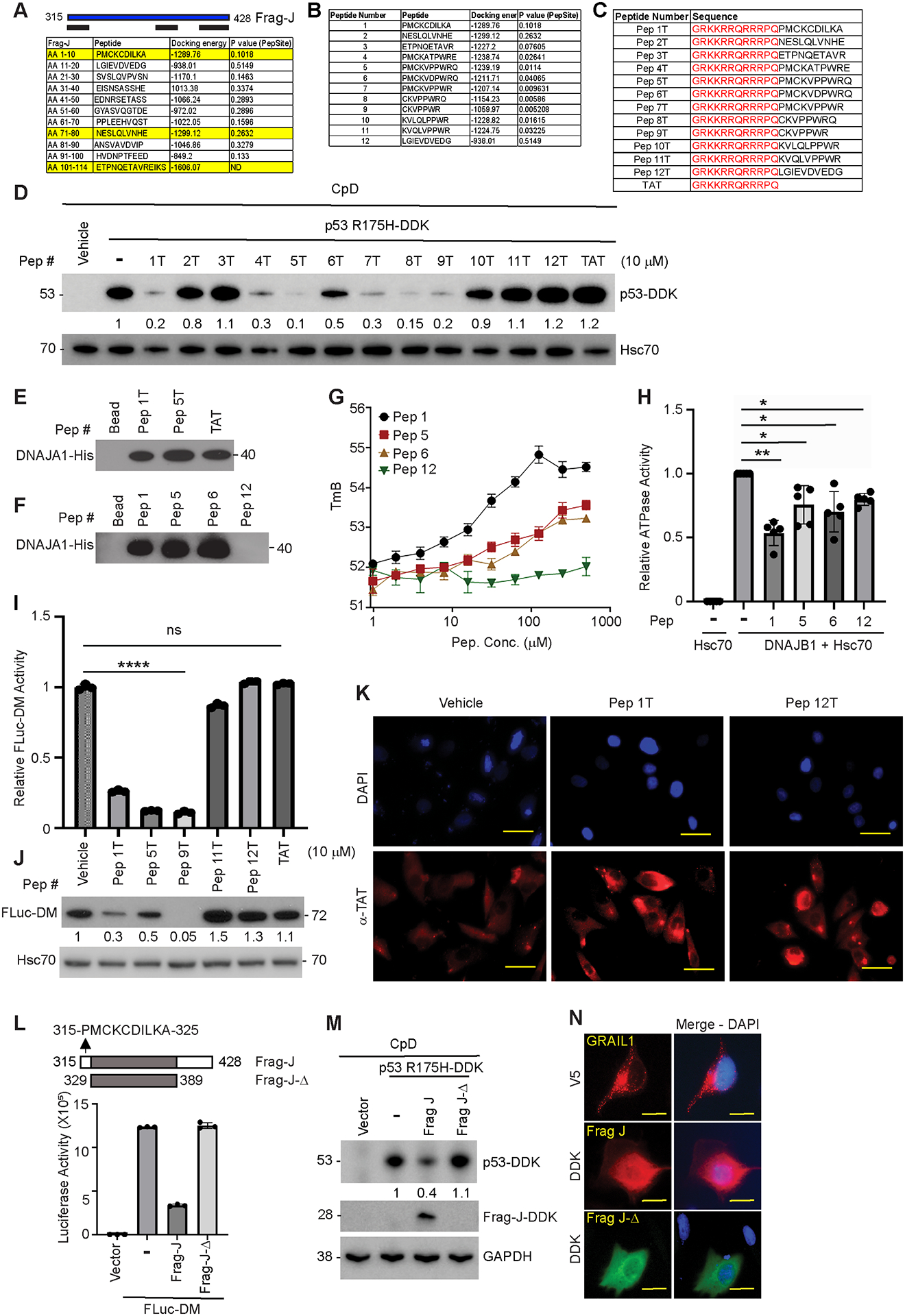
Frequent (>70%) TP53 mutations often promote its protein stabilization, driving esophageal adenocarcinoma (EAC) development linked to poor survival and therapy resistance. We previously reported that during Barrett's esophagus progression to EAC, an isoform switch occurs in the E3 ubiquitin ligase RNF128 (aka GRAIL-gene related to anergy in lymphocytes), enriching isoform 1 (hereby GRAIL1) and stabilizing the mutant p53 protein. Consequently, GRAIL1 knockdown degrades mutant p53. But, how GRAIL1 stabilizes the mutant p53 protein remains unclear. In search for a mechanism, here, we performed biochemical and cell biology studies to identify that GRAIL has a binding domain (315-PMCKCDILKA-325) for heat shock protein 40/DNAJ. This interaction can influence DNAJ chaperone activity to modulate misfolded mutant p53 stability. As predicted, either the overexpression of a GRAIL fragment (Frag-J) encompassing the DNAJ binding domain or a cell-permeable peptide (Pep-J) encoding the above 10 amino acids can bind and inhibit DNAJ-Hsp70 co-chaperone activity, thus degrading misfolded mutant p53. Consequently, either Frag-J or Pep-J can reduce the survival of mutant p53 containing dysplastic Barrett's esophagus and EAC cells and inhibit the growth of patient-derived organoids of dysplastic Barrett's esophagus in 3D cultures. The misfolded mutant p53 targeting and growth inhibitory effects of Pep-J are comparable with simvastatin, a cholesterol-lowering drug that can degrade misfolded mutant p53 also via inhibiting DNAJA1, although by a distinct mechanism. Implications: We identified a novel ubiquitin ligase-independent, chaperone-regulating domain in GRAIL and further synthesized a first-in-class novel misfolded mutant p53 degrading peptide having future translational potential.


Conflict of interest statement

Conflict of Interest Statement: The authors declare that they have no conflicts of interest with the contents of this article.

Figures

Figure 1.
Figure 1.. GRAIL1 knockdown degrades misfolded mutant p53 to reduce clonogenic survival of EAC cells.
(A) Three EAC cell lines with different p53 mutations (OE33 – p53C135Y; Flo1 – p53C277F; OE19 – N310fs26X) were transfected with either control, TP53, or GRAIL1 (two different ones) siRNAs. Twenty-four hours after siRNA transfection, cells were trypsinized, and plated for clonogenic survival. Data presented as mean ± SEM from three independent sets of studies. Two tailed unpaired student’s t-test *p< 0.05; **p<0.01; ***p<0.001. (B) Due to the lack of GRAIL isoform specific antibodies, particularly against GRAIL2, we performed qRT-PCR to determine relative GRAIL1 transcript levels in the above three EAC cell lines following TP53 and GRAIL1 knockdown. Forty-eight hours post-transfection, total RNA was isolated and GRAIL isoform specific primers were used as described earlier (4). Data showing GRAIL1 transcript quantification. GRAIL2 transcript was undetected in these cell lines (not shown). (C) Proteins were isolated 48 hours post-siRNA transfection and subjected to immunoblotting using indicated antibodies. GRAIL1 loss had minimal impact on C277F-folded mutant p53 in Flo1 cells, which was least responsive showing minimal change in survival fraction.
Figure 2.
Figure 2.. A C-terminal fragment (Frag-J) of GRAIL1 is effective in promoting misfolded mutant p53 degradation.
(A) Domain structure of 1 showing location of Ring Finger (RF) ubiquitin ligase domain and the C-terminal fragment (Frag-J). It is further shows in 368-SSGYAS-373 residues, a phosphodegron for b-TrCP1 (4). We cloned and tested the effects of DDK-tagged 114 amino acid (515–428) of GRAIL1 (Frag-J) and its Ser to Ala AAGYAS mutant of Frag-J (Frag-Jm). SS – Signal sequence; PA: Protease associated; TM – Transmembrane; CC – Coiled coil; RF – RING Finger. (B) Left panel: p53R175H mutant was co-overexpressed with either Frag-J or Frag-Jm in CpD cells. Cell lysates were prepared 24 hours post-transfection and immunoblotted using indicated antibodies. Right panel: the effect of Frag-Jm was lesser on p53R248Q folded mutant. (C) CpD cells were transfected with p53R175H in the presence or absence of Frag-Jm. Twenty hours post-transfection, cells were treated with either proteasomal (MG132: 2 μg/ml) or lysosomal (3-MA, 25 mM) inhibitors for 4 hours. Cell lysates were then subjected to immunoblotting as shown. (D, E) Frag-Jm was co-overexpressed along with p53R175H in CpD cells. Twenty-four hours post-transfection, cells were treated with 50 μg/ml of cycloheximide (CHX) for indicated time points. Cell lysates were then subjected to immunoblotting using indicated antibodies. For determination of half-lives, band intensities were quantified using Image J assuming the 0-hour time point as ‘1’ (arbitrary unit, a.u.). Plot represents data from three independent studies. (F) Frag-Jm was overexpressed in mutant p53 driven BE (CpD) and EAC (OE33) cell lines. Forty-eight hours post-transfection, cell lysates were prepared and subjected to immunoblotting for indicated antibodies. (G) Twenty-four hours post-transfection, CpD and OE33 cells overexpressing Frag-Jm were subjected to clonogenic survival assay as described in materials and methods. Data presented as mean ± SEM from three independent experiments. Two tailed unpaired student’s t-test *p< 0.05; **p<0.01.
Figure 3.
Figure 3.. Frag-J interacts with DNAJA1 to lock complex with Hsp70 and inhibit chaperone activity.
(A) CpD cells were co-transfected with DNAJA1-GFP and either Frag-J or Frag-Jm. Twenty-four hours post-transfection, cell lysates were prepared and subjected to immunoprecipitation using anti-GFP antibody and immunoblotted using indicated antibodies. Total cell lysates were also subjected to immunoblotting for detection of expression of transgenes and GAPDH was used as a loading control. (B, C) To determine Frag-J/Jm effects on chaperone activity, indicated cell lines (CpA, CpD and p53 isogenic NCI-H460 paired cell line) were co-transfected with Frag-J and Fluc-DM-GFP reporter construct. Twenty-four hours post-transfection, cell lysates were prepared and subjected to luciferase assay using a kit as described in methods. Results showing data from three independent studies. (D) Cell lysates from NCI-H460 isogenic cell lines which were either grown at normal growth condition (37°C) or received heat shock (42°C for 2 hours), were subjected to immunoblotting using indicated antibodies. (E) We have performed Hsp40-Hsp70 ATPase assay (described in methods) in the presence of increasing concentrations of Frag-J (recombinant protein purified from bacterial system) as shown. (F and G) Chaperone activity was quantified in CpA and CpD cells following co-transfection of Fluc-DM and full length (FL) GRAIL1 as described above. Cell lysates from the same experiment were then subjected to immunoblotting using indicated antibodies. (H) p53R175H misfolded mutant p53 was overexpressed in the presence or absence of either wild-type or ligase-dead (C280A) GRAIL1-V5 as indicated. Twenty-four hours post-transfection, cell lysates were prepared and subjected to immunoblotting using indicated antibodies.
Figure 4.
Figure 4.. Development of a Frag-J mimicking DNAJA1 interacting and chaperone activity inhibiting peptide.
(A) GRAIL1 Frag-J (315–428) showing identification of key fragments (yellow highlighted) encompassing 1–10, 71–80, and 101–114 along with corresponding docking energy (based on Hex 8.0.0) and Pepsite analyses using three independent crystal structures as described. (B) Design of 12 peptides (Pep 1 to Pep 12) based on the three identified areas and their predicted docking and interacting probabilities. (C) For cell permeabilization and cellular studies Pep 1–12 were tagged with 12 amino acid long HIV-TAT sequence (numbered as Pep 1T to Pep 12T). TAT only was used as a negative control. (D) CpA cells overexpressing p53R175H mutant were treated with 10 μM of indicated TAT-tagged peptides and cell lysates were prepared 24h post-peptide treatment. Cell lysates were then subjected to immunoblotting for mutant p53 and Hsc70 was used as a loading control. (E, F) Peptide pull-down assay using NHS-activated Sepharose beads were performed either with TAT (Pep 1T, 5T, TAT in panel E) or without TAT (Pep 1, 5, 6, 12 in panel F). (G) Thermal stability assay (TSA) using TAT-less peptides in the presence of His-tagged DNAJA1 as detailed in methods. (H) In vitro ATPase assay was performed using recombinant, purified DNAJB1 and Hsp70 in the presence and absence of 250 μM of indicated peptides. DNAJA1-Hsp70 co-complex activity was normalized to ‘1’ to determine peptide effects on chaperone activity. (I) Effect of TAT-tagged peptides on in vivo chaperone activity was determined using Fluc-DM reporter assay as described in Fig. 3. (J) Cell lysates were prepared from the above experiment and cell lysates were subjected to immunoblotting as indicated. (K) CpA cells were treated with 10 μM of TAT-containing peptides (Pep 1T and 12T) as indicated. Thirty minutes post-treatment, cells were washed twice with PBS and then fixed with 10% buffered formalin. Fixed cells were then stained with αTAT antibody and cellular entry of peptides were visualized using immunofluorescence staining. DAPI was used to stain nuclei. Untreated cells were used as a negative control. Scale bar, 10 μm. (L) A 60 amino acid long deletion construct called Frag-J-Δ (329–389 residues of GRAIL1) lacking the N-terminal 315-PMCKCDILKA-325 amino acid and end of C-terminal domain, was co-overexpressed with Fluc-DM reporter and 24h post-transfection cell lysates were tested for luciferase activity and compared with Frag-J (315–428 amino acid). (M) Similar experiments were performed to study Frag-J and Frag-J-Δ effects on mutant p53R175H. Cell lysates were subjected to immunoblotting and GAPDH was used as a loading control. (N) Cells from above experiment (grown on cover glass) were fixed using 10% buffered formalin and subjected to immunofluorescence staining using DDK (Frag-Jm and Frag-J-Δ) and V5 (for full length GRAIL1) as indicated. Scale bar, 10 μm.
Figure 5.
Figure 5.. Synthesis and characterization of Pep-J (Pep 1T) capable of degrading mutant p53 to reduce clonogenic survival of mutant p53-driven dysplastic BE and EAC cells.
(A, B) Wild-type p53 containing CpA and mutant p53-driven CpD Barrett’s cell lines were plated at clonal density. After overnight incubation, cells were treated with indicated TAT-tagged peptides at different concentrations (up to 30 μM) and subjected to clonogenic survival assay as described. Data presented as mean ± SEM from three independent experiments. (C) CpA and CpD cells were treated with 10 mM of indicated TAT-tagged peptides and cell lysates were prepared after 24h post-treatment and immunoblotted using indicated antibodies. (D-F) EAC cells (OE19, OE33, and Flo1) were plated at clonal density and 24h post-plating cells were treated with indicated concentrations of different TAT-tagged peptides. Survival fractions were calculated considering untreated cells as ‘1’. Data presented as mean ± SEM from three independent experiments. (G) OE19, OE33 and Flo1 cells were treated with 10 μM of indicated TAT-tagged peptides. Forty-eight hours port-treatment, cell lysates were prepared and subjected to immunoblot analysis as indicated.
Figure 6.
Figure 6.. Pep-J reduces mutant p53 levels and inhibits growth in patient-derived organoids (PDOs).
(A) TP53 exon sequencing of a newly established patient derived organoid (HT410-BE) isolated from a BE patient showing the presence of a missense mutation at valine 143 position to alanine (V143A). (B) Phase contrast images of HT410-BE organoids (passage 22) taken 24 hours post-treatment with either DMSO, simvastatin (5 mM), Pep-J (50 μM) or scrambled Pep-J (50 μM) as indicated. Scale bar, 200 μm. (C) HT410-BE lysates were prepared at 48h post-peptide/statin treatment as indicated and compared with DMSO treated cells. Immunoblotting was performed using indicated antibodies. (D) Immunofluorescence image showing dysplastic BE organoids stained with α-p53 (green), EpCam (red) and DAPI (blue). Presence of nuclear localized p53 is indicative of missense mutation. Organoids treated with simvastatin, Pep-J and Scr. Pep-J-1 are shown. (E) Immunofluorescence images from different treatment groups were subjected to ImageJ/Fiji based analysis to determine intensity. We have measured on 50 individual cells from each treatment group. Data presented as mean ± SEM. (F) In vivo live cell imaging was performed on PDOs as described in methods. Two days following plating on Matrigel, PDOs were treated either with DMSO, simvastatin (5 μM) or Pep-J (50 μM) as indicated and imaged every day for up to 9 days. Based on captured videos, changes in organoid diameter (n=50 for each treatment group) were determined using ImageJ/Fiji and mean diameter ± SEM and plotted to demonstrate treatment effects on PDO growth. R2 values were used to assess goodness of fit.
Figure 7.
Figure 7.
Model showing that in cancer cells with higher mevalonate-5-phosphate (MVP – blue) levels, the DNAJA1-Hsp70 chaperone complex is active in promoting stabilization of misfolded mutant p53. We found that GRAIL, a transmembrane (yellow) containing endosomal E3 ligase, encodes a domain (315-PMCKCDILKA-325) at its C-terminal end (red) that can interact with DNAJ family of chaperones, if accessible. The full-length GRAIL1, due to its distinct cellular compartmental localization, does not interact with DNAJs. However, a 114 amino acid long GRAIL fragment (Frag-J ---) or even a 10 amino acid long peptide (Pep-J ) can bind to DNAJA1 and lock the DNAJA1-Hsp70 co-chaperone complex inhibiting activity. Consequently, either the Frag-J or Pep-J causes misfolded mutant p53 degradation (possibly via CHIP mediated degradation). Statins, cholesterol-lowering drugs, and potent inducer of misfolded mutant p53 degradation, also target DNAJA1, however via lowering the MVP levels to compromise DNAJA1’s ability in recognizing misfolded proteins. Together, we identified a novel DNAJ binding domain in GRAIL that was used to develop a novel, first-in-class peptide capable of degrading misfolded mutant p53 with comparable efficacy like statins.

References

    1. Cancer Genome Atlas Research N, Analysis Working Group: Asan U, Agency BCC, Brigham, Women’s H, Broad I, et al. Integrated genomic characterization of oesophageal carcinoma. Nature 2017;541(7636):169–75 doi 10.1038/nature20805. - DOI - PMC - PubMed
    1. Dulak AM, Stojanov P, Peng S, Lawrence MS, Fox C, Stewart C, et al. Exome and whole-genome sequencing of esophageal adenocarcinoma identifies recurrent driver events and mutational complexity. Nature genetics 2013;45(5):478–86 doi 10.1038/ng.2591. - DOI - PMC - PubMed
    1. Secrier M, Li X, de Silva N, Eldridge MD, Contino G, Bornschein J, et al. Mutational signatures in esophageal adenocarcinoma define etiologically distinct subgroups with therapeutic relevance. Nature genetics 2016;48(10):1131–41 doi 10.1038/ng.3659. - DOI - PMC - PubMed
    1. Ray D, Ray P, Ferrer-Torres D, Wang Z, Nancarrow D, Yoon HW, et al. Isoforms of RNF128 Regulate the Stability of Mutant P53 in Barrett’s Esophageal Cells. Gastroenterology 2020;158(3):583–97 e1 doi 10.1053/j.gastro.2019.10.040. - DOI - PMC - PubMed
    1. Ray P, Nancarrow DJ, Ferrer-Torres D, Wang Z, San Martinho M, Hinton T, et al. UBCH5 Family Members Differentially Impact Stabilization of Mutant p53 via RNF128 Iso1 During Barrett’s Progression to Esophageal Adenocarcinoma. Cellular and molecular gastroenterology and hepatology 2021. doi 10.1016/j.jcmgh.2021.08.003. - DOI - PMC - PubMed

Publication types

MeSH terms

Supplementary concepts